


Article Figures & Data
Figures
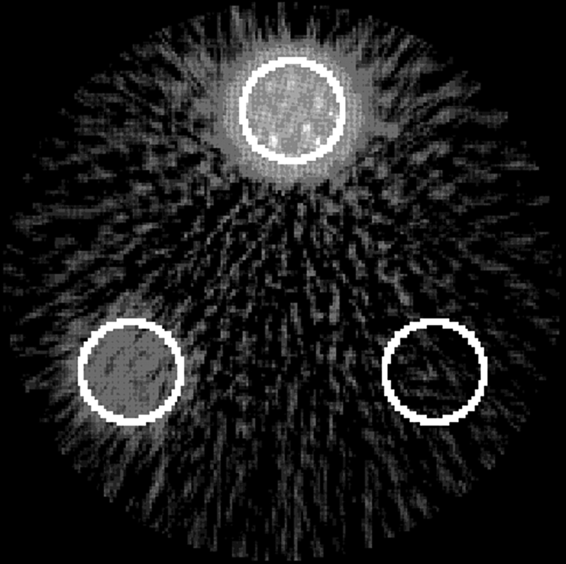
- FIGURE 1.
Cross-section of phantom reconstructed with FBP. The 3 ROIs are outlined in white.
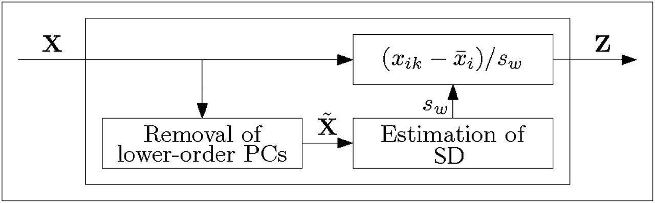
- FIGURE 2.
Schematic of higher-order PC noise prenormalization.
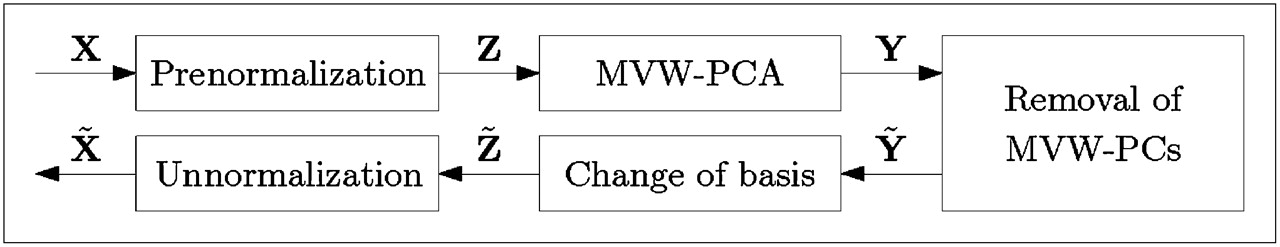
- FIGURE 3.
Removal of masked volumewise (MVW) PCs from X.
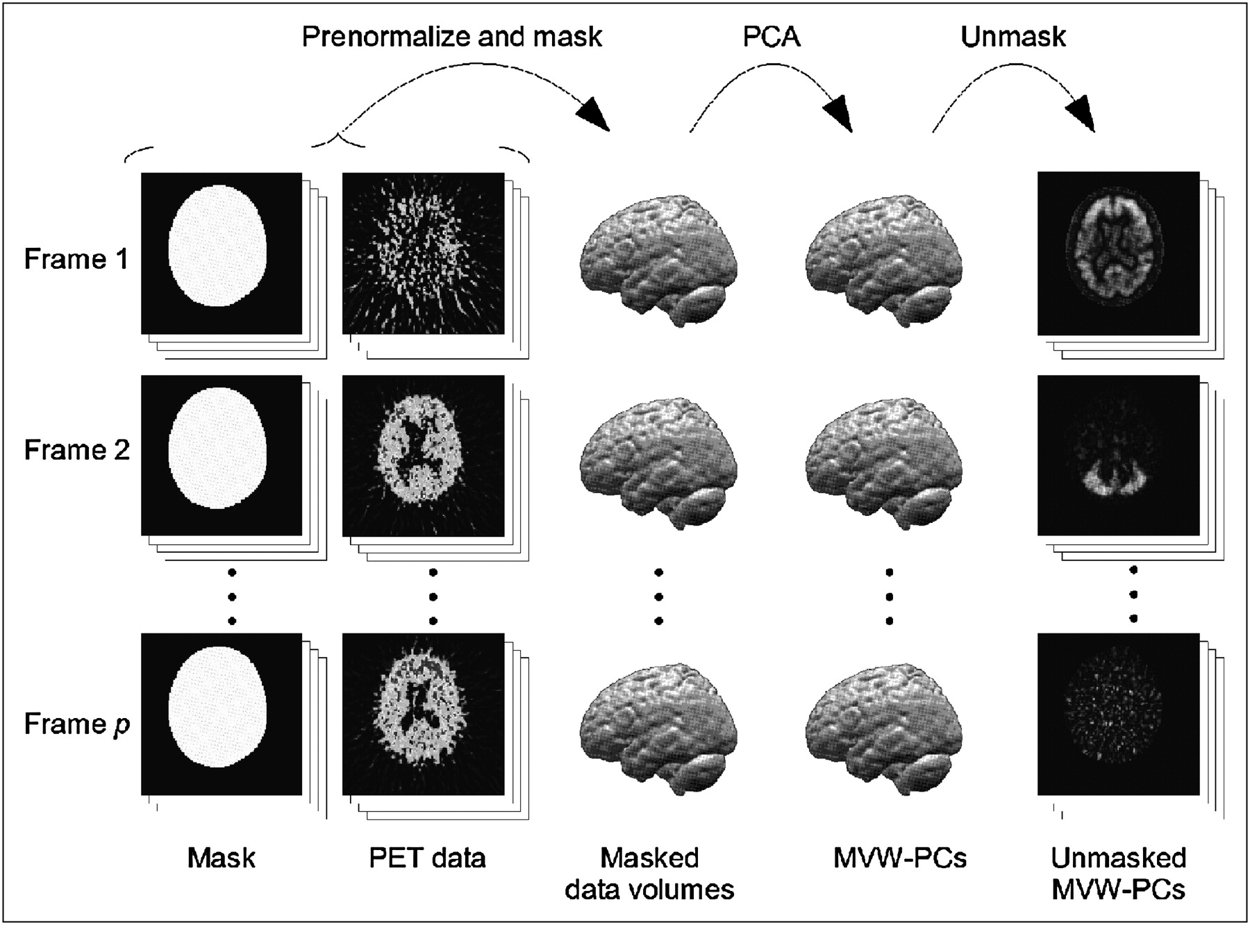
- FIGURE 4.
Illustration of masked volumewise PCA procedure.
- FIGURE 5.
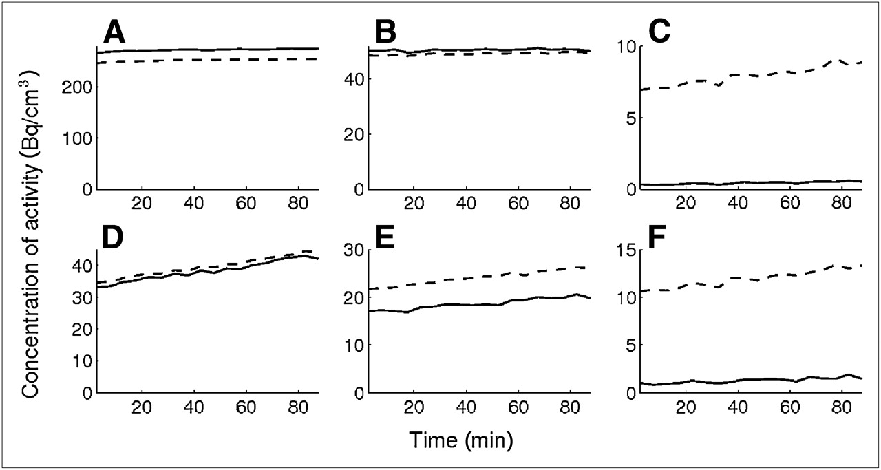
Arithmetic mean (A–C) and sample SD (D–F) for FBP (dashed line) and OSEM (solid line) over time in the 3 ROIs: high-activity ROI (A and D), low-activity ROI (B and E), and no-activity ROI (C and F).
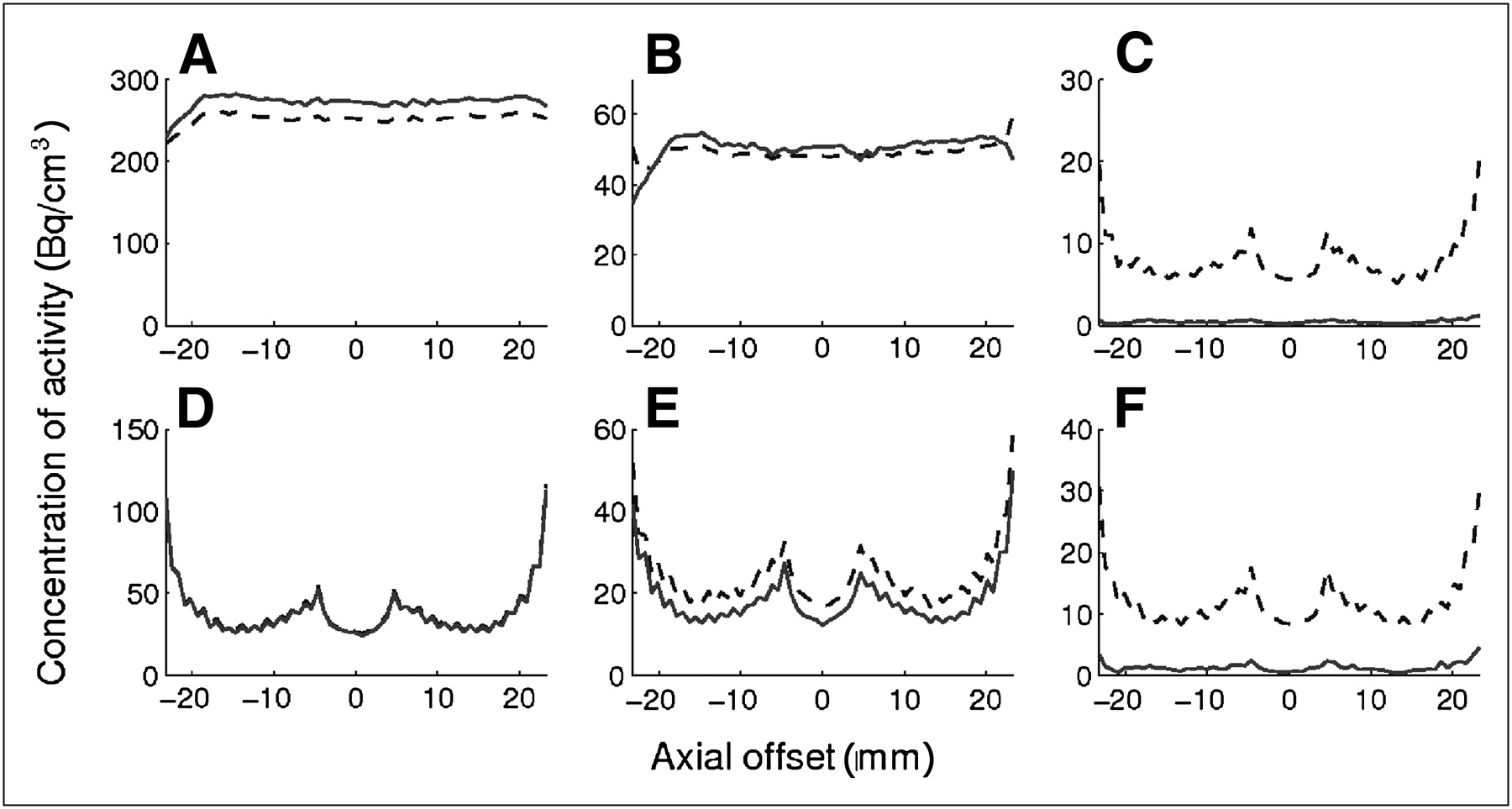
- FIGURE 6.
Arithmetic mean (A–C) and sample SD (D–F) for FBP (dashed line) and OSEM (solid line) for different axial offsets in the 3 ROIs: high-activity ROI (A and D), low-activity ROI (B and E), and no-activity ROI (C and F).
- FIGURE 7.
Scaled plots of SD used by higher-order PC, from full-body study using 18F-FDG (A) and brain study using 11C-Pittsburgh compound B (B). Higher-order PC 1 (□), higher-order PC 2 (⋄), and higher-order PC 3 (▵Δ) are compared with background noise (○). Index of higher-order PC corresponds to number of removed masked volumewise PCs in prenormalization step.
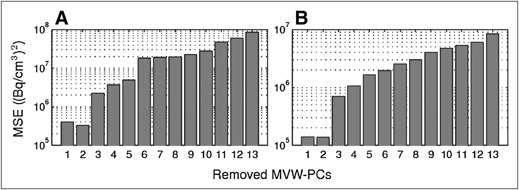
- FIGURE 8.
MSE between SD of background noise and scaled SD used by higher-order PC noise prenormalization: result from full-body study (A) and result from brain study (B). Logarithmic scale is used.
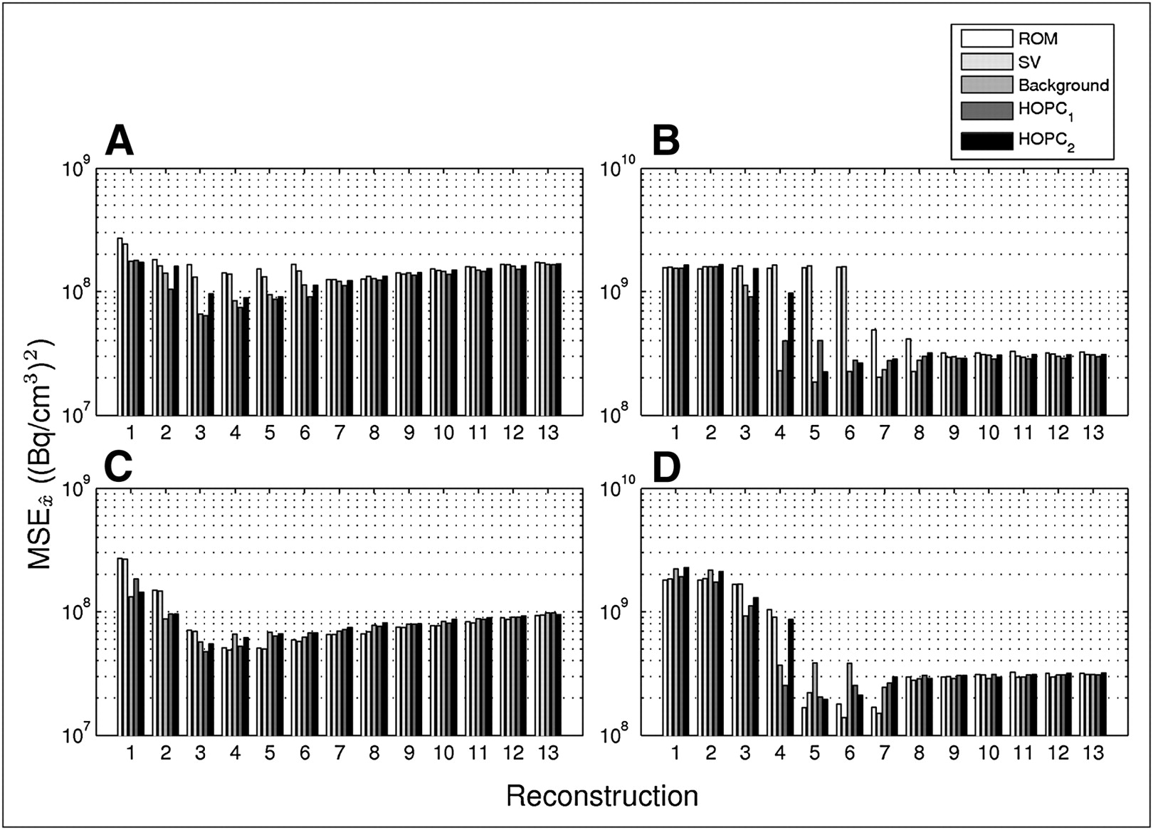
- FIGURE 9.
MSE in different reconstructions for different prenormalizations, compared with optimal signal
 . FBP is used in A and B whereas OSEM is used in C and D. Measurements are from adrenal gland VOI (A and C) and stomach VOI (B and D). Index of higher-order PC corresponds to number of removed masked volumewise PCs in prenormalization step. Logarithmic scale is used.
 - FIGURE 10.
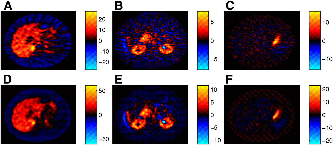
Slices from first 3 masked volumewise PCs from datasets reconstructed with FBP (A–C) and OSEM (D–F). Most adrenal gland and general tracer behavior is described by masked volumewise PC 1 (A and D). Masked volumewise PC 2 (B and E) describes early tracer accumulation in kidneys, whereas tracer concentration in stomach can be separated with masked volumewise PC 3 (C and F).
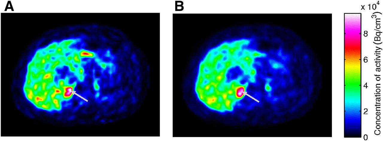
- FIGURE 11.
One slice in original (A) and dimension-reduced (B) dataset showing adrenal gland (arrows).


Jump to section
Related Articles
Cited By...
- No citing articles found.