


Article Figures & Data
Figures
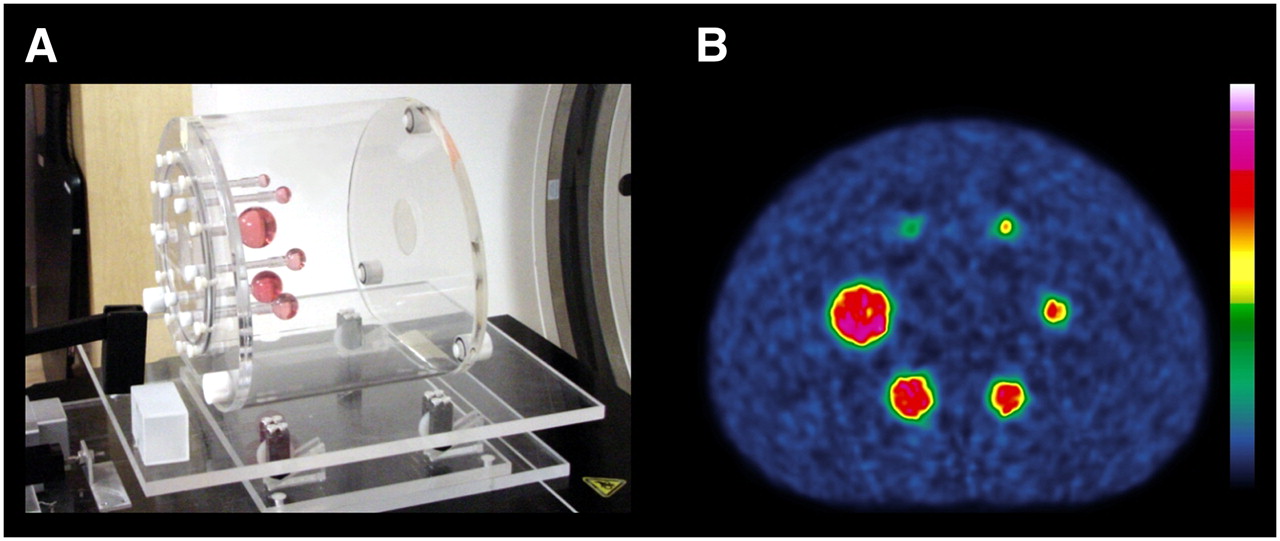
- FIGURE 1.
(A) NEMA/IEC 2001 phantom (Data Spectrum) on plastic platform. (B) PET mid-transaxial slice through static acquisition in 2D mode with 9.7 S/B ratio.
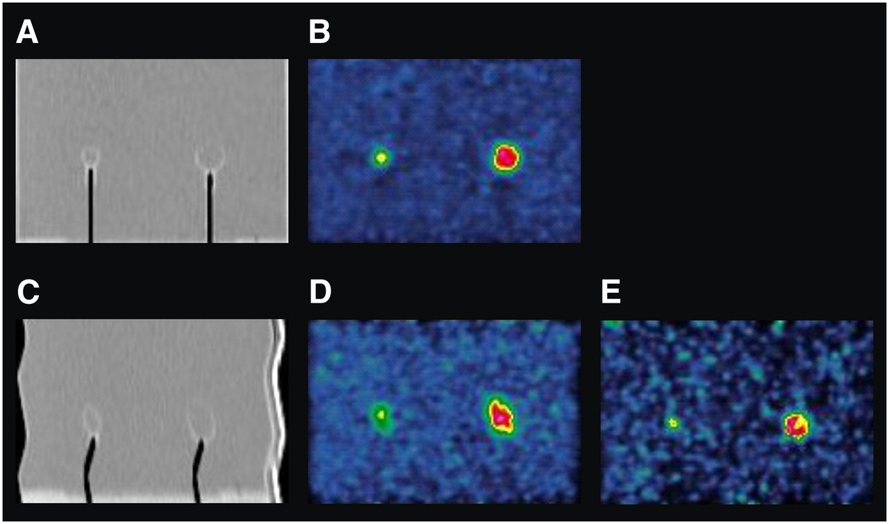
- FIGURE 2.
(A and B) Sagittal images acquired through 13- and 22-mm spheres with phantom at rest in 2D mode and with 9.7 S/B ratio: CT (A) and PET (B). (C–E) Images acquired with same phantom in motion: CT (C), nongated PET (D), and gated (1 of 10 time bins) PET (E). All PET images have been scaled relative to each other. On CT images, air in stems of spheres appears black.
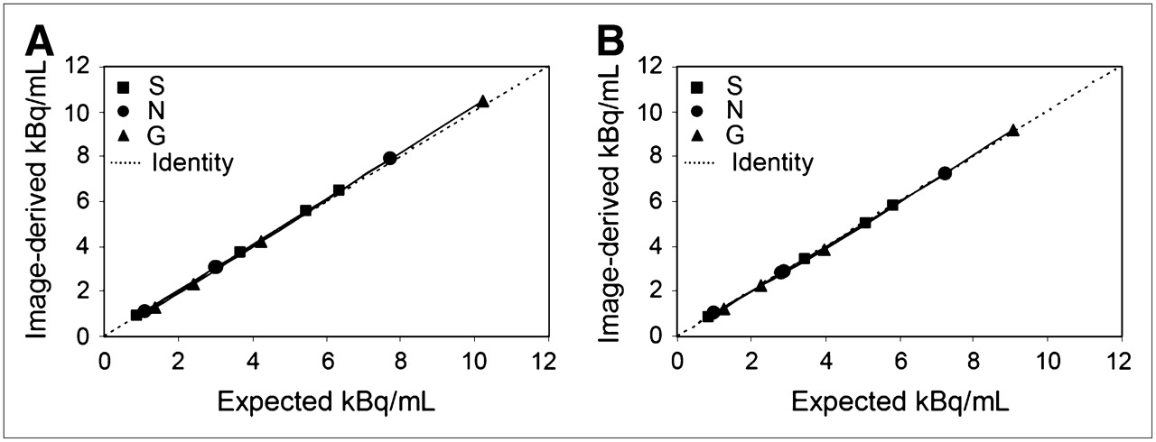
- FIGURE 3.
Image-derived and expected RC in phantom background, in 2D (A) and 3D (B) modes of PET acquisition. The 3 types of scans were static (S), motion nongated (N), and gated (G), and each series was fitted with linear regression.
- FIGURE 4.
Comparison of image-derived and expected sphere RC for data analysis methods: maximum pixel (A), mean pixel (B), and threshold (C). Data are for largest sphere (37 mm) in 2D mode of PET acquisition for each scan type—static (S), motion nongated (N), and gated (G)—and each series was fitted with linear regression.
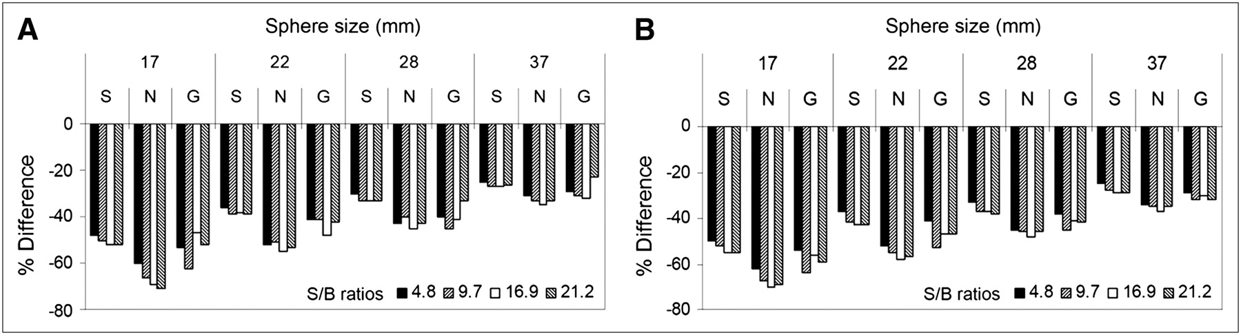
- FIGURE 5.
Underestimation of image-derived RC from expected value, in 2 modes of PET acquisition: 2D (A) and 3D (B). Data are shown for all sphere sizes (17, 22, 28, and 37 mm), S/B ratios (4.8, 9.7, 16.9, and 21.2), and scan types (static [S], motion nongated [N], and gated [G]).
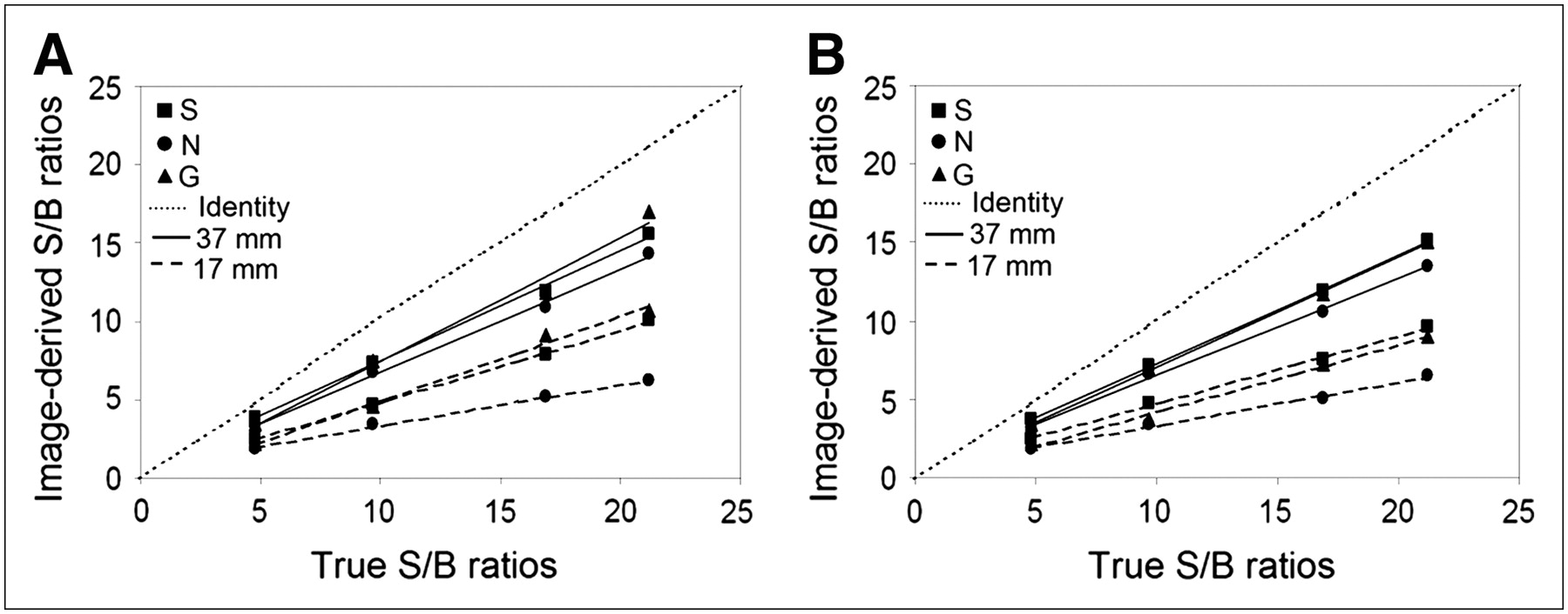
- FIGURE 6.
Relationship between image-derived and true S/B ratios in 2D (A) and 3D (B) modes of PET acquisition. Data are shown for 2 sphere sizes (17 and 37 mm) and all scan types (static [S], motion nongated [N], and gated [G]), and each series was fitted with linear regression.
- FIGURE 7.
Relationship between number of voxels and RC in static (A) and gated (B) scan types. Data are from 56-mm volume of interest around largest sphere (37 mm) in S/B ratio of 9.7 and in 2D mode of PET acquisition. Indicated on histograms are measured maximum, mean, and background values of RC from within volume of interest; also marked is expected RC in sphere.


Tables
Site kBq/mL SUV or S/B Centimeters Patient (tumors) Mean 36.5 20.7 4.1 Range 7.4–82 2.9–57 1.7–8 Phantom (spheres) Mean 39.1 13.2 2.1 Range 19–62 4.8–21 1.0–3.7


