


Article Figures & Data
Figures
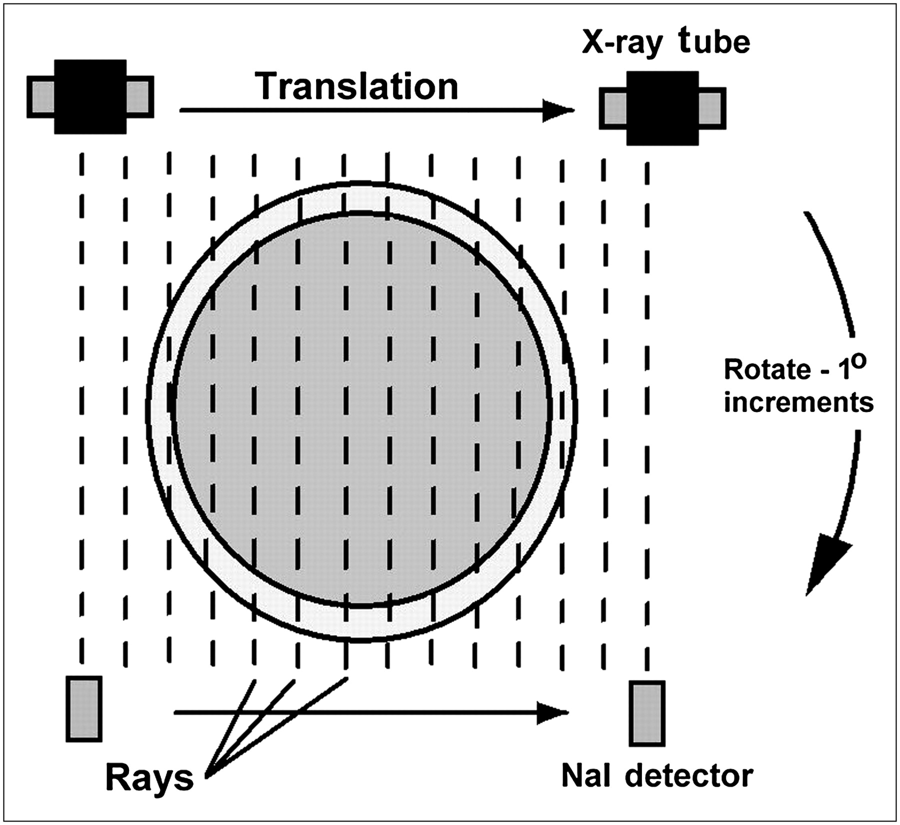
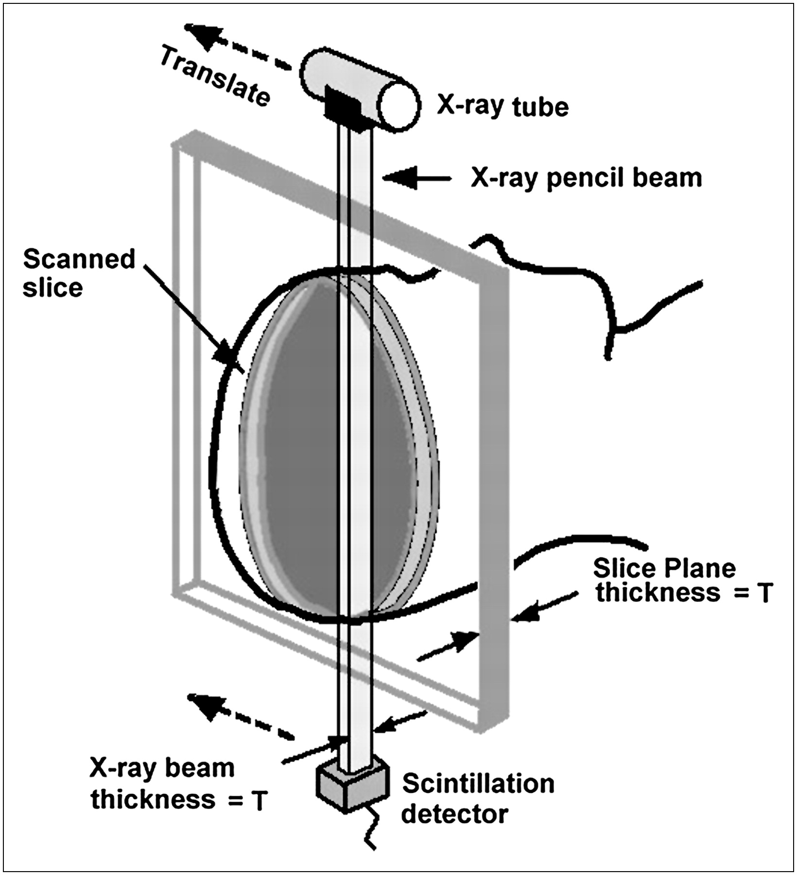
- FIGURE 1.
CT arrangement. Axial slice through patient is swept out by narrow (pencil-width) x-ray beam as linked x-ray tube–detector apparatus scans across patient in linear translation. Translations are repeated at many angles. Thickness of narrow beam is equivalent to slice thickness.
- FIGURE 2.
x-Ray transmission measurements. Measurements are obtained at many points during translation motion of tube and detector. x-Ray path corresponding to each measurement is designated a ray, and set of rays measured during translation is designated a view. Views are collected at many angles (in 1° increments in this example) to acquire sufficient data for image reconstruction.
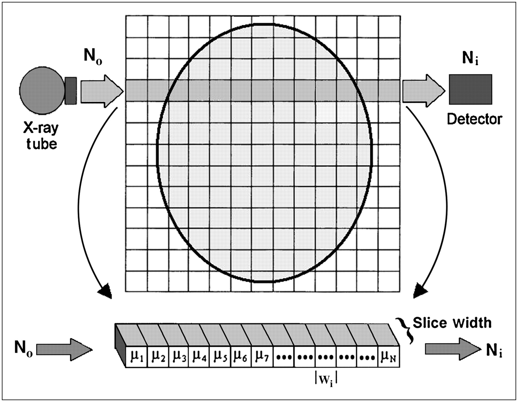
- FIGURE 3.
Reconstruction matrix. Hounsfield envisioned scanned slice as being composed of matrix of small boxes of tissue called voxels, each with attenuation coefficient μ. x-Ray transmission measurements (Ni) can be expressed as sum of attenuation values occurring in voxels along path of ray for Ni.
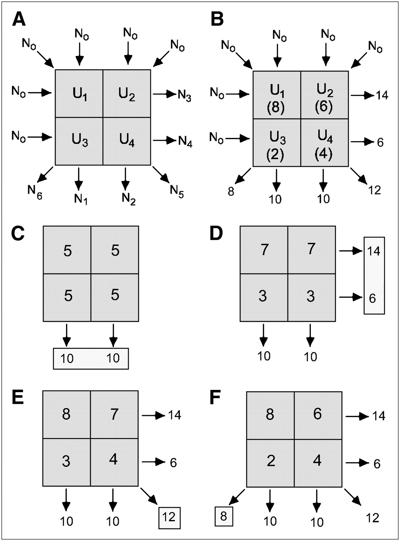
- FIGURE 4.
ART. (A) ART algorithm for 4-voxel “patient.” (B) Attenuation measurements. (C) Starting estimate is constructed by dividing measurements from first view equally along their ray paths. (D–F) This estimate is iteratively adjusted to match measurements for each consecutive view, stopping when transmission measurements predicted by current estimate match all actual measurements to within some preset tolerance.
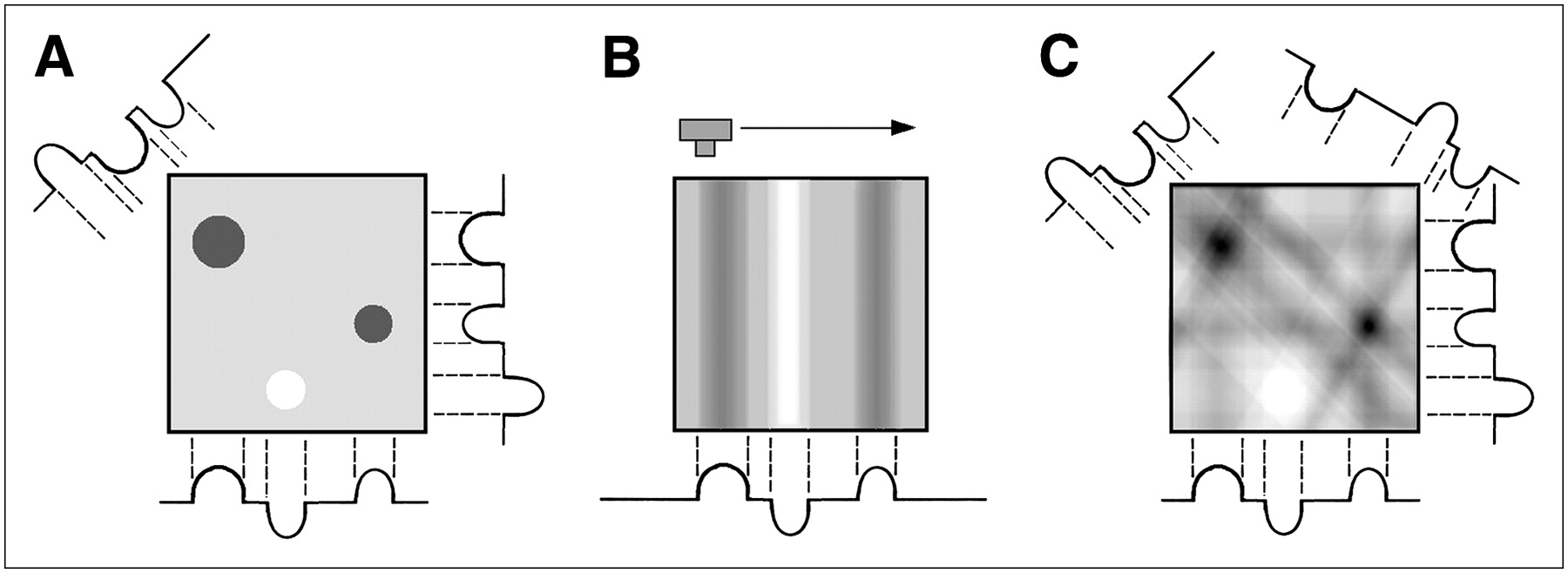
- FIGURE 5.
(A) Backprojection reconstruction for simple phantom containing 3 objects with different attenuation values. (B) For each view, attenuation values are simply divided evenly along their ray paths. Summing backprojected views from several angles builds image. (C) Four views of phantom are summed. Although this method is efficient, images reconstructed with backprojection exhibit considerable blurriness.
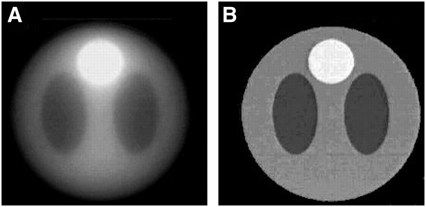
- FIGURE 6.
FBP. Mathematic phantom image reconstructed without (A) and with (B) filtering. FBP effectively reconstructs high-quality images. Adapted from S. Napel.
- FIGURE 7.
Second-generation data collection. (A) Transmissions of multiple narrow beams (3, in this case) were simultaneously acquired by multiple detectors during each translation. (B–D) Small angle between narrow beams allowed each detector to acquire complete separate view at different angle. Number of required translations was correspondingly reduced by factor of 1/(number of detectors).
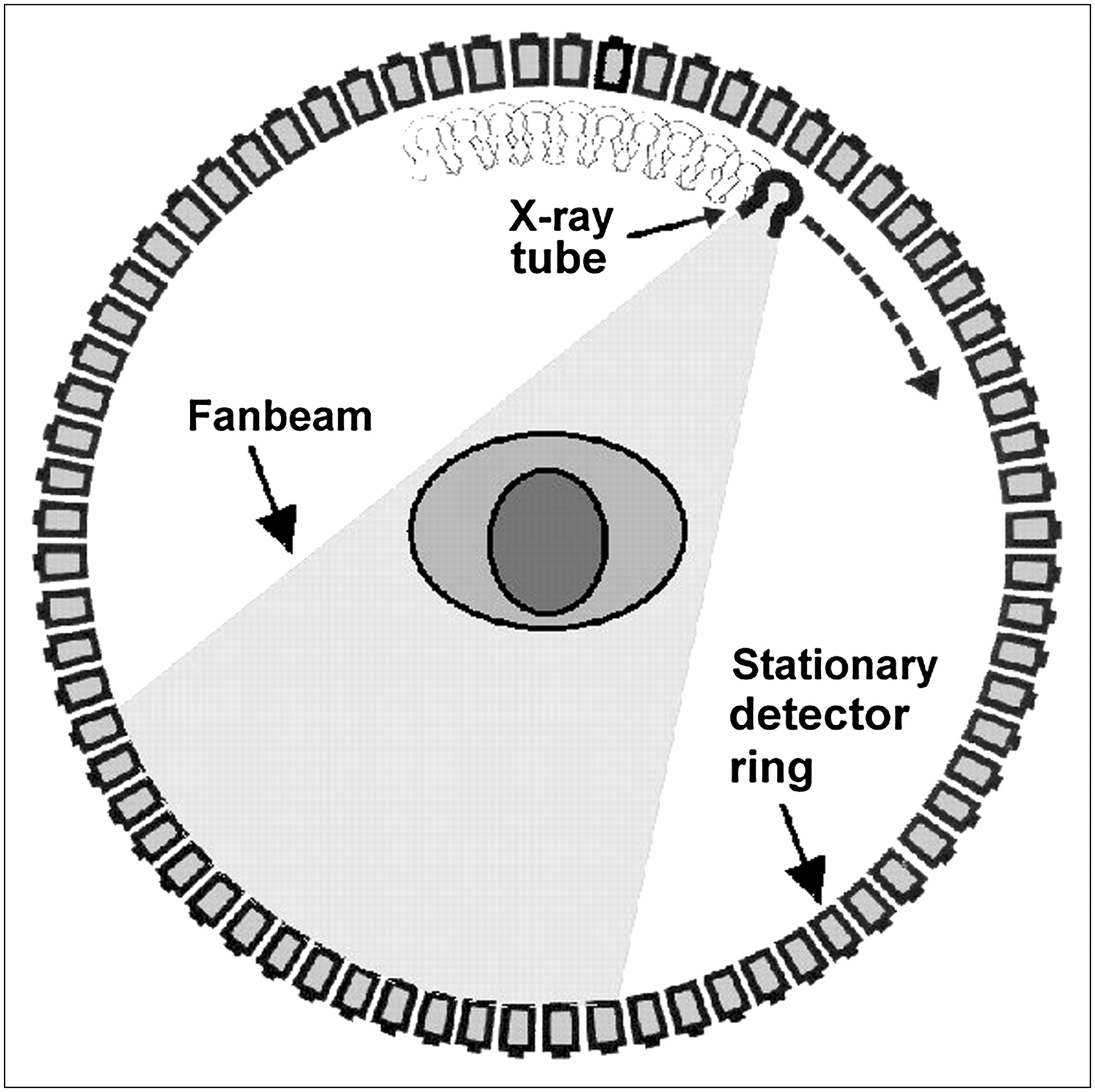
- FIGURE 8.
Third-generation geometry. Time-consuming and mechanically complex translation motion was eliminated by opening x-rays into fanbeam. Large array of detectors measured data across width of fan. Tube and detectors were rigidly linked and underwent single rotational motion.
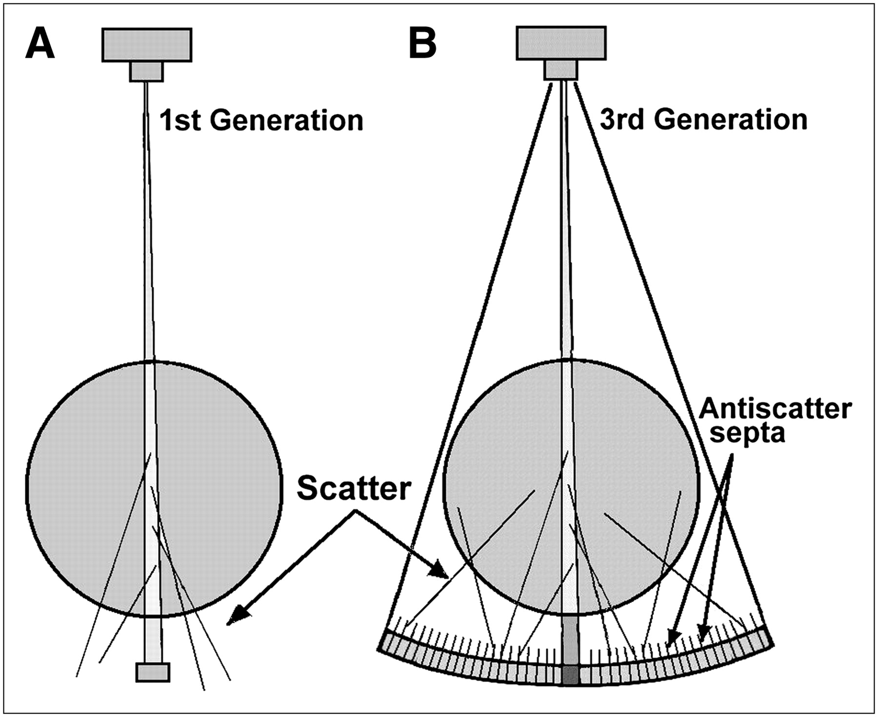
- FIGURE 9.
Scatter with CT fanbeam. Use of fanbeam increases scatter production at any moment (more tissue is irradiated), and more scatter can reach detectors. Amount of scatter produced is still much smaller than that in radiography because fanbeam is only ∼1 cm thick. Scatter can be eliminated in third-generation CT with scatter removal septa, which act like nearly ideal grid. (A) First-generation CT. (B) Third-generation CT.
- FIGURE 10.
Fourth-generation scan geometry. Fixed detector ring in original design was quite large, because tube rotated inside ring. Later designs moved tube outside ring and tilted ring out of way of x-ray beam as x-ray tube swept by.
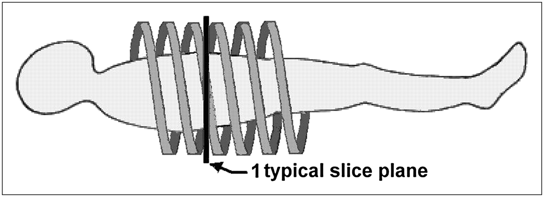
- FIGURE 11.
Helical CT. Improved body CT was made possible with advent of helical CT (or spiral CT). Patient table is moved smoothly through gantry as rotation and data collection continue. Resulting data form spiral (or helical) path relative to patient; slices at arbitrary locations may be reconstructed from these data.
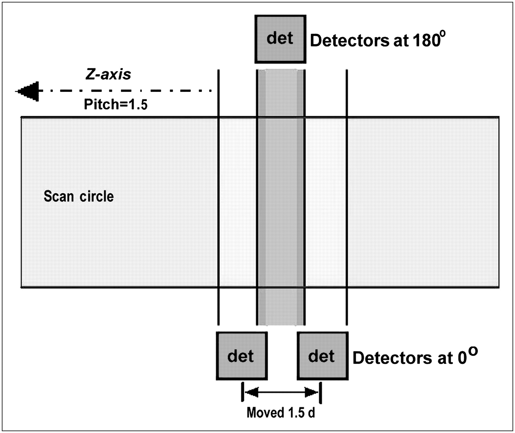
- FIGURE 12.
Helical CT sample spacing and interpolation. If data for desired slice of thickness d (dark gray bar in figure) are interpolated between equivalent rays from adjacent helical rotations (loops) with pitch of 1.5, samples will be 1.5 d apart along z-axis (e.g., 10.5 mm apart for 7-mm thickness). Larger spacing means greater chance that interpolated estimate is in error. If 180°-opposed rays are included, measurements average half as far apart (and are more likely to actually lie within slice). det = detector.


Additional Files
Supplemental Data
Files in this Data Supplement:


Jump to section
- Article
- Abstract
- LIMITATIONS OF CONVENTIONAL RADIOGRAPHY
- BASIC PRINCIPLES OF CT: FIRST GENERATION OF CT
- CT IMAGE RECONSTRUCTION
- CT IMAGE PRESENTATION
- FIRST-GENERATION EMI CT SCANNER
- REDUCING SCAN TIME: SECOND GENERATION OF CT
- THIRD GENERATION OF CT
- CT PERFORMANCE CRITERIA
- FOURTH-GENERATION SCANNERS
- ELECTRON-BEAM CT (EBCT)
- SLIP RING SCANNERS AND HELICAL CT
- CT FLUOROSCOPY
- APPENDIX
- Footnotes
- References
- Figures & Data
- Supplemental
- Info & Metrics
Related Articles
Cited By...
- Automatic semantic segmentation of the osseous structures of the paranasal sinuses
- High Accuracy Machine Learning Model for Sarcopenia Severity Diagnosis based on Sit-to-stand Motion Measured by Two Micro Motion Sensors
- High Accuracy Machine Learning Model for Sarcopenia Severity Diagnosis based on Sit-to-stand Motion Measured by Two Micro Motion Sensors
- Sensitivity of modern multislice CT for subarachnoid haemorrhage at incremental timepoints after headache onset: a 10-year analysis
- Radiation Protection in Computed Tomography
- Accurate Assessment of Aortic Stenosis: A Review of Diagnostic Modalities and Hemodynamics
- Comparative assessment of three image reconstruction techniques for image quality and radiation dose in patients undergoing abdominopelvic multidetector CT examinations
- Neuroradiology Back to the Future: Head and Neck Imaging
- Neuroradiology Back to the Future: Spine Imaging
- Assessment of Patient Exposure to X-Radiation from SPECT/CT Scanners
- Principles of CT: Multislice CT
- Principles of CT: Radiation Dose and Image Quality