


Abstract
99mTc-Dimercaptosuccinic acid (DMSA) scintigraphy is a frequently used diagnostic test to assess the presence and severity of cortical damage. The aim of this study is to investigate the variability in the interpretation of 99mTc-DMSA scans, evaluate the usefulness of oblique images, and assess their impact on scan interpretation. Methods: Two experienced nuclear medicine physicians independently interpreted 100 99mTc-DMSA scans (197 kidneys) 4 times. Interpretation was twice based on posterior projection images and twice based on posterior and posterior oblique projection images. For each kidney, the observers had to choose between the following results: normal, abnormal, and indeterminate. The indices of variability used were the percentage of agreement, κ-statistic, and marginal homogeneity. Results: Intraobserver and interobserver reproducibility (κ-values) varied between 0.683 and 0.708 and between 0.609 and 0.671, respectively, for posterior images. Disagreement in 99mTc-DMSA scan interpretation occurred in 18% of kidneys within observers and in 21% of kidneys between observers when only posterior images were used. Oblique views changed the interpretation in 14% and 11.5% of kidneys for the first and second observers, respectively. The use of oblique views increased the agreement rate within and between observers (κ-values, 0.725–0.812 and 0.768–0.732, respectively; mean agreement, 86.5 and 87.25, respectively). Conclusion: Oblique views were found useful in approximately 13% of kidneys and affected inter- and intraobserver variability. Our results suggest that oblique views should be used routinely in children with clinically suspected urinary tract infection to reliably interpret images.
Scintigraphy with 99mTc-dimercaptosuccinic acid (DMSA) is used to identify children who have renal cortical damage, and clinical decisions are often influenced by the scan results (1–5). Although procedure guidelines suggest that optimal imaging should include right and left posterior oblique views in addition to posterior images, the utility of oblique views has been questioned (6). There are also not enough reliable data in the literature on the utility of oblique views in the interpretation of scintigraphic reports.
This work was undertaken to evaluate the level of inter- and intraobserver variability in 99mTc-DMSA scintigraphy and to identify the impact of posterior oblique views on scan interpretation in the pediatric population.
MATERIALS AND METHODS
A total of 197 kidneys, which were randomly selected from our database, were retrospectively evaluated. Patient age varied between 3 mo and 17 y. All scans were displayed on gray-scale, clear-based, hard-copy radiographic films. Static renal scintigraphy was performed 3 h after the administration of 99mTc-DMSA. The dose was scaled on a body weight basis using an adult dose of 185 MBq (5 mCi) (range, 37–185 MBq [1–5 mCi]). Scintigraphic images (128 × 128 matrix) were acquired over 5 min from posterior and posterior oblique projections using a γ-camera equipped with a high-resolution collimator and using a 1.23 zoom factor.
Patients with congenital malformations, renal failure, and low-quality images were excluded from the study.
All films were viewed by 2 experienced nuclear medicine specialists. Apart from being informed that urinary tract infection was the indication for 99mTc-DMSA scintigraphy, the nuclear medicine physicians were given no other clinical information. Films were read on 4 separate occasions—all independently to ensure that no bias was introduced. The first interpretation was based on the posterior images. The second was based on both the posterior and the posterior oblique images. The third, 2 wk later, was a reinterpretation of the same cases in the same sequence. Images were classified as normal, abnormal, or indeterminate (Figs. 1–3⇓). 99mTc-DMSA scan findings were defined as abnormal if a defect in renal contour or an area of photon deficiency was present in the renal cortex.
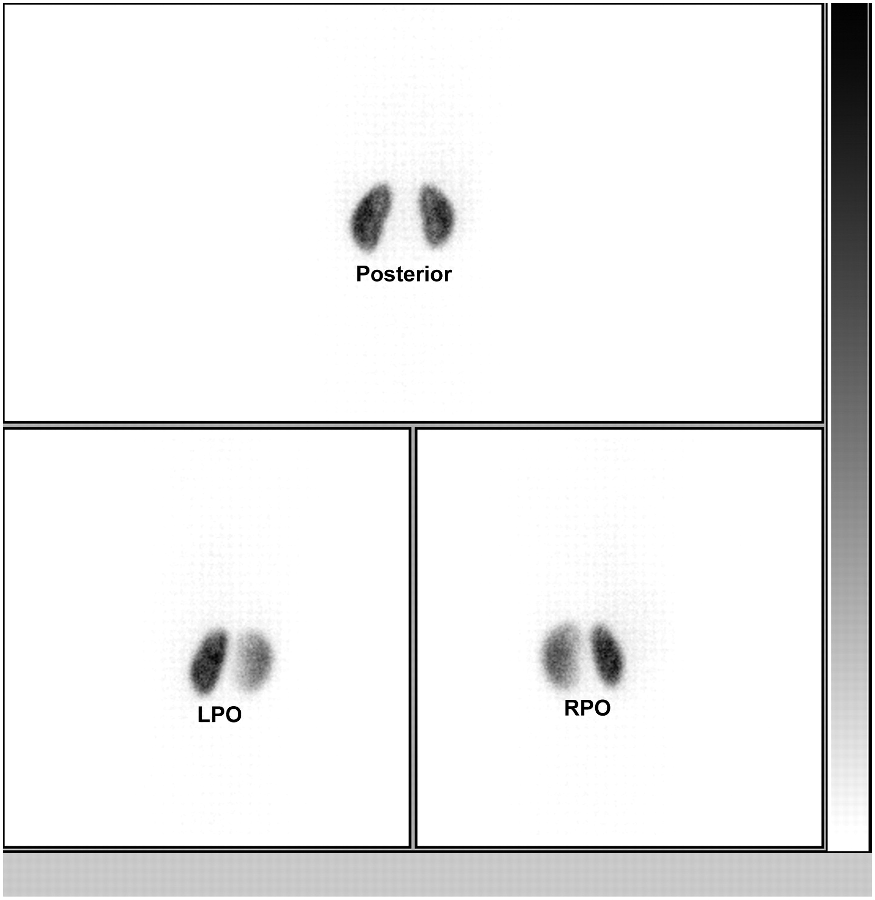
Normal findings on planar 99mTc-DMSA scintigraphy. LPO = left posterior oblique; RPO = right posterior oblique.

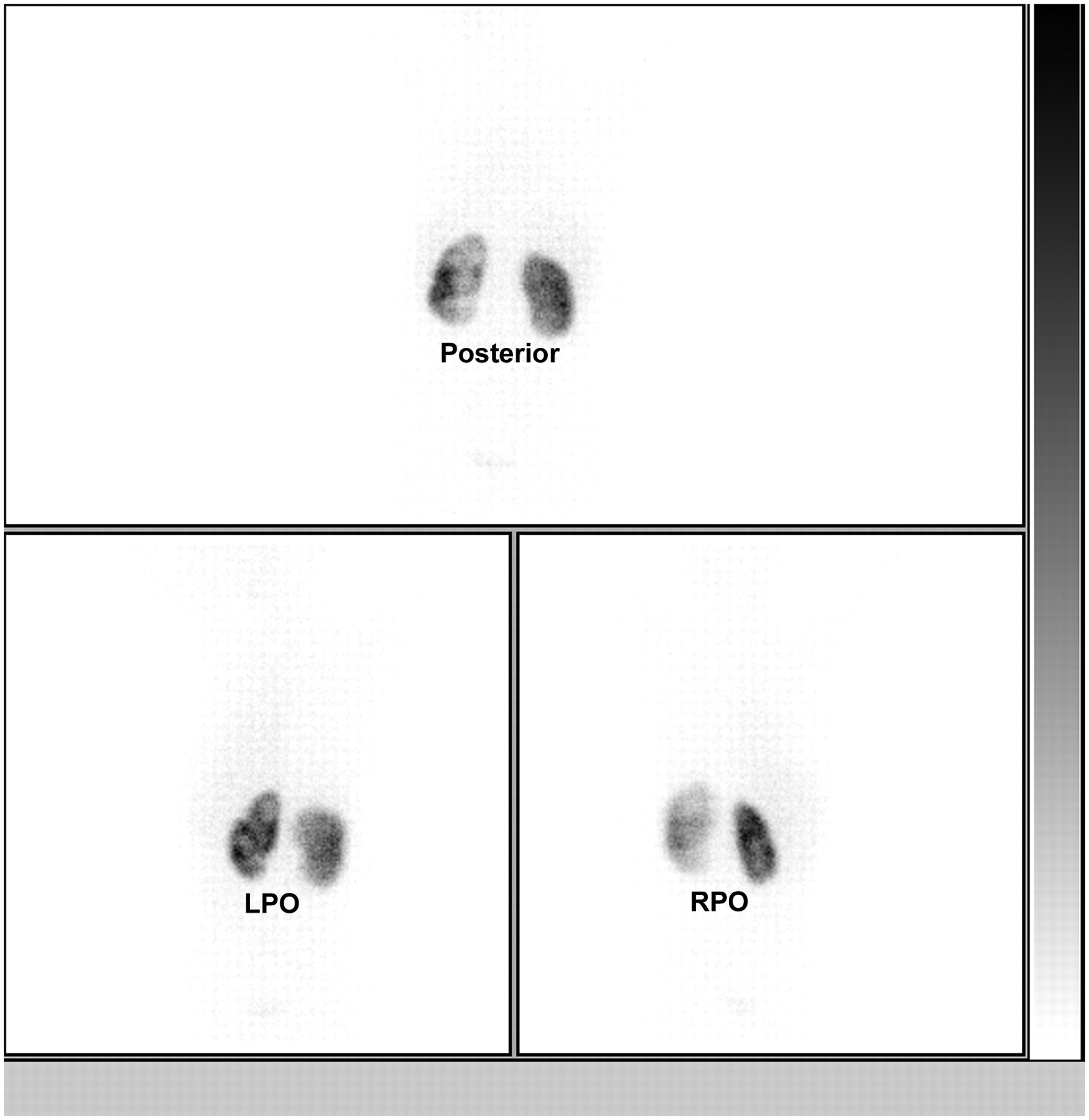
Abnormal findings on 99mTc-DMSA scintigraphy. Posterior, left posterior oblique (LPO), and right posterior oblique (RPO) images reveal slight enlargement of left kidney, with cortical defects. Although right kidney appears normal on posterior image, cortical defect is seen in lower pole on RPO view.

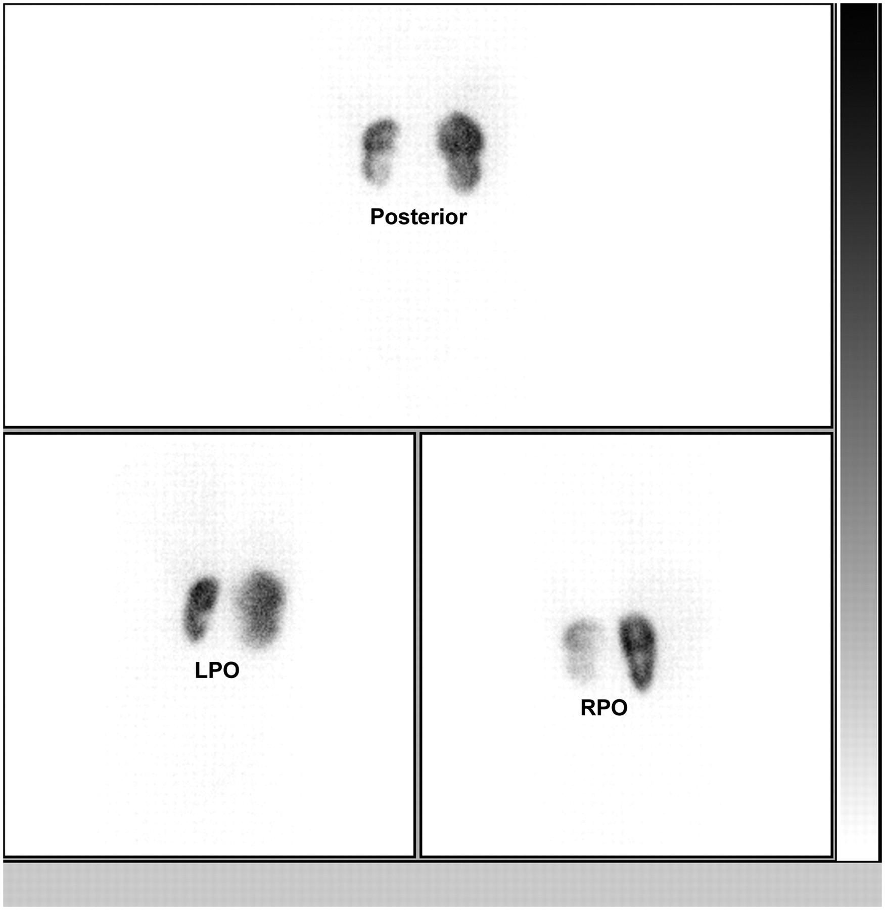
99mTc-DMSA scintigraphy showing small left kidney with parenchymal scarring. Right side is considered indeterminate on posterior and posterior oblique views because of midzone impression seen in upper pole–lower pole junction suggestive of duplex kidney. Because no evidence of duplex kidney was present, right kidney was considered indeterminate for acute pyelonephritis or cortical scar. LPO = left posterior oblique; RPO = right posterior oblique.
Variability in the interpretation of 197 kidneys was assessed from the readings of 2 nuclear medicine physicians. The percentage of agreement and κ-statistics were used to measure inter- and intraobserver variability. In all calculations, reproducibility was estimated on the basis of complete agreement between observers where the number of possible answers was 3 (normal, abnormal, and indeterminate). The percentage of agreement was defined as an index that gave a good measure of how well any posterior distribution agreed with the prior distribution. This rather simple statistic has the merit of being easy to understand. A measure generally thought to be more robust than the simple percentage agreement calculation is Cohen's κ-coefficient (7), because it adjusts for agreement attributable to chance and can range from −1 to +1, the latter representing perfect agreement. The κ-statistic was used to measure interobserver variability (inconsistency of interpretation among different physicians) and intraobserver variability (the failure of a physician to be consistent in independent readings of the same scan). In practical terms, values above 0.61 were taken to indicate a high standard of agreement and a level at which the data could be accepted as reproducible (8).
Scan results were compared using a marginal homogeneity test for matched pairs. A P value of less than 0.05 was considered significant.
RESULTS
Table 1 shows the results given by 2 observers for 197 kidneys. Adding oblique images changed the results for the first and second observers in 32 kidneys (16%) and 27 kidneys (14%), respectively, in the first interpretation and in 24 kidneys (12%) and 20 kidneys (10%), respectively, in the second interpretation. The proportion of indeterminate results was significantly reduced when oblique images were used for interpretation (P = 0.023, P = 000, P = 0.036), except for the second observer in the second interpretation (P = 0.307).
Impact of Oblique Images for DMSA Scan Interpretation
The κ-statistics for inter- and intraobserver variability is given in Tables 2 and 3. The reproducibility of image interpretation was good and was slightly better when posterior oblique images were used. When only posterior images were used, complete agreement was achieved in 151 kidneys (77%) (κ = 0.609) in the first interpretation and in 159 kidneys (81%) (κ = 0.671) in the second. When both the posterior and the posterior oblique views were used, complete agreement was achieved in 173 kidneys (88%) (κ = 0.768) and 167 kidneys (85%) (κ = 0.732) in the first and second interpretations, respectively.
κ-Statistics for Intraobserver Variability of DMSA Scintigraphy
κ-Statistics for Interobserver Variability of DMSA Scintigraphy
DISCUSSION
Although renal cortical scintigraphy with 99mTc-DMSA is widely used, primarily for the evaluation of renal parenchymal damage, conflicting opinions have been expressed about its reproducibility (9–13). Some investigators advocate the addition of pinhole images and SPECT (14); however, motion artifacts due to the long acquisition time may be a problem (15). Recent results demonstrated that high-resolution planar imaging, pinhole imaging, and SPECT can provide similar results in demonstrating renal cortical defects (16).
This study demonstrated a high level of agreement in the interpretation of 99mTc-DMSA scans showing urinary tract infection. The κ-values varied between 0.683 and 0.708 for intraobserver variability and between 0.609 and 0.671 for interobserver variability, which was classified as good when only posterior views were used. With the addition of oblique projections, the κ-values improved slightly (0.725–0.812 and 0.768–0.732, respectively). A higher level of intraobserver consistency suggested that observers are of clearer mind about the definition of renal parenchymal damage. Oblique views, when used with posterior images, provide a firm basis for clinical decision making, especially in patients who have photon-deficient areas due to dilated calices or questionable contours. The interpretation of 99mTc-DMSA scintigraphy from only the posterior view, compared with oblique views also, will underestimate the number of normal scans. This effect is relevant in patients with urinary tract infection, because a strategy to perform voiding cystourethrography in only patients with renal lesions has been proposed (5).
The degree of reproducibility may also depend on the severity and extent of lesions. Abnormalities seen during the acute phase of infection may be more striking and less challenging. In this study, the scans were randomly selected and represented the routine clinical workload. Areas of parenchymal damage appeared as hypoactive, with or without deformity of the contours. Difficulties remain in the interpretation of subtle cases and are related to the lack of an established definition for anatomic variants.
CONCLUSION
These results indicate that technically well-performed studies on a planar high-resolution collimator can yield valuable information in children with either clinically suspected acute pyelonephritis or renal scarring. In our series, at least 10% of patients with urinary tract infection could have been sent for voiding cystourethrography on the basis of equivocal 99mTc-DMSA results. Although this study was limited by the lack of a standard of reference against which to judge the performance of both views, our results indicated that the lesions were more confidently diagnosed on oblique images.
Footnotes
-
COPYRIGHT © 2007 by the Society of Nuclear Medicine, Inc.
References
- Received for publication August 27, 2006.
- Accepted for publication February 12, 2007.


