
Main menu

  • Home
  • Content
    • Current
      • JNMT Supplement
    • Ahead of print
    • Past Issues
    • Continuing Education
    • JNMT Podcast
    • SNMMI Annual Meeting Abstracts
  • Subscriptions
    • Subscribers
    • Rates
    • Journal Claims
    • Institutional and Non-member
  • Authors
    • Submit to JNMT
    • Information for Authors
    • Assignment of Copyright
    • AQARA Requirements
  • Info
    • Reviewers
    • Permissions
    • Advertisers
    • Corporate & Special Sales
  • About
    • About Us
    • Editorial Board
    • Contact Information
  • More
    • Alerts
    • Feedback
    • Help
    • SNMMI Journals
  • SNMMI
    • JNMT
    • JNM
    • SNMMI Journals
    • SNMMI

User menu

  • Subscribe
  • My alerts
  • Log in
  • My Cart

Search

  • Advanced search
Journal of Nuclear Medicine Technology
  • SNMMI
    • JNMT
    • JNM
    • SNMMI Journals
    • SNMMI
  • Subscribe
  • My alerts
  • Log in
  • My Cart
Journal of Nuclear Medicine Technology

Advanced Search

  • Home
  • Content
    • Current
    • Ahead of print
    • Past Issues
    • Continuing Education
    • JNMT Podcast
    • SNMMI Annual Meeting Abstracts
  • Subscriptions
    • Subscribers
    • Rates
    • Journal Claims
    • Institutional and Non-member
  • Authors
    • Submit to JNMT
    • Information for Authors
    • Assignment of Copyright
    • AQARA Requirements
  • Info
    • Reviewers
    • Permissions
    • Advertisers
    • Corporate & Special Sales
  • About
    • About Us
    • Editorial Board
    • Contact Information
  • More
    • Alerts
    • Feedback
    • Help
    • SNMMI Journals
  • Watch or Listen to JNMT Podcast
  • Visit SNMMI on Facebook
  • Join SNMMI on LinkedIn
  • Follow SNMMI on Twitter
  • Subscribe to JNMT RSS feeds
OtherIMAGING

Substitution of Oral 18F-FDG for Intravenous 18F-FDG in PET Scanning

Narendra Nair, Archi Agrawal and Rajnath Jaiswar
Journal of Nuclear Medicine Technology June 2007, 35 (2) 100-104; DOI: https://doi.org/10.2967/jnmt.106.036129
Narendra Nair
  • Find this author on Google Scholar
  • Find this author on PubMed
  • Search for this author on this site
Archi Agrawal
  • Find this author on Google Scholar
  • Find this author on PubMed
  • Search for this author on this site
Rajnath Jaiswar
  • Find this author on Google Scholar
  • Find this author on PubMed
  • Search for this author on this site
  • Article
  • Figures & Data
  • Info & Metrics
  • PDF
Loading

Article Figures & Data

Figures

  • FIGURE 1. 
    • Download figure
    • Open in new tab
    • Download powerpoint
    FIGURE 1. 

    Patient 1, 59-y-old man with right lung carcinoma. Both intravenous (A) and oral (B) studies show right-mid-zone mass and right hilar lymph node. MIP = maximum intensity projection.
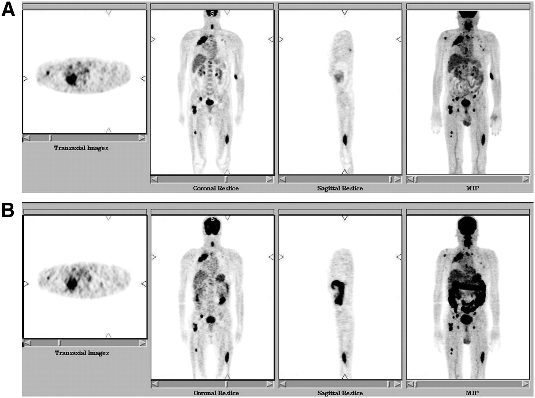
  • FIGURE 2. 
    • Download figure
    • Open in new tab
    • Download powerpoint
    FIGURE 2. 

    Patient 2, 60-y-old man with right lung carcinoma and multiple skeletal metastases. Both intravenous (A) and oral (B) studies show right-upper-lobe lesion, with metastases in vertebrae, right shoulder, pelvic bone, and both femurs. MIP = maximum intensity projection.
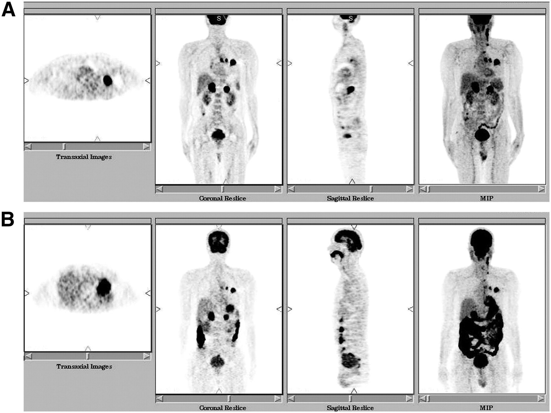
  • FIGURE 3. 
    • Download figure
    • Open in new tab
    • Download powerpoint
    FIGURE 3. 

    Patient 3, 47-y-old man with left lung carcinoma. Also seen are mediastinal lymph nodes and bilateral adrenal metastases. The lesions match on both intravenous (A) and oral (B) studies. MIP = maximum intensity projection.
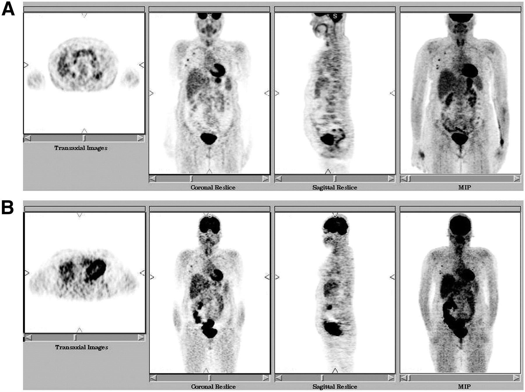
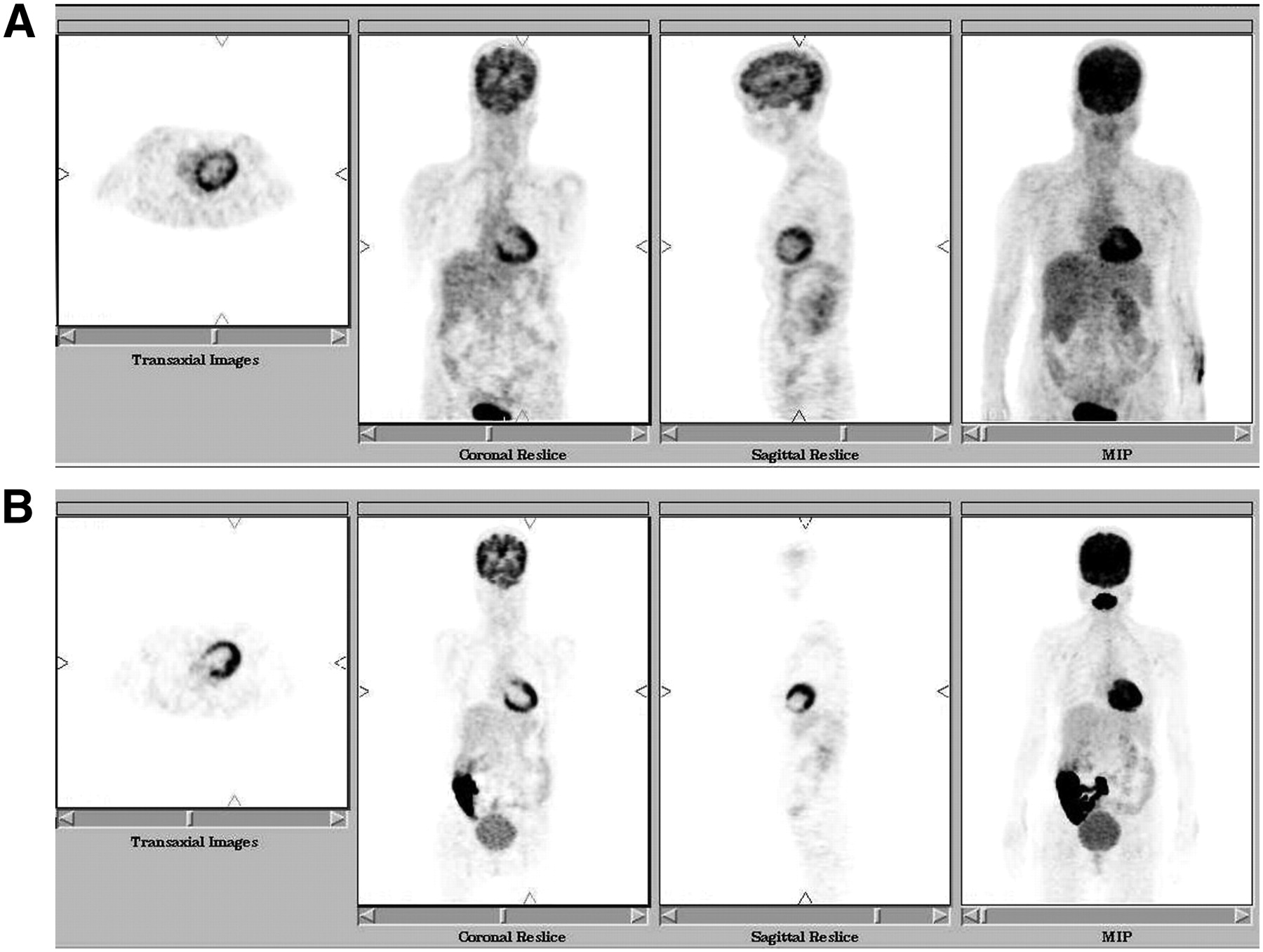
  • FIGURE 4. 
    • Download figure
    • Open in new tab
    • Download powerpoint
    FIGURE 4. 

    Patient 5, 55-y-old woman with non-Hodgkin's lymphoma after treatment. Intravenous study (A) shows tiny right axillary lymph nodes, which are identifiable on oral study too (B). MIP = maximum intensity projection.
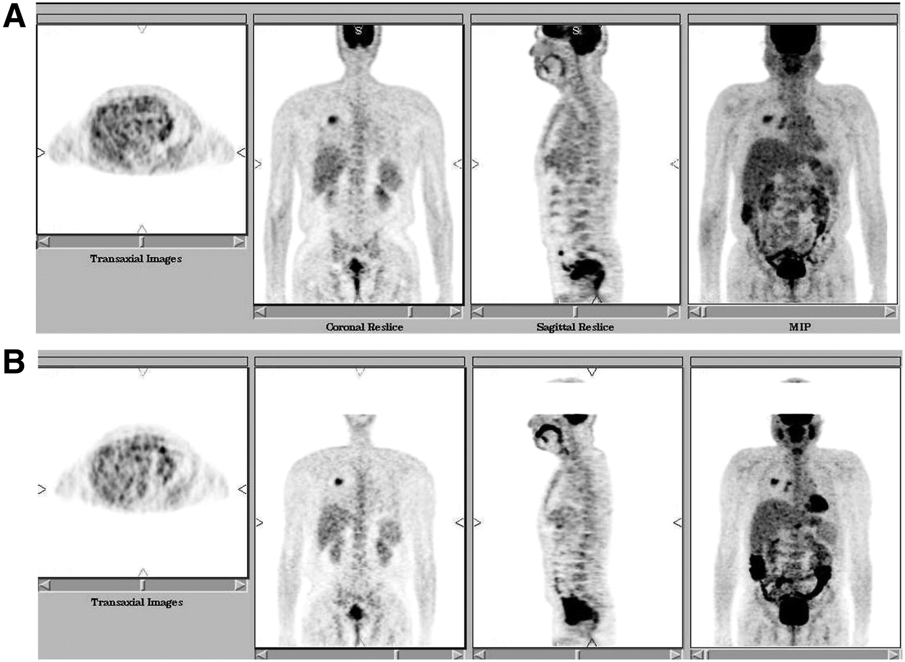
  • FIGURE 5. 
    • Download figure
    • Open in new tab
    • Download powerpoint
    FIGURE 5. 

    Patient 6, 45-y-old woman in whom myocardial viability was being assessed. Both intravenous (A) and oral (B) studies show similar results. MIP = maximum intensity projection.
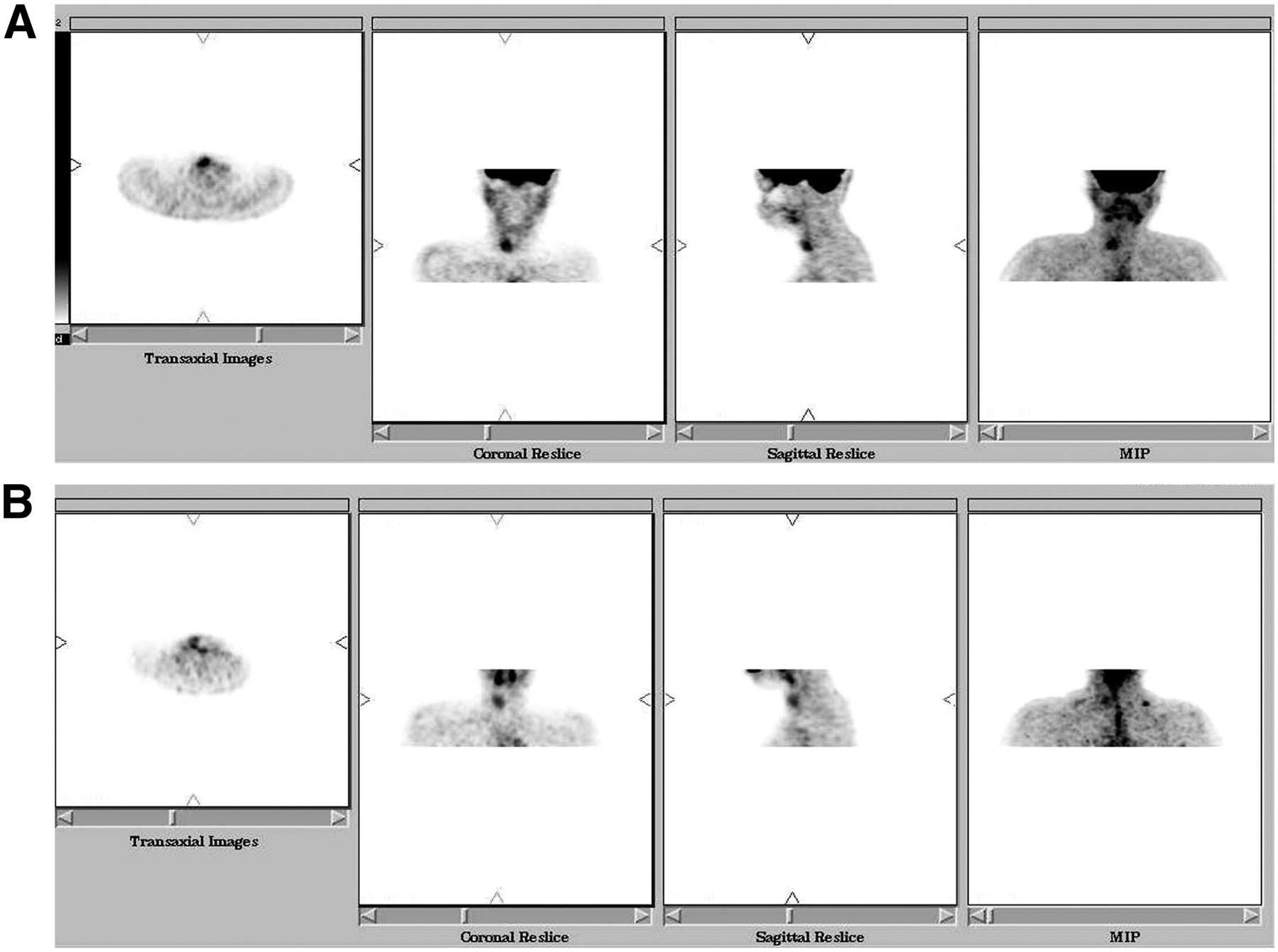
  • FIGURE 6. 
    • Download figure
    • Open in new tab
    • Download powerpoint
    FIGURE 6. 

    Patient 7, 16-y-old girl with solitary thyroid nodule in right lobe. 18F-FDG uptake is seen on both intravenous (A) and oral (B) studies. MIP = maximum intensity projection.

PreviousNext
Back to top

In this issue

Journal of Nuclear Medicine Technology: 35 (2)
Journal of Nuclear Medicine Technology
Vol. 35, Issue 2
June 2007
  • Table of Contents
  • About the Cover
  • Index by author
Print
Download PDF
Article Alerts
Sign In to Email Alerts with your Email Address
Email Article

Thank you for your interest in spreading the word on Journal of Nuclear Medicine Technology.

NOTE: We only request your email address so that the person you are recommending the page to knows that you wanted them to see it, and that it is not junk mail. We do not capture any email address.

Enter multiple addresses on separate lines or separate them with commas.
Substitution of Oral 18F-FDG for Intravenous 18F-FDG in PET Scanning
(Your Name) has sent you a message from Journal of Nuclear Medicine Technology
(Your Name) thought you would like to see the Journal of Nuclear Medicine Technology web site.
Citation Tools
Substitution of Oral 18F-FDG for Intravenous 18F-FDG in PET Scanning
Narendra Nair, Archi Agrawal, Rajnath Jaiswar
Journal of Nuclear Medicine Technology Jun 2007, 35 (2) 100-104; DOI: 10.2967/jnmt.106.036129

Citation Manager Formats

  • BibTeX
  • Bookends
  • EasyBib
  • EndNote (tagged)
  • EndNote 8 (xml)
  • Medlars
  • Mendeley
  • Papers
  • RefWorks Tagged
  • Ref Manager
  • RIS
  • Zotero
Share
Substitution of Oral 18F-FDG for Intravenous 18F-FDG in PET Scanning
Narendra Nair, Archi Agrawal, Rajnath Jaiswar
Journal of Nuclear Medicine Technology Jun 2007, 35 (2) 100-104; DOI: 10.2967/jnmt.106.036129
Twitter logo Facebook logo LinkedIn logo Mendeley logo
  • Tweet Widget
  • Facebook Like
  • Google Plus One
Bookmark this article

Jump to section

  • Article
    • Abstract
    • MATERIALS AND METHODS
    • RESULTS
    • DISCUSSION
    • CONCLUSION
    • Footnotes
    • References
  • Figures & Data
  • Info & Metrics
  • PDF

Related Articles

  • No related articles found.
  • PubMed
  • Google Scholar

Cited By...

  • Human Radiation Dosimetry for Orally and Intravenously Administered 18F-FDG
  • Dynamic Observation of 18F-FDG Uptake After Oral Administration in a Healthy Subject
  • Google Scholar

More in this TOC Section

  • Early 10-Minute Postinjection [18F]F-FAPI-42 uEXPLORER Total-Body PET/CT Scanning Protocol for Staging Lung Cancer Using HYPER Iterative Reconstruction
  • Single- Versus Dual-Time-Point Imaging for Transthyretin Cardiac Amyloid Using 99mTc-Pyrophosphate
  • Does Arthrography Improve Accuracy of SPECT/CT for Diagnosis of Aseptic Loosening in Patients with Painful Knee Arthroplasty: A Systematic Review and Metaanalysis
Show more Imaging

Similar Articles

SNMMI

© 2025 SNMMI

Powered by HighWire