


Article Figures & Data
Figures
- FIGURE 1.
Illustration of complex lymphatic system of head and neck region.
- FIGURE 2.
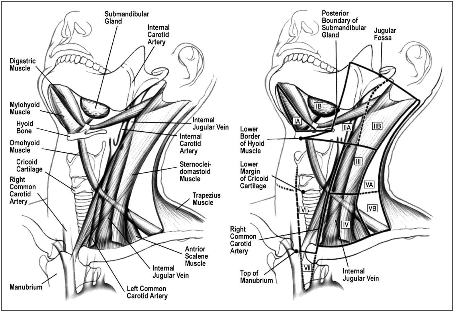
Level system of cervical lymph node classification. (Reprinted with permission of (11).)
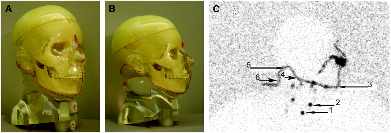
- FIGURE 3.
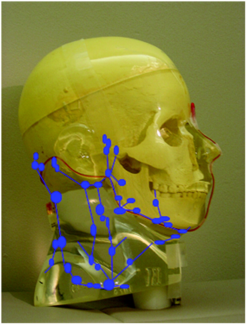
(A and B) Proper placement of 57Co markers before lateral marker image is obtained. Flexible marker should be taped at facial inflection points. Disk markers should be placed at thyroid cartilage and suprasternal notch. (C) Reference marker placement. Proper placement of flexible marker taped at facial inflection points to obtain reference points. Reference points are suprasternal notch (1), thyroid cartilage (2), chin (3), mandibular angle (4), mastoid (5), and occipital area (6).
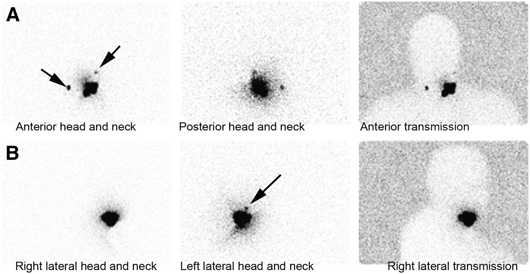
- FIGURE 4.
Two lymph nodes are visualized on anterior and posterior head and neck images obtained with and without transmission, but on left lateral image, only 1 lymph node is visualized because of masking from radiotracer at injection site. Left lateral image for anatomic referencing is therefore not useful in this case.
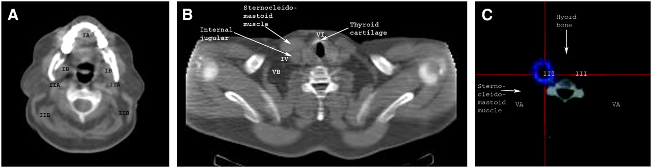
- FIGURE 5.
Axial CT anatomy of most important structures needed for lymph node level classification.
- FIGURE 6.
Referencing of imaging-based lymph node level classification on axial CT imaging (A and B) and fused SPECT/CT (C).
- FIGURE 7.
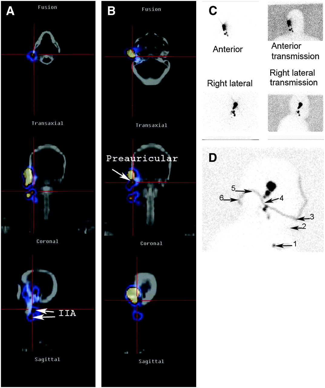
Preauricular melanoma. (A and B) On SPECT/CT, level IIA lymph node (A) and preauricular node (B) are precisely localized. (C and D) Localization is approximate on planar image (C) because optimal linear marker image (D) references suprasternal notch (1), thyroid cartilage (2), chin (3), mandibular angle (4), mastoid (5), and occipital area (6).
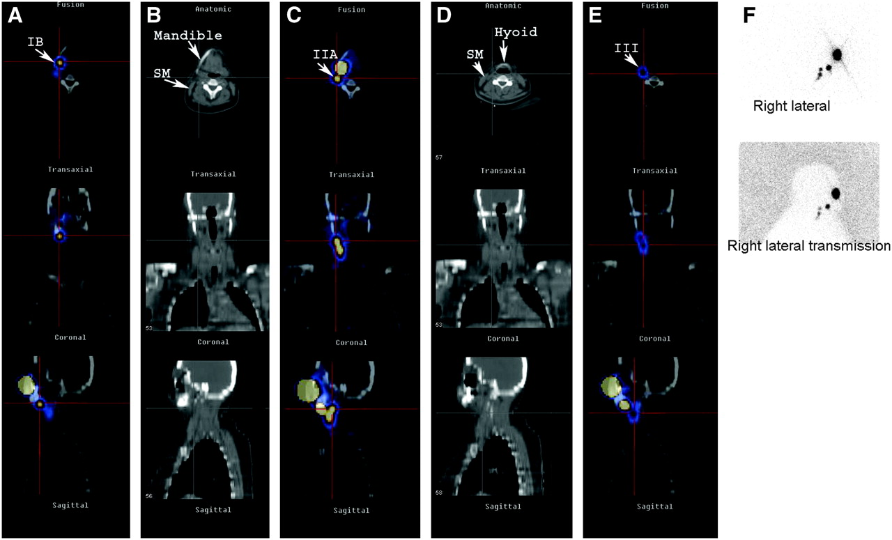
- FIGURE 8.
Right cheek melanoma. SPECT/CT can accurately differentiate sublevels by showing CT landmarks of mandible, hyoid bone, and sternocleidomastoid muscle (B and D). Level IB lymph node (A) is located above hyoid bone and below mandible, whereas level IIA node (C) is located at mandibular angle above hyoid bone and anterior to sternocleidomastoid muscle. On planar imaging (F), jugular levels are not well defined, whereas SPECT/CT (E) encompasses structures needed to classify level III node.
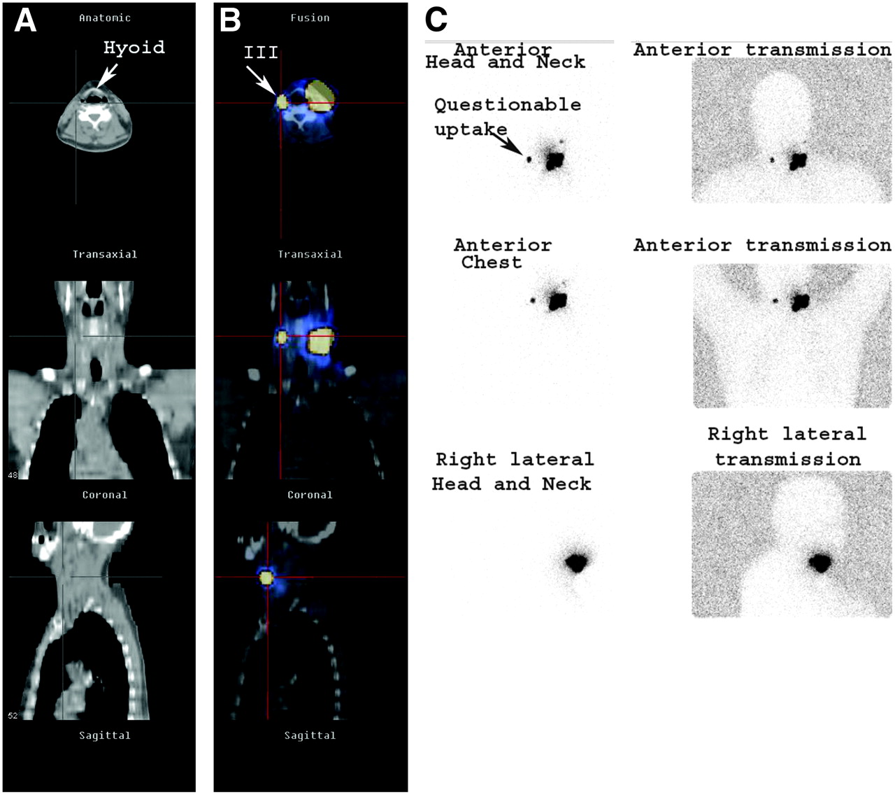
- FIGURE 9.
Left neck melanoma. Contamination is very common during subcutaneous radionuclide injection. Therefore, when an unusual contralateral lymph node is visualized on planar imaging (C), the possibility of contamination should be considered. SPECT/CT is helpful in ruling out contamination. In this case, SPECT/CT precisely demonstrated that unusual contralateral node was level III node (B) residing between hyoid bone and sternocleidomastoid muscle (A).
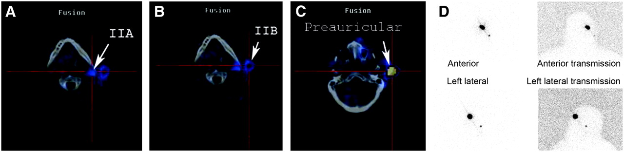
- FIGURE 10.
Sebaceous cell carcinoma of left upper eyelid: Planar imaging (D) demonstrated only 1 node, whereas SPECT/CT demonstrated 4 nodes, possibly because of slight delay in imaging time. Level IIA (A), level IIB (B), and preauricular (C) lymphatic chains are shown.


