Index by author
Cover image

About the Cover
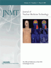
From left to right, volume-rendered CT, PET, and fused PET/CT images of a man with newly diagnosed non-Hodgkin's lymphoma who underwent imaging for staging before treatment. The images were obtained 60 min after 677 MBq (18.3 mCi) of 18F-FDG had been injected. A large mass is seen on the left side of the neck, with left axillary involvement. Courtesy of Eileen J. Ehret, CNMT, Stacey Simon, CNMT, and Daniel Pakuszewski, CNMT, PET Imaging of Willowbrook, Houston, Texas.


