
Main menu

  • Home
  • Content
    • Current
      • JNMT Supplement
    • Ahead of print
    • Past Issues
    • Continuing Education
    • JNMT Podcast
    • SNMMI Annual Meeting Abstracts
  • Subscriptions
    • Subscribers
    • Rates
    • Journal Claims
    • Institutional and Non-member
  • Authors
    • Submit to JNMT
    • Information for Authors
    • Assignment of Copyright
    • AQARA Requirements
  • Info
    • Reviewers
    • Permissions
    • Advertisers
    • Corporate & Special Sales
  • About
    • About Us
    • Editorial Board
    • Contact Information
  • More
    • Alerts
    • Feedback
    • Help
    • SNMMI Journals
  • SNMMI
    • JNMT
    • JNM
    • SNMMI Journals
    • SNMMI

User menu

  • Subscribe
  • My alerts
  • Log in
  • My Cart

Search

  • Advanced search
Journal of Nuclear Medicine Technology
  • SNMMI
    • JNMT
    • JNM
    • SNMMI Journals
    • SNMMI
  • Subscribe
  • My alerts
  • Log in
  • My Cart
Journal of Nuclear Medicine Technology

Advanced Search

  • Home
  • Content
    • Current
    • Ahead of print
    • Past Issues
    • Continuing Education
    • JNMT Podcast
    • SNMMI Annual Meeting Abstracts
  • Subscriptions
    • Subscribers
    • Rates
    • Journal Claims
    • Institutional and Non-member
  • Authors
    • Submit to JNMT
    • Information for Authors
    • Assignment of Copyright
    • AQARA Requirements
  • Info
    • Reviewers
    • Permissions
    • Advertisers
    • Corporate & Special Sales
  • About
    • About Us
    • Editorial Board
    • Contact Information
  • More
    • Alerts
    • Feedback
    • Help
    • SNMMI Journals
  • Watch or Listen to JNMT Podcast
  • Visit SNMMI on Facebook
  • Join SNMMI on LinkedIn
  • Follow SNMMI on Twitter
  • Subscribe to JNMT RSS feeds
OtherCASE STUDY

18F-FDG PET Hemispheric Cerebral Hypometabolic Activity: Patient Motion Artifact

Carlos A. Garcia, Giuseppe Esposito, Elmo R. Acio and Douglas Van Nostrand
Journal of Nuclear Medicine Technology June 2006, 34 (2) 86-87;
Carlos A. Garcia
  • Find this author on Google Scholar
  • Find this author on PubMed
  • Search for this author on this site
Giuseppe Esposito
  • Find this author on Google Scholar
  • Find this author on PubMed
  • Search for this author on this site
Elmo R. Acio
  • Find this author on Google Scholar
  • Find this author on PubMed
  • Search for this author on this site
Douglas Van Nostrand
  • Find this author on Google Scholar
  • Find this author on PubMed
  • Search for this author on this site
  • Article
  • Figures & Data
  • Info & Metrics
  • PDF
Loading

Abstract

A 51-y-old man underwent 18F-FDG PET for disease restaging of non-Hodgkin's lymphoma. Initial attenuation correction images demonstrated unilateral hemispheric cerebral hypometabolic activity. The remainder of the scan was otherwise unremarkable. Non–attenuation correction images demonstrated a patient motion artifact attributable to lateral movement of the patient's head during image acquisition. Subsequent repeat 18F-FDG PET showed normal cerebral cortical activity.

  • 18F-FDG
  • motion artifact
  • cerebral hypometabolic activity

The following case report illustrates the importance of preventing motion of the patient's head during 18F-FDG PET, because of the consequent motion artifact resembling decreased cerebral metabolism.

CASE REPORT

A 51-y-old man with a diagnosis of non-Hodgkin's lymphoma underwent 18F-FDG PET for disease restaging. Initial attenuation correction images demonstrated unilateral hemispheric cerebral hypometabolic activity (Fig. 1). The ipsilateral scalp also demonstrated hypometabolic activity (Fig. 1). The remainder of the scan was otherwise unremarkable. Non–attenuation correction images demonstrated lateral displacement of the patient's head during image acquisition (Fig. 2). As a result of this movement, the images presented a motion artifact simulating a unilateral decrease in cerebral metabolic activity. The patient was rescheduled for repeat 18F-FDG PET. Subsequent images revealed normal cerebral cortical activity (Fig. 3).

FIGURE 1. 
  • Download figure
  • Open in new tab
  • Download powerpoint
FIGURE 1. 

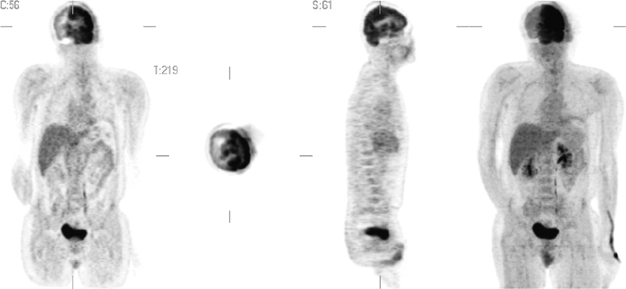
Coronal, transaxial, sagittal, and maximum-intensity-projection 3-dimensional 18F-FDG PET attenuation correction images demonstrating unilateral hemispheric cerebral hypometabolic activity. Decreased activity also was seen in the ipsilateral scalp.

FIGURE 2. 
  • Download figure
  • Open in new tab
  • Download powerpoint
FIGURE 2. 

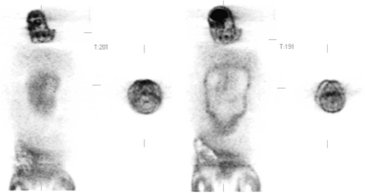
Coronal and transaxial 18F-FDG PET non–attenuation correction images demonstrating patient lateral head motion at sequential stages.

FIGURE 3. 
  • Download figure
  • Open in new tab
  • Download powerpoint
FIGURE 3. 

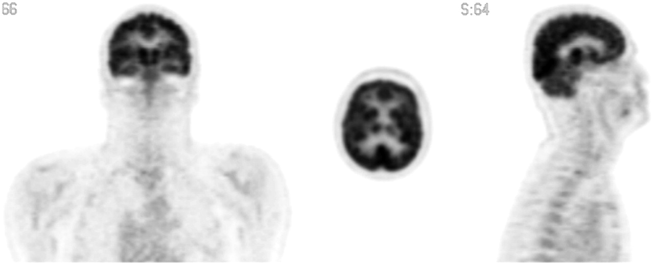
Coronal, transaxial, and sagittal attenuation correction images from repeat scan demonstrating normal cerebral cortical activity.

DISCUSSION

Attenuation correction is needed in PET not only for accurate quantification of the radioactivity detected by the PET scanner but also for proper visualization of different anatomic structures. The accuracy of attenuation correction depends on the proper alignment of transmission and emission scans. Stabilization of the head, either with a brace or lateral cushioning, may aid in reducing lateral motion during the scanning process. Movement that occurs between the emission and the transmission scans will produce inaccurate quantification and, for severe patient motion, even image artifacts. The effect of motion on the accuracy of attenuation correction has been studied with phantom simulations (1). Displacements of as little as 5 mm may cause errors of as much as 14% in measured activity. In PET scans of real brains, a 5-mm mismatch between emission and transmission scans can cause an ∼10% error in a 1-cm-thick cortical region of interest, and a 10-mm mismatch can cause a 25% error (2). With computer realignment of emission and transmission images, the errors can be corrected. However, for severe patient motion, the realignment may be less satisfactory and may leave residual inaccuracies in the attenuation correction. In these cases, attenuation correction can be calculated for the brain without a transmission scan by use of a standard attenuation coefficient. However, this method is not accurate for areas in which there is an interface of soft tissues with air or bony structures, such as in the body, particularly the chest. At present, methods to improve the accuracy of motion correction are being explored (3–5).

References

  1. 1.↵
    Huang SC, Hoffman EJ, Phelps ME, Kuhl DE. Quantitation in positron emission tomography: 2. Effect of inaccurate attenuation correction. J Comput Assist Tomogr. 1979;3:804–814.
    OpenUrlPubMed
  2. 2.↵
    Andersson JL, Vagnhammar BE, Schneider H. Accurate attenuation correction despite movement during PET imaging. J Nucl Med. 1995;36:670–678.
    OpenUrlAbstract/FREE Full Text
  3. 3.↵
    Buhler P, Just U, Will E, Kotzerke J, van den Hoff J. An accurate method for correction of head movement in PET. IEEE Trans Med Imaging. 2004;23:1176–1185.
    OpenUrlCrossRefPubMed
  4. 4.
    van den Heuvel OA, Boellaard R, Veltman DJ, Mesina C, Lammertsma AA. Attenuation correction of PET activation studies in the presence of task-related motion. Neuroimage. 2003;19:1501–1509.
    OpenUrlPubMed
  5. 5.↵
    Bloomfield PM, Spinks TJ, Reed J, et al. The design and implementation of a motion correction scheme for neurological PET. Phys Med Biol. 2003;48:959–978.
    OpenUrlCrossRefPubMed
  • Received for publication March 20, 2006.
  • Accepted for publication March 27, 2006.
PreviousNext
Back to top

In this issue

Journal of Nuclear Medicine Technology: 34 (2)
Journal of Nuclear Medicine Technology
Vol. 34, Issue 2
June 2006
  • Table of Contents
  • About the Cover
  • Index by author
Print
Download PDF
Article Alerts
Sign In to Email Alerts with your Email Address
Email Article

Thank you for your interest in spreading the word on Journal of Nuclear Medicine Technology.

NOTE: We only request your email address so that the person you are recommending the page to knows that you wanted them to see it, and that it is not junk mail. We do not capture any email address.

Enter multiple addresses on separate lines or separate them with commas.
18F-FDG PET Hemispheric Cerebral Hypometabolic Activity: Patient Motion Artifact
(Your Name) has sent you a message from Journal of Nuclear Medicine Technology
(Your Name) thought you would like to see the Journal of Nuclear Medicine Technology web site.
Citation Tools
18F-FDG PET Hemispheric Cerebral Hypometabolic Activity: Patient Motion Artifact
Carlos A. Garcia, Giuseppe Esposito, Elmo R. Acio, Douglas Van Nostrand
Journal of Nuclear Medicine Technology Jun 2006, 34 (2) 86-87;

Citation Manager Formats

  • BibTeX
  • Bookends
  • EasyBib
  • EndNote (tagged)
  • EndNote 8 (xml)
  • Medlars
  • Mendeley
  • Papers
  • RefWorks Tagged
  • Ref Manager
  • RIS
  • Zotero
Share
18F-FDG PET Hemispheric Cerebral Hypometabolic Activity: Patient Motion Artifact
Carlos A. Garcia, Giuseppe Esposito, Elmo R. Acio, Douglas Van Nostrand
Journal of Nuclear Medicine Technology Jun 2006, 34 (2) 86-87;
Twitter logo Facebook logo LinkedIn logo Mendeley logo
  • Tweet Widget
  • Facebook Like
  • Google Plus One
Bookmark this article

Jump to section

  • Article
    • Abstract
    • CASE REPORT
    • DISCUSSION
    • References
  • Figures & Data
  • Info & Metrics
  • PDF

Related Articles

  • No related articles found.
  • PubMed
  • Google Scholar

Cited By...

  • No citing articles found.
  • Google Scholar

More in this TOC Section

  • Importance of Gated Imaging in Both Phases of Myocardial Perfusion SPECT: Myocardial Stunning After Dipyridamole Infusion
  • Penile Implant on Bone Scan Imaging: A Case Study
Show more CASE STUDY

Similar Articles

SNMMI

© 2025 SNMMI

Powered by HighWire