


Abstract
The accreditation programs of the American College of Radiology (ACR) are the most established and widely proven for all imaging modalities. For facilities committed to quality imaging, the ACR Nuclear Medicine and PET Accreditation Program provides a solid foundation for a continuous quality improvement program through a peer review and educational process. This article provides general information describing the goals and development of the ACR accreditation programs. The ACR Nuclear Medicine and PET Accreditation Program evaluates the qualifications of personnel, equipment, image quality, and quality control measures. It is believed that these are the primary factors that have an impact on the quality of patient imaging. This article describes the application process, including the clinical examinations that are required and the quality control and performance tests that are expected from each facility. Outcomes and pass/fail statistics are discussed, as are common pitfalls that may cause deficiencies. Upon completion of this article, the reader should be able to describe the application process, the components of the ACR accreditation program, the most common causes for failure, and the site survey process.
The American College of Radiology (ACR) is a national organization serving more than 32,000 radiology professionals, including diagnostic and interventional radiologists, radiation oncologists, nuclear medicine physicians, medical physicists, and clinical researchers. The ACR's mission is to serve patients and society by maximizing the value of radiology, nuclear medicine, radiation oncology, interventional radiology, and medical physics by advancing the science of radiology, improving the quality of patient care, and providing continuing education for radiology and allied health professionals.
The ACR has a long history, dating back to 1963, of leadership in quality assurance and accreditation activities that assess the quality of imaging facilities. The Mammography Accreditation Program was introduced in 1987. In 1992, the Federal Government mandated that all mammography facilities be accredited. As a result of the federal legislation, the U.S. Food and Drug Administration approved the ACR as the only national accrediting body for mammography. Since then, 7 additional modality-specific accreditation programs, including those for nuclear medicine and PET, have been developed on the basis of the mammography model.
GOALS
The goal of the ACR Nuclear Medicine and PET Accreditation Program is to set quality standards for facilities and to help them continuously improve the quality of care given to patients. The voluntary accreditation programs offer nuclear medicine and PET physicians an opportunity for comprehensive review and evaluation of their nuclear medicine and PET facilities, personnel qualifications, image quality, equipment, quality control procedures, and quality assurance programs through a confidential, peer review mechanism. The program is designed to be educational in nature, focusing on the primary factors that have an impact on the quality of clinical images and the quality of patient care.
DEVELOPMENT OF NUCLEAR MEDICINE AND PET ACCREDITATION PROGRAM
The concept of the Nuclear Medicine Accreditation Program was approved by the ACR Council in September 1994. The program is directed by the ACR Committee on Nuclear Medicine Accreditation of the Commission on Quality and Safety. The program was developed and continues to be monitored by experts in the field of nuclear medicine and PET. These experts are drawn from all geographic areas of the country, from both private and public sectors, and from large- and small-practice settings (1). The ACR accreditation programs are based on the ACR Practice Guidelines and Technical Standards. These are developed through a consensus process based on peer review literature and frequently are developed jointly with other medical specialty organizations. The Nuclear Medicine Accreditation Program was launched at the Radiologic Society of North America in 1999. The increased use of PET and concerns about the use of less-than-optimal equipment prompted the development of the PET module that was implemented in 2002.
APPLICATION
In an attempt to streamline the application process, the ACR developed the Diagnostic Modality Accreditation Program (2). This program includes all of the voluntary accreditation programs, with the exception of that for radiation oncology, in 1 application. This design is particularly useful for physician groups that seek accreditation in multiple modalities and that practice at numerous sites. It reduces the paperwork burden and offers the facility a 10% discount if the facility has accreditation for 3 or more modalities or pending accreditation. The Diagnostic Modality Accreditation Program application is available online at www.acr.org, or a copy of the application (paper or CD version) may be requested by contacting the ACR at 1-800-770-0145.
The application is a 2-step process. In the first step, the group submits required information about the practice, including policies, procedures, personnel qualifications, and modality equipment, as well as the appropriate fees. Once the application has been deemed complete, it is processed, and the testing package is sent to the group. The testing package includes specific instructions for submission of clinical images, clinical test image data sheets for summarizing clinical image techniques, phantom instruction booklets, a quality assurance questionnaire, dated bar-coded labels for all images, and a tutorial CD that provides helpful information about improving image quality.
The second step of the process involves the submission of clinical and phantom images. Information is collected on the quality control and quality assurance programs in place. Additional information is collected on follow-up procedures, data collection, reporting, radiopharmaceutical procedures, radiation safety, and laboratory safety. Facilities are required to submit copies of their most recent state or Nuclear Regulatory Commission audits, including a written response to any violations. Facilities also must submit an annual physicist report, which documents compliance with all quality control tests and corrective action if required. All testing materials must be collected and processed according to the instructions provided in the testing package. The facility has 90 d to complete testing; the due date is shown on the bar-coded labels.
Assembling a team to complete the application process will ensure that everyone in the department contributes to the accreditation effort. The supervising and interpreting physicians, the qualified medical physicist, and the nuclear medicine technologists all have important roles. The accreditation process will be successful if all members contribute their knowledge and skills.
PERSONNEL QUALIFICATIONS
Personnel interpreting or performing nuclear medicine and PET studies must meet specific education and experience qualifications (Tables 1 and 2) (3). In addition, there are specific recommendations for continuing medical education (CME) for physicians, continuing education for technologists and physicists, and continuing experience for physicians. A supervising nuclear medicine or PET physician must be designated to have the primary responsibility for nuclear medicine or PET at the facility. Each physician and each medical physicist are required to complete and sign an attestation signifying compliance with training, licensing, and CME requirements. In addition, the supervising physician must agree that no imaging procedures will be performed by personnel who do not meet the specified requirements. Each facility should maintain detailed documentation supporting the qualifications of personnel at the site. Although applicants are not required to submit the documentation during the accreditation process, it will be reviewed for compliance during any site survey (described later in the article).
Requirements for Physicians
Requirements for Technologists and Physicists
CLINICAL EXAMINATIONS
Nuclear medicine accreditation requires that all units (cameras or scanners) be evaluated in order for the facility to be granted accreditation. Facilities may choose from 1 or more of 3 modules for nuclear medicine, including general nuclear medicine (planar), SPECT, and nuclear cardiology imaging. A facility is required to apply for all modules performed at the site. Facilities also may select 1 or more of 3 submodules for PET, including oncology and brain and cardiac imaging. Again, a facility is required to apply for all PET submodules performed at the site; however, failure to apply for PET accreditation will not affect current nuclear medicine accreditation. If the departments are separate, either department may apply for accreditation for the modules performed.
The ACR Committee on Nuclear Medicine Accreditation has determined that certain examinations best illustrate the ability of a facility to produce images of acceptable quality in a given module. The facility must submit 2 different examinations for each module (3), as follows: module 1 (general nuclear medicine [planar imaging])—whole body or spot bone (required) and hepatobiliary, perfusion lung, or multigated acquisition (MUGA); module 2 (SPECT)—bone SPECT (required) and bone SPECT, brain SPECT, hepatic blood pool, liver SPECT, or myocardial perfusion; module 3 (nuclear cardiology imaging) (requires 2 examinations even if a myocardial perfusion examination is submitted for SPECT)—myocardial perfusion examination (required) and cardiac function examination (e.g., MUGA or gated SPECT); and module 4 (PET/coincidence imaging) (3 submodules)—oncology (the site must submit 2 examinations, 1 of which must be abnormal; the examinations can be any combination of the following: whole body, with and without measured attenuation correction, or chest and abdomen, with and without measured attenuation correction, if routinely used; the examinations may be from the same patient), brain (the site must submit 2 examinations, 1 of which must be abnormal, with attenuation correction), and cardiac (the site must submit 2 examinations, 1 of which must be abnormal, with and without measured attenuation correction, if available).
For modules 1, 2, and 3, facilities should submit normal examinations; however, abnormal studies may be submitted if no high-quality normal examinations are available.
All examinations should be submitted in the standard format used for image interpretation at the facility. The examinations should be consistent with the ACR Practice Guidelines and Technical Standards. A dated physician's report that clearly states the type of examination performed and pertinent clinical history must accompany each examination. A corresponding clinical test image data sheet for summarizing the specific techniques used, including the radiopharmaceutical and dose injected, scan time, and acquisition parameters, must be completed for each examination. In addition, the facility must submit a copy of the written procedure used for each examination.
The clinical examinations and all related submitted materials are sent to qualified physician reviewers who are experts in nuclear medicine and PET. These reviewers are required to participate in a training program on the image review process. As with all of the ACR accreditation programs, the primary assumption of the clinical image reviewers is that the images chosen by the facility represent examples of their best work. Multiple factors are assessed when the reviewers examine the image quality of patient examinations. These include report and film identification, radiopharmaceutical biodistribution, image acquisition, processing, and display, as well as adequate anatomic coverage. The supervising physician and the chief technologist should work together to carefully select images that represent the best-quality patient care that the facility provides.
PERFORMANCE TEST AND QUALITY CONTROL
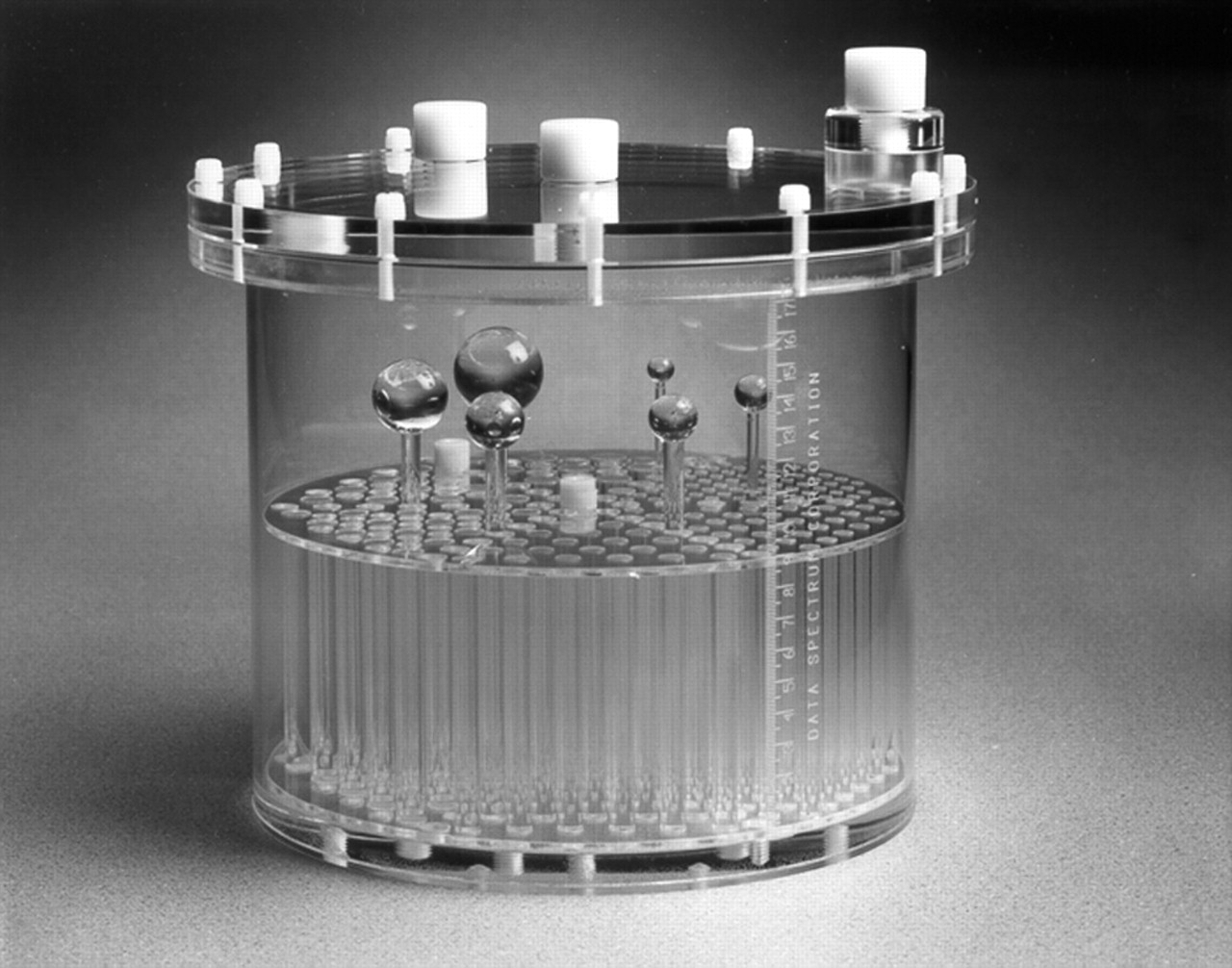
The Subcommittee on Nuclear Medicine Physics determined which technical parameters must be evaluated to assess γ-camera capabilities (3). These measures are intentionally stringent, because the program assesses compliance with requirements of the Nuclear Regulatory Commission and appropriate state regulatory agencies. Facilities must submit planar and SPECT (if performed at the site) images for review with the ACR-approved phantom, the Jaszczak Deluxe Flangeless ECT phantom (Data Spectrum), on all units. The phantom is a cylinder with an internal radius of 10.8 cm. The lower portion of the cylinder contains 6 sets of acrylic rods arranged in a pie-shaped pattern and with the following diameters: 4.8, 6.4, 7.9, 9.5, 11.1, and 12.7 mm. The upper section contains 6 solid spheres with the following diameters: 9.5, 12.7, 15.9, 19.1, 25.4, and 31.8 mm (Fig. 1). The phantom acquisition requirements may differ from those normally used by the facility but were designed to minimize variability in images submitted by different facilities. Phantom images are scored by a panel of qualified medical physicists who have participated in review process training. The physics subcommittee has set standards for uniformity, noise, spatial resolution, and contrast.

Jaszczak Deluxe Flangeless ECT Phantom.
Performance tests must be performed at least annually on all nuclear medicine units. The prescribed testing is a comprehensive set of individual measurements that would adequately detect any significant changes in the performance of the units (Table 3). The testing can be performed by a qualified medical physicist, a qualified nuclear medicine technologist, or a medical physicist in training, with oversight by a qualified medical physicist. The test results must be reviewed by the medical physicist and documented in an annual survey report.
Required Annual Performance Tests
The nuclear medicine technologist is responsible for verifying the day-to-day instrument operations and performing additional tests quarterly (Table 4). These requirements are consistent with the ACR Technical Standard for Medical Nuclear Physics Performance Monitoring of Nuclear Medicine Imaging Equipment (4) and comply with the requirements and recommendations of the Joint Commission on the Accreditation of Healthcare Organizations, as well as state and federal agencies. Protocols for daily camera tests should include thresholds for failure of the quality control tests.
Technologist Quality Control Tests
Documentation of compliance with all quality control tests and corrective action is required as part of the accreditation process. An annual survey report by the facility's qualified medical physicist must be submitted and thereafter must be available for review should an on-site visit be conducted.
Facilities must submit phantom testing with the ACR-approved phantom on each PET scanner. This is the same Jaszczak Deluxe Flangeless ECT phantom used for SPECT testing but with a modified faceplate. The faceplate has various cylinders that can be filled: thin-walled cylinders (8, 12, 16, and 25 mm in diameter); 2 additional 25-mm cylinders, 1 for air and 1 for “cold” water; and a Teflon (DuPont) cylinder (Fig. 2). Phantom testing instructions are provided with the testing materials. Again, a qualified medical physicist must review and score the images by using the standards recommended by the physics subcommittee for uniformity, noise, spatial resolution, and contrast. At this time, there is no prescribed PET quality control testing; however, testing should be performed in accordance with the ACR Technical Standard for Medical Nuclear Physics Performance Monitoring of PET Imaging Equipment (5). Each facility is required to submit a summary of the quality control and frequency of testing currently being done on each PET unit. In the near future, the ACR Committee on Nuclear Medicine Accreditation will establish quality control requirements based on these collected data.
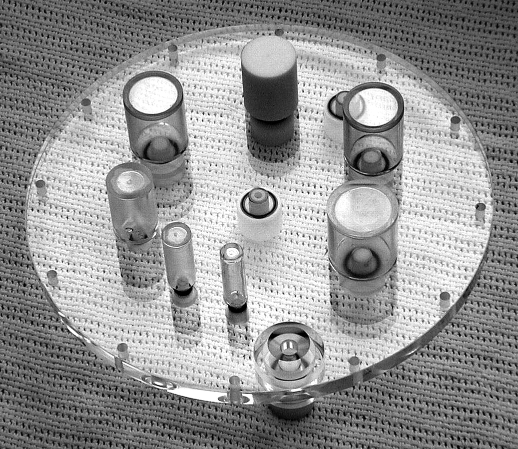
Modified faceplate used with Jaszczak Deluxe Flangeless ECT Phantom for PET phantom acquisition.
OUTCOMES
After all stages of the evaluation are completed, the ACR issues for each unit a final report that includes specific assessments, defines areas for improvement, and provides recommendations about the performance of nuclear medicine or PET studies. Facilities that successfully meet all of the criteria will be awarded a 3-y accreditation, a certificate, and a decal for each approved unit specific to the modules performed on that unit. The ACR will provide facilities with marketing tools and press release assistance. For referring physician and patient information, accredited nuclear medicine and PET facilities will be listed on the ACR Web site.
A facility that receives notification of a deficiency will receive specific recommendations for improvement. These recommendations, as a quality improvement tool, provide guidance so that the facility can meet the criteria after corrective action and reapplication. The facility has the option to appeal, withdraw the unit or module, or repeat testing. A facility that does not meet the initial evaluation criteria will be required to repeat only those items that were deficient, for example, phantom or clinical images. Facilities that reapply after notification of a deficiency must submit their request along with the appropriate fees.
The feedback received by the facilities from the final report enables most sites to correct deficiencies and achieve accreditation, as shown in Table 5.
Pass Rates on First Round and Repeat Attempts,* June 2005
COMMON PITFALLS
The most common deficiency for clinical images is failure to label images as to laterality and orientation. The ACR Committee on Nuclear Medicine Accreditation has determined that all images must be labeled. This requirement is necessary to reduce the number of serious treatment errors resulting from the lack of appropriate labeling and to address quality patient care issues raised by the recent focus on patient safety in medicine. Some of the other reasons for deficiencies include, but are not limited to, incomplete submission of images, incomplete clinical test image data sheets, and failure to follow the submitted written procedures. For successful submission, it is imperative that facility personnel carefully read and follow testing instructions. If the site has questions, the ACR has highly qualified staff available to assist and can be reached at 1-800-770-0145.
In order to correct phantom deficiencies, it is recommended that, before acquiring phantom images, facilities should determine that the center of rotation and high-count floods are current. In addition, it is important to ensure good mixing of the phantom after filling. Phantom positioning is critical, and the use of a bubble level is recommended. Examples of adequate phantom images are shown in the tutorial CD.
Table 6 shows the pass/fail rates for both clinical and phantom images during the initial cycle.
Pass/Fail Rates for Clinical and Phantom Images During Initial Cycle
SITE SURVEYS
The ACR has several validation mechanisms in place, including validation film checks, which are done through the mail and random site surveys. The ACR conducts random site surveys at a percentage of their accredited facilities each year. The ACR reserves the right to conduct these site surveys at any time during or after the accreditation process. These surveys are done to validate information submitted to the program and to monitor continuing compliance with accreditation standards. In addition, the ACR uses this mechanism to provide an educational opportunity, through direct feedback and constructive criticism for quality improvement, to facilities participating in the accreditation program. The site surveys also are used to provide assistance in areas in which facilities may need guidance.
The facility is notified 2 wk before the visit to allow adequate time for the facility to adjust the schedules of appropriate staff members to meet with the survey team and to adjust patient scheduling, if necessary. The survey team comprises a physician and a physicist, both of whom are active reviewers in the Nuclear Medicine and PET Accreditation Program, and an ACR staff member, typically a registered nuclear medicine technologist.
The physician randomly selects a minimum of 10 clinical examinations for review to assess whether the facility has maintained high-quality imaging. The physician also reviews the facility's quality assurance program. The physicist supervises the acquisition and processing of the images with the ACR-approved phantom by a qualified nuclear medicine technologist or physicist. The physicist also reviews the facility's quality control program, all log books, and the medical physicist's annual survey report. The ACR staff member verifies all application information, including personnel qualifications and credentials, continuing education, licensing, and equipment. In addition, the staff member reviews the facility's policies and procedures and the employee orientation program specific to nuclear medicine or PET. After the review is complete, the survey team meets with the supervising physician, the chief nuclear medicine technologist, and the medical physicist for a brief exit interview, offering guidance and feedback. A final report is issued to the facility within 6 wk of the visit to outline the findings and to explain any required corrective action.
The ACR has determined that most sites have maintained the quality that is expected of an accredited facility; however, some sites have needed to improve their quality control measures. Some sites, although carrying out the required daily testing, did not communicate to the physicist or to service personnel that the results were outside the specified parameters; therefore, appropriate corrective measures were not taken. Feedback from the facilities after the site surveys has been positive. Many of the facilities believed that the visits were educational and helped staff members to improve the overall quality of their department.
STATISTICS
Since 1999, 370 nuclear medicine facilities and 182 PET facilities have applied for the ACR Nuclear Medicine and PET Accreditation Program. Table 7 shows the total numbers of those facilities that have been accredited, that are currently active, and that are currently accredited. The graph in Figure 3 shows the total number of units that have applied, by year of application.

Number of units by year that have applied for accreditation (including renewals) as of 30 November 2005.
Numbers of Facilities That Have Been Accredited, That Are Currently Active, and That Are Currently Accredited
CONCLUSION
The most important goal of accreditation is to improve patient care. The ACR Nuclear Medicine and PET Accreditation Program is an excellent quality improvement tool. It is an opportunity for comprehensive review and evaluation of a facility through peer review. A facility's designation as accredited, after voluntary evaluation of a practice, means that the site has achieved and maintained a level of practice that promotes the delivery of the highest-quality health care. Accreditation demonstrates to referring physicians, patients, and payers that a facility provides high-quality patient care and is committed to meeting the highest standards.
Acknowledgments
The ACR is a major national medical specialty association dedicated to improving the quality of patient care. For more information, contact the ACR at 1891 Preston White Dr., Reston, VA 20191-4397; telephone, 800-227-5463; fax, 703-295-6776; web site, www.acr.org.
Footnotes
-
↵* NOTE: FOR CE CREDIT, YOU CAN ACCESS THIS ACTIVITY THROUGH THE SNM WEB SITE (http://www.snm.org/ce_online) THROUGH MARCH 2007.
- Received for publication September 9, 2005.
- Accepted for publication January 9, 2006.


Jump to section
Related Articles
Cited By...
- A Path to Qualification of PET/MRI Scanners for Multicenter Brain Imaging Studies: Evaluation of MRI-Based Attenuation Correction Methods Using a Patient Phantom
- Measurement of PET Quantitative Bias In Vivo
- Automated Quantitative Analysis of American College of Radiology PET Phantom Images
- Qualification of National Cancer Institute-Designated Cancer Centers for Quantitative PET/CT Imaging in Clinical Trials
- Qualification of PET Scanners for Use in Multicenter Cancer Clinical Trials: The American College of Radiology Imaging Network Experience
- Achieving Quality in Cardiovascular Imaging II: Proceedings From the Second American College of Cardiology-Duke University Medical Center Think Tank on Quality in Cardiovascular Imaging
- Achieving Quality in Cardiovascular Imaging: Proceedings From the American College of Cardiology-Duke University Medical Center Think Tank on Quality in Cardiovascular Imaging