
Main menu

  • Home
  • Content
    • Current
      • JNMT Supplement
    • Ahead of print
    • Past Issues
    • Continuing Education
    • JNMT Podcast
    • SNMMI Annual Meeting Abstracts
  • Subscriptions
    • Subscribers
    • Rates
    • Journal Claims
    • Institutional and Non-member
  • Authors
    • Submit to JNMT
    • Information for Authors
    • Assignment of Copyright
    • AQARA Requirements
  • Info
    • Reviewers
    • Permissions
    • Advertisers
    • Corporate & Special Sales
  • About
    • About Us
    • Editorial Board
    • Contact Information
  • More
    • Alerts
    • Feedback
    • Help
    • SNMMI Journals
  • SNMMI
    • JNMT
    • JNM
    • SNMMI Journals
    • SNMMI

User menu

  • Subscribe
  • My alerts
  • Log in
  • Log out
  • My Cart

Search

  • Advanced search
Journal of Nuclear Medicine Technology
  • SNMMI
    • JNMT
    • JNM
    • SNMMI Journals
    • SNMMI
  • Subscribe
  • My alerts
  • Log in
  • Log out
  • My Cart
Journal of Nuclear Medicine Technology

Advanced Search

  • Home
  • Content
    • Current
    • Ahead of print
    • Past Issues
    • Continuing Education
    • JNMT Podcast
    • SNMMI Annual Meeting Abstracts
  • Subscriptions
    • Subscribers
    • Rates
    • Journal Claims
    • Institutional and Non-member
  • Authors
    • Submit to JNMT
    • Information for Authors
    • Assignment of Copyright
    • AQARA Requirements
  • Info
    • Reviewers
    • Permissions
    • Advertisers
    • Corporate & Special Sales
  • About
    • About Us
    • Editorial Board
    • Contact Information
  • More
    • Alerts
    • Feedback
    • Help
    • SNMMI Journals
  • Watch or Listen to JNMT Podcast
  • Visit SNMMI on Facebook
  • Join SNMMI on LinkedIn
  • Follow SNMMI on Twitter
  • Subscribe to JNMT RSS feeds
OtherIMAGING

Selected Cases Demonstrating the Value of Furosemide-Primed 18F-FDG PET in Identifying Adrenal Involvement

Narendra Nair and Sandip Basu
Journal of Nuclear Medicine Technology September 2005, 33 (3) 166-171;
Narendra Nair
  • Find this author on Google Scholar
  • Find this author on PubMed
  • Search for this author on this site
Sandip Basu
  • Find this author on Google Scholar
  • Find this author on PubMed
  • Search for this author on this site
  • Article
  • Figures & Data
  • Info & Metrics
  • PDF
Loading

Article Figures & Data

Figures

  • FIGURE 1.
    • Download figure
    • Open in new tab
    • Download powerpoint
    FIGURE 1.

    Whole-body PET images of 49-y-old woman in whom adenocarcinoma of right ovary was diagnosed in 1992 and high-grade leiomyosarcoma of left ovary was diagnosed in 2003. She underwent hysterectomy and right salpingo-oophorectomy for the former and left salpingo-oophorectomy and omentectomy for the latter. Postoperatively, she was treated with external radiotherapy and ifosfamide (Holoxan; Baxter Oncology)-based chemotherapy. Her recent abdominal CT findings were normal, and tumor markers were within normal limits. PET was performed to search for any residual disease.
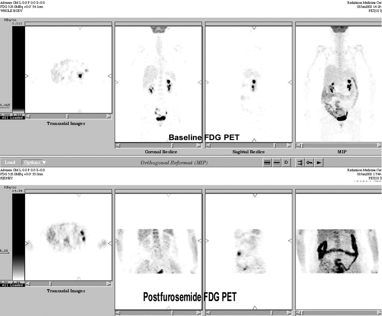
  • FIGURE 2.
    • Download figure
    • Open in new tab
    • Download powerpoint
    FIGURE 2.

    18F-FDG PET images of 4-y-old boy in whom, 3 y previously, left adrenal neuroblastoma was diagnosed and treated with left adrenalectomy. Histopathologic examination revealed differentiated, stroma-poor neuroblastoma, and all sampled retroperitoneal nodes were free of disease. Bone marrow was not involved. Patient was not subjected to chemotherapy or radiotherapy in view of localized disease. The 24-h urinary vanillylmandelic acid level had recently been rising and was 19.5 mg/g of creatinine (reference range, 1–10 mg/g) at time of PET.
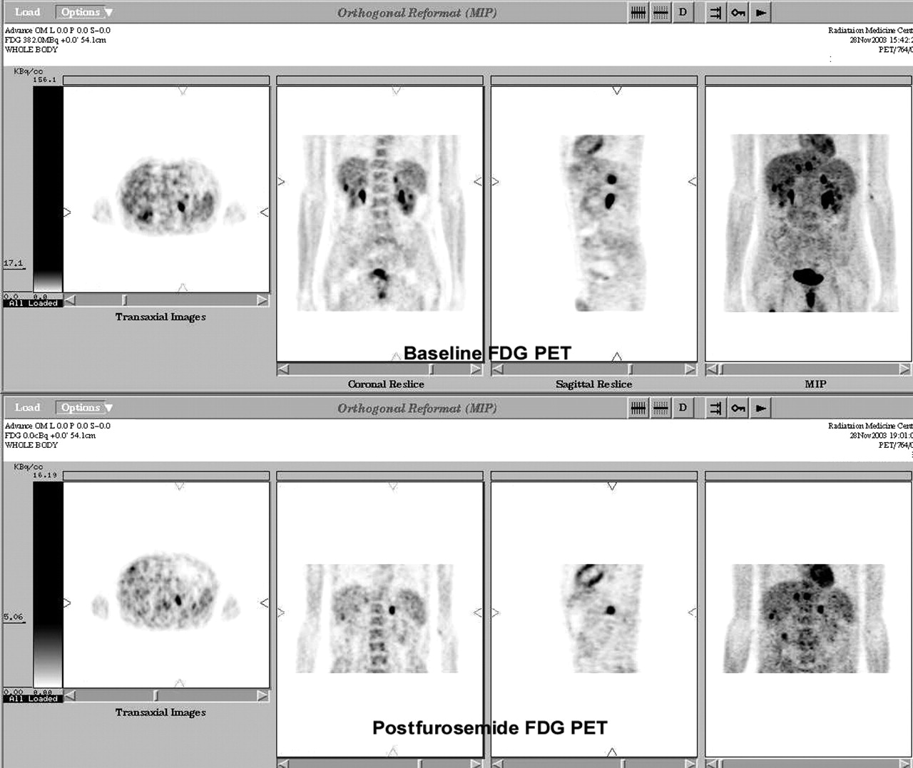
  • FIGURE 3.
    • Download figure
    • Open in new tab
    • Download powerpoint
    FIGURE 3.

    18F-FDG PET images of 64-y-old man with 2.5-mo history of right-sided chest pain, cough, and hemoptysis. Thoracic CT revealed 5.5 × 3.8 cm spiculated mass in right upper lobe and extending to hilum. Cytology of CT-guided fine-needle aspirate was suggestive of adenocarcinoma. He was referred for metastatic survey with PET.
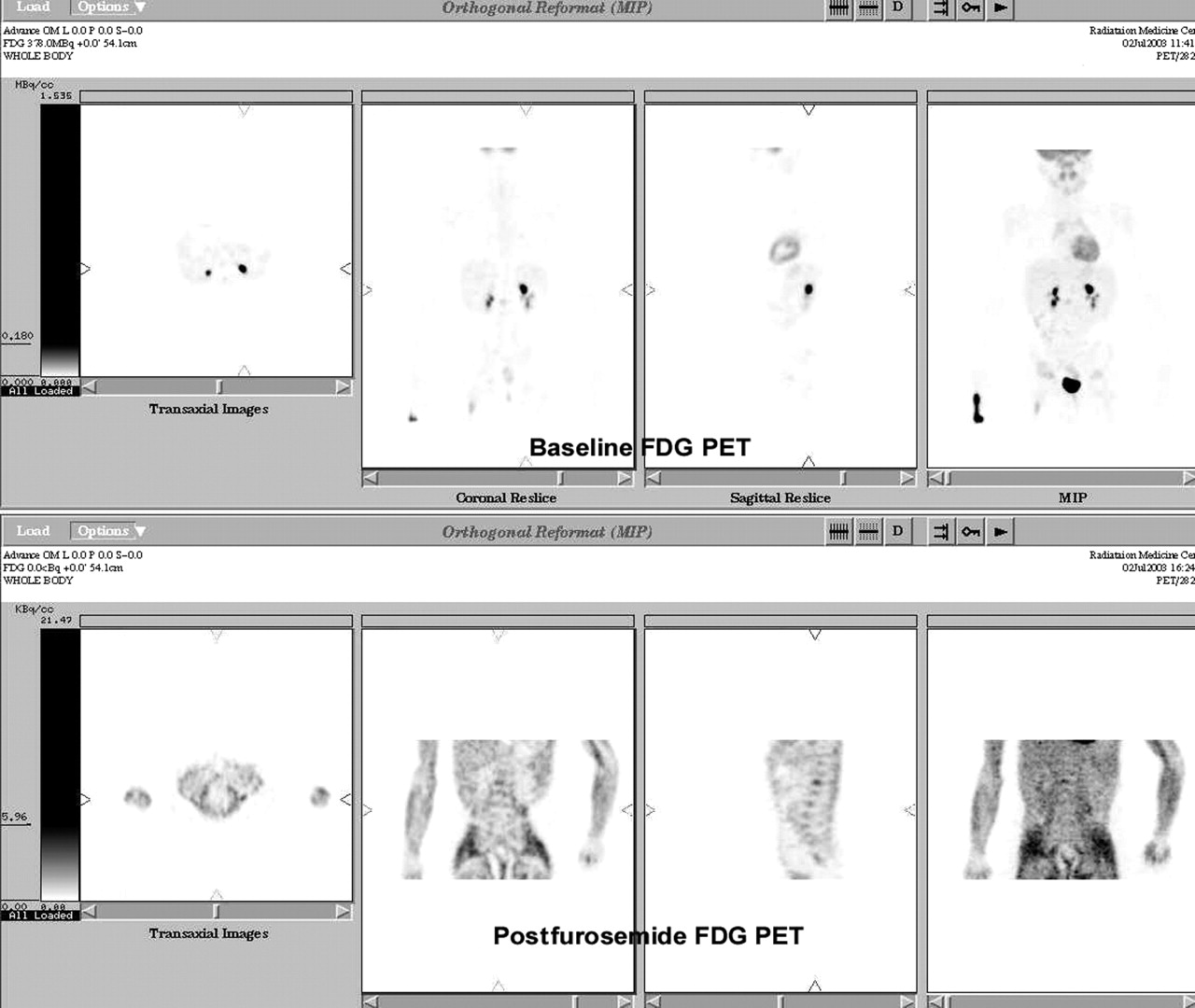
  • FIGURE 4.
    • Download figure
    • Open in new tab
    • Download powerpoint
    FIGURE 4.

    18F-FDG PET images of 15-y-old boy in whom non-Hodgkin’s lymphoma had been diagnosed at the age of 6 y and who had been disease free and asymptomatic for the past 8 y. Recent abdominal CT performed as part of routine follow-up had shown hypodense, minimally enhancing 1-cm area in right kidney most likely representing a cystic lesion. However, PET was performed because, considering patient’s history, a lymphomatous deposit could not be ruled out.
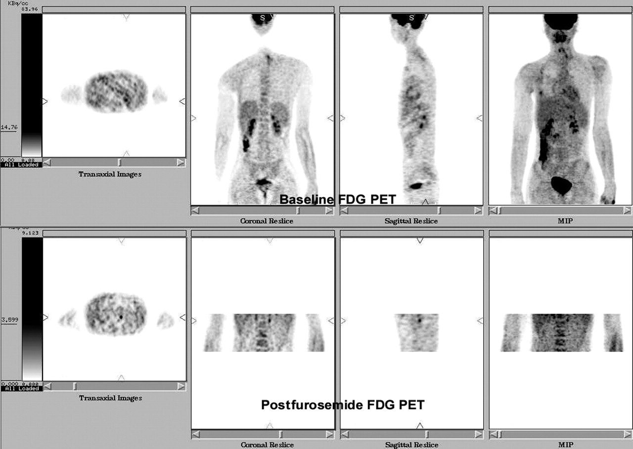
  • FIGURE 5.
    • Download figure
    • Open in new tab
    • Download powerpoint
    FIGURE 5.

    18F-FDG PET images of 15-y-old boy with primitive neuroectodermal tumor of right chest wall. PET was performed to evaluate the disease after surgery and chemotherapy. Recent thoracic CT had shown evidence of partial collapse and consolidation of anterior segment of right upper and middle lobes, with thickening of right minor fissure.

PreviousNext
Back to top

In this issue

Journal of Nuclear Medicine Technology: 33 (3)
Journal of Nuclear Medicine Technology
Vol. 33, Issue 3
September 1, 2005
  • Table of Contents
  • About the Cover
  • Index by author
Print
Download PDF
Article Alerts
Sign In to Email Alerts with your Email Address
Email Article

Thank you for your interest in spreading the word on Journal of Nuclear Medicine Technology.

NOTE: We only request your email address so that the person you are recommending the page to knows that you wanted them to see it, and that it is not junk mail. We do not capture any email address.

Enter multiple addresses on separate lines or separate them with commas.
Selected Cases Demonstrating the Value of Furosemide-Primed 18F-FDG PET in Identifying Adrenal Involvement
(Your Name) has sent you a message from Journal of Nuclear Medicine Technology
(Your Name) thought you would like to see the Journal of Nuclear Medicine Technology web site.
Citation Tools
Selected Cases Demonstrating the Value of Furosemide-Primed 18F-FDG PET in Identifying Adrenal Involvement
Narendra Nair, Sandip Basu
Journal of Nuclear Medicine Technology Sep 2005, 33 (3) 166-171;

Citation Manager Formats

  • BibTeX
  • Bookends
  • EasyBib
  • EndNote (tagged)
  • EndNote 8 (xml)
  • Medlars
  • Mendeley
  • Papers
  • RefWorks Tagged
  • Ref Manager
  • RIS
  • Zotero
Share
Selected Cases Demonstrating the Value of Furosemide-Primed 18F-FDG PET in Identifying Adrenal Involvement
Narendra Nair, Sandip Basu
Journal of Nuclear Medicine Technology Sep 2005, 33 (3) 166-171;
Twitter logo Facebook logo LinkedIn logo Mendeley logo
  • Tweet Widget
  • Facebook Like
  • Google Plus One
Bookmark this article

Jump to section

  • Article
    • Abstract
    • MATERIALS AND METHODS
    • RESULTS
    • DISCUSSION
    • CONCLUSION
    • Footnotes
    • REFERENCES
  • Figures & Data
  • Info & Metrics
  • PDF

Related Articles

  • No related articles found.
  • PubMed
  • Google Scholar

Cited By...

  • Could Different Hydration Protocols Affect the Quality of 18F-FDG PET/CT Images?
  • Forced Diuresis Improves the Diagnostic Accuracy of 18F-FDG PET in Abdominopelvic Malignancies
  • Google Scholar

More in this TOC Section

  • Early 10-Minute Postinjection [18F]F-FAPI-42 uEXPLORER Total-Body PET/CT Scanning Protocol for Staging Lung Cancer Using HYPER Iterative Reconstruction
  • Single- Versus Dual-Time-Point Imaging for Transthyretin Cardiac Amyloid Using 99mTc-Pyrophosphate
  • Does Arthrography Improve Accuracy of SPECT/CT for Diagnosis of Aseptic Loosening in Patients with Painful Knee Arthroplasty: A Systematic Review and Metaanalysis
Show more Imaging

Similar Articles

SNMMI

© 2025 SNMMI

Powered by HighWire