


Article Figures & Data
Figures
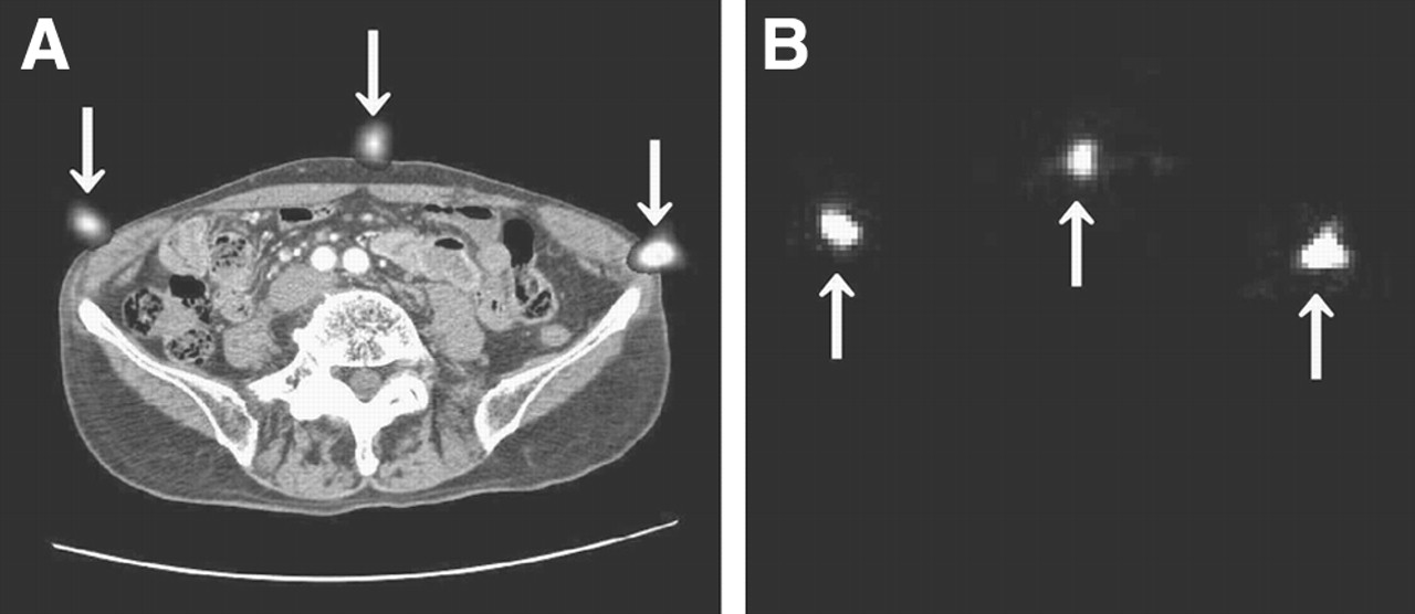
- FIGURE 1.
Landmarks (arrows) on axial slices of CT (A) and SPECT (B).
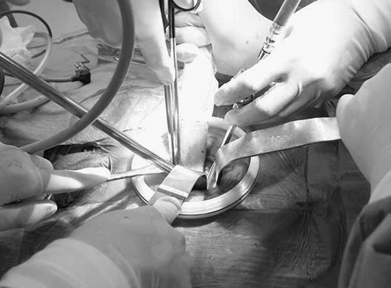
- FIGURE 2.
Endoscopic lymphadenectomy by means of minilaparotomy.
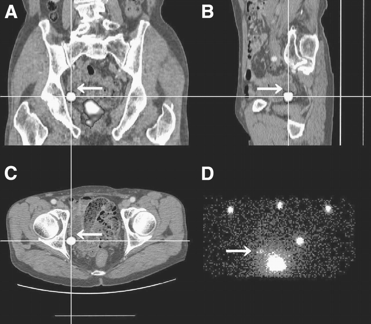
- FIGURE 3.
Fusion of SPECT and CT images indicating left external iliac lymph node (arrow) in case 2. (A) Coronal slice. (B) Sagittal slice. (C) Axial slice. (D) Anterior planar lymphoscintigraphic image.
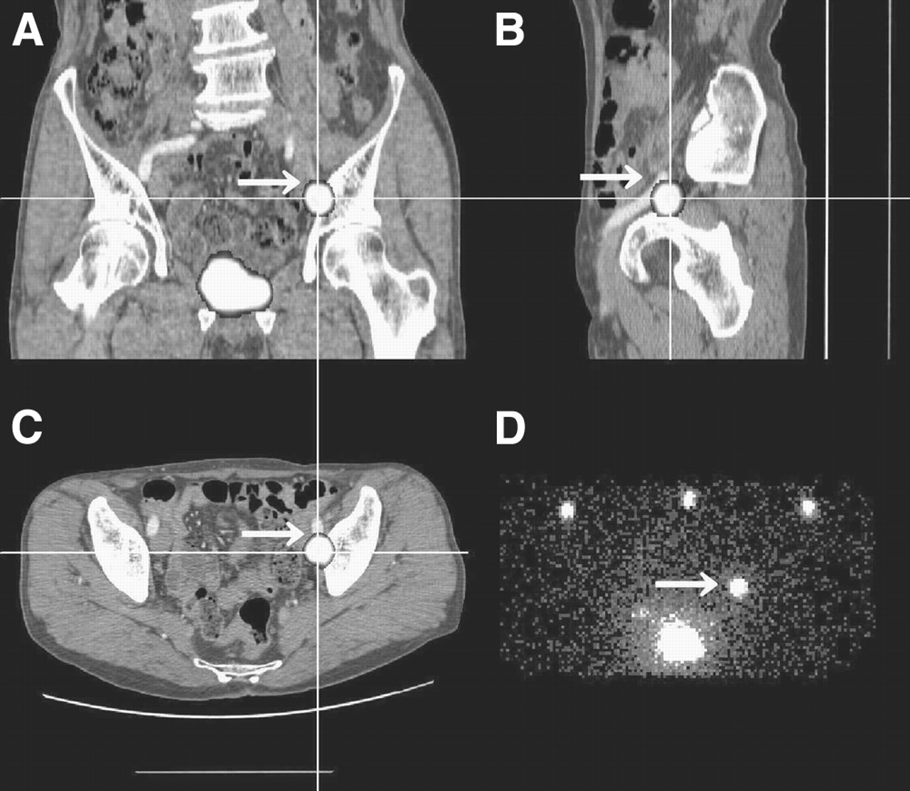
- FIGURE 4.
Fusion of SPECT and CT images indicating right obturator lymph node (arrow) in case 2. (A) Coronal slice. (B) Sagittal slice. (C) Axial slice. (D) Anterior planar lymphoscintigraphic image.


Tables
Case Size, mm × mm (cps), of the following lymph node*: External iliac Internal iliac Obturator R L R L R L 1 (252) (1,740) (110) 2 6 × 4 (717)† 10 × 7 (10) 3 6 × 4 (34), 3 × 2 (4) 4 8 × 5 (522) 5 5 × 5 (196) 6 × 4 (12) 6 (1,673) (105) (220) 7 5 × 3 (3)‡ 8 9 × 6 (1)‡ 4 × 3 (2)‡ 9 9 × 4 (62) 2 × 2 (27) 5 × 4 (698) 4 × 3 (372) 10 5 × 5 (10), 7 × 5 (41) 8 × 7 (179), 10 × 8 (695) 4 × 4 (853) 7 × 5 (684) 11 15 × 10 (430) 8 × 7 (3,556) 3 × 3 (349), 4 × 3 (56) 2 × 2 (11) Total 7 6 5 4 4 5


Jump to section
Related Articles
Cited By...
- Value of SPECT/CT for Detection and Anatomic Localization of Sentinel Lymph Nodes Before Laparoscopic Sentinel Node Lymphadenectomy in Prostate Carcinoma
- Sentinel lymph node biopsy using dynamic lymphoscintigraphy combined with ultrasound-guided fine needle aspiration in penile carcinoma
- Localization of Metastases from Malignant Pheochromocytoma in Patients Undergoing 131I-MIBG Therapy with Manually Fused 123I-MIBG SPECT and CT Images
- The Additional Value of SPECT/CT in Lymphatic Mapping in Breast Cancer and Melanoma