
Main menu

  • Home
  • Content
    • Current
      • JNMT Supplement
    • Ahead of print
    • Past Issues
    • Continuing Education
    • JNMT Podcast
    • SNMMI Annual Meeting Abstracts
  • Subscriptions
    • Subscribers
    • Rates
    • Journal Claims
    • Institutional and Non-member
  • Authors
    • Submit to JNMT
    • Information for Authors
    • Assignment of Copyright
    • AQARA Requirements
  • Info
    • Reviewers
    • Permissions
    • Advertisers
    • Corporate & Special Sales
  • About
    • About Us
    • Editorial Board
    • Contact Information
  • More
    • Alerts
    • Feedback
    • Help
    • SNMMI Journals
  • SNMMI
    • JNMT
    • JNM
    • SNMMI Journals
    • SNMMI

User menu

  • Subscribe
  • My alerts
  • Log in
  • Log out
  • My Cart

Search

  • Advanced search
Journal of Nuclear Medicine Technology
  • SNMMI
    • JNMT
    • JNM
    • SNMMI Journals
    • SNMMI
  • Subscribe
  • My alerts
  • Log in
  • Log out
  • My Cart
Journal of Nuclear Medicine Technology

Advanced Search

  • Home
  • Content
    • Current
    • Ahead of print
    • Past Issues
    • Continuing Education
    • JNMT Podcast
    • SNMMI Annual Meeting Abstracts
  • Subscriptions
    • Subscribers
    • Rates
    • Journal Claims
    • Institutional and Non-member
  • Authors
    • Submit to JNMT
    • Information for Authors
    • Assignment of Copyright
    • AQARA Requirements
  • Info
    • Reviewers
    • Permissions
    • Advertisers
    • Corporate & Special Sales
  • About
    • About Us
    • Editorial Board
    • Contact Information
  • More
    • Alerts
    • Feedback
    • Help
    • SNMMI Journals
  • Watch or Listen to JNMT Podcast
  • Visit SNMMI on Facebook
  • Join SNMMI on LinkedIn
  • Follow SNMMI on Twitter
  • Subscribe to JNMT RSS feeds
OtherIMAGING

Localization of Metastases from Malignant Pheochromocytoma in Patients Undergoing 131I-MIBG Therapy with Manually Fused 123I-MIBG SPECT and CT Images

Hiroto Kizu, Teruhiko Takayama, Hiroyuki Tsushima, Atsushi Noguchi, Kenichi Nakajima, Masahisa Onoguchi and Seigo Kinuya
Journal of Nuclear Medicine Technology December 2008, 36 (4) 186-188; DOI: https://doi.org/10.2967/jnmt.108.051656
Hiroto Kizu
  • Find this author on Google Scholar
  • Find this author on PubMed
  • Search for this author on this site
Teruhiko Takayama
  • Find this author on Google Scholar
  • Find this author on PubMed
  • Search for this author on this site
Hiroyuki Tsushima
  • Find this author on Google Scholar
  • Find this author on PubMed
  • Search for this author on this site
Atsushi Noguchi
  • Find this author on Google Scholar
  • Find this author on PubMed
  • Search for this author on this site
Kenichi Nakajima
  • Find this author on Google Scholar
  • Find this author on PubMed
  • Search for this author on this site
Masahisa Onoguchi
  • Find this author on Google Scholar
  • Find this author on PubMed
  • Search for this author on this site
Seigo Kinuya
  • Find this author on Google Scholar
  • Find this author on PubMed
  • Search for this author on this site
  • Article
  • Figures & Data
  • Info & Metrics
  • PDF
Loading

Abstract

131I-metaiodobenzylguanidine (MIBG) has been used as a therapeutic agent for pheochromocytoma. Tumor localization and precise staging are essential for therapy with high-dose 131I-MIBG. The sites and extent of 123I-MIBG uptake are usually estimated to predict the effectiveness of therapy before administration. However, conventional scintigraphic images provide insufficient anatomic information. Therefore, we tried to manually superimpose 123I-MIBG SPECT and CT images using free software.

  • pheochromocytoma
  • SPECT
  • registration
  • 123I-MIBG
  • x-ray CT

The recent introduction of a hybrid SPECT/CT system has facilitated the process of image fusion (1), but the availability of such specialized equipment is still limited. Therefore, we tried to construct SPECT/CT fusion images without using such equipment (2–6). This study investigated the feasibility and validity of producing SPECT/CT fusion images for accurate localization of metastases from malignant pheochromocytoma.

MATERIALS AND METHODS

This study enrolled 7 patients referred to the Kanazawa University Hospital for therapy with high-dose 131I-metaiodobenzylguanidine (MIBG) for recurrence or metastases from malignant pheochromocytoma (7–9). All patients underwent CT and 123I-MIBG imaging to evaluate the sites and extent of 131I-MIBG uptake. On imaging of CT and scintigraphy, external markers were placed on the same skin surfaces: the center of the sternal notch, the xiphoid process, and bilaterally on the iliac crests (Fig. 1). The markers consisted of 4 small plastic bullets with a diameter of 8 mm, and for SPECT the markers contained 0.3 MBq of 99mTc-pertechnetate. Imaging datasets from the 2 modalities were coregistered and manually superimposed using free software (MRIcro; www.mricro.com) (10).

FIGURE 1. 
  • Download figure
  • Open in new tab
  • Download powerpoint
FIGURE 1. 

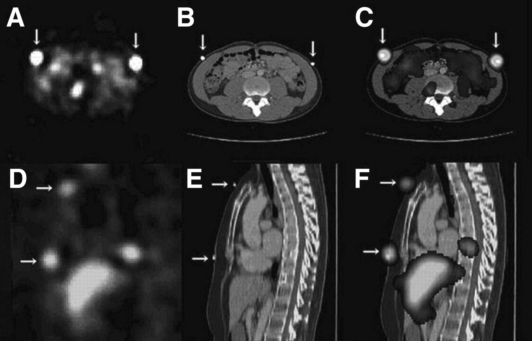
SPECT images (A, axial; D, sagittal) and CT images (B, axial; E, sagittal) show external markers bilaterally on iliac crests (arrows), sternal notch, and xiphoid process (arrows). In fused images (C, axial; F, sagittal), all CT markers are completely included in SPECT markers, representing good registration.

RESULTS

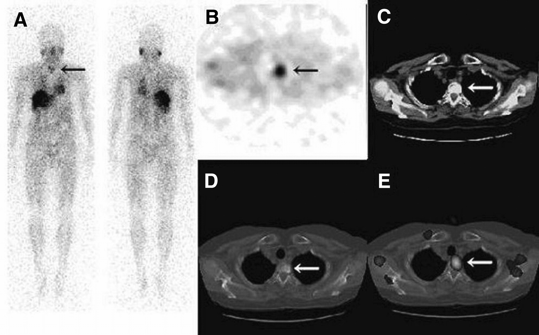
Table 1 shows the rate of metastasis detection by the different imaging modalities. Overall, 123I-MIBG scintigraphy detected 56 metastatic lesions, comprising 35 in bone, 10 in the chest, and 11 in the abdomen. In patient 1, bone metastasis was overlooked on CT images, despite abnormal uptake at the upper lung level on scintigraphy (Fig. 2). Fused images demonstrated that the site of abnormal uptake corresponded to a metastasis on the T2 vertebra, which was confirmed with MRI. In contrast, CT was superior to scintigraphy in detecting small lesions in the lung field and liver. In our experience, poor registrations occurred more frequently in the lower chest and upper abdomen. Registration of bone lesions was performed correctly because of the limited effect of respiratory movement on those regions.

FIGURE 2. 
  • Download figure
  • Open in new tab
  • Download powerpoint
FIGURE 2. 

(A) 123I-MIBG whole-body scintigraphy of 72-y-old woman (left, anterior; right, posterior) shows area (arrow) of abnormally increased tracer uptake at level of upper chest. (B) Axial SPECT image clearly shows increased tracer uptake, but sites were indeterminate because of lack of anatomic structures. (C and D) Axial CT images do not depict any abnormality under routine imaging conditions, with heterogeneous density under optimal window level (arrows). (E) Fused image shows abnormal uptake (arrow) in metastatic T2 vertebra, with good registration.

View this table:
  • View inline
  • View popup
TABLE 1

Detection of Metastases by Different Imaging Modalities in the 7 Patients

DISCUSSION

Compared with hybrid SPECT/CT systems, the procedure in the present study has some advantages. Fusion of images can easily be obtained without such specialized equipment, and the patient's exposure to radiation is reduced. We used the data from CT to construct fusion images, which were used to search for metastases. But, the procedure in the present study has some disadvantages. Manual fusion requires a few minutes for data transfer and processing, and external markers need to be correctly placed on the body surface to avoid misregistration between CT and scintigraphic images.

CONCLUSION

Image fusion was successful, showed the relationship between the structural CT data and the functional SPECT data, and precisely localized metastatic pheochromocytoma.

Footnotes

  • COPYRIGHT © 2008 by the Society of Nuclear Medicine, Inc.

References

  1. 1.↵
    Schillaci O, Fillippi L, Danieli R, Simonetti G. Single-photon emission computed tomography/computed tomography in abdominal disease. Semin Nucl Med. 2007;37:48–61.
    OpenUrlCrossRefPubMed
  2. 2.↵
    Israel O, Keidar Z, Iosilevsky G, Bettman L, Sachs J, Frenkel A. The fusion of anatomic and physiologic imaging in the management of patients with cancer. Semin Nucl Med. 2001;31:191–205.
    OpenUrlCrossRefPubMed
  3. 3.
    Kizu H, Takayama T, Fukuda M, et al. Fusion of SPECT and multidetector CT images for accurate localization of pelvic sentinel lymph nodes in prostate cancer patient. J Nucl Med Technol. 2005;33:78–82.
    OpenUrlAbstract/FREE Full Text
  4. 4.
    Tang HR, Da Sliva AJ, Matthay KK, et al. Neuroblastoma imaging using a combined CT scanner-scintillation camera and 131I-MIBG. J Nucl Med. 2001;42:237–247.
    OpenUrlAbstract/FREE Full Text
  5. 5.
    Perault C, Schvartz C, Wampach H, Liehn JC, Delisle MJ. Thoracic and abdominal SPECT-CT image fusion without external markers in endocrine carcinomas. J Nucl Med. 1997;38:1234–1242.
    OpenUrlAbstract/FREE Full Text
  6. 6.↵
    Yamamoto Y, Nishiyama Y, Monden T, Matsumura Y, Satoh K, Ohkawa M. Clinical usefulness of fusion of 131I-SPECT and CT images in patients with differentiated thyroid carcinoma. J Nucl Med. 2003;44:1905–1910.
    OpenUrlAbstract/FREE Full Text
  7. 7.↵
    Loh KC, Fitzgerald PA, Matthay KK, Yeo PP, Price DC. The treatment of malignant pheochromocytoma with iodine-131 metaiodobenzylguanidine (131I-MIBG): a comprehensive review of 116 reported patients. J Endocrinol Invest. 1997;20:648–658.
    OpenUrlPubMed
  8. 8.
    Monsieurs M, Brans B, Bacher K, Dierckx R, Thierens H. Patient dosimetry for 131I-MIBG therapy for neuroendocrine tumours based on 123I-MIBG scans. Eur J Nucl Med Mol Imaging. 2002;29:1581–1587.
    OpenUrlCrossRefPubMed
  9. 9.↵
    Rose B, Matthay KK, Price D, et al. High-dose 131I-metaiodobenzylguanidine therapy for 12 patients with malignant pheochromocytoma. Cancer. 2003;98:239–248.
    OpenUrlCrossRefPubMed
  10. 10.↵
    Rorden C, Brett M. Stereotaxic display of brain lesions. Behav Neurol. 2000;12:191–200.
    OpenUrlPubMed
  • Received for publication February 11, 2008.
  • Accepted for publication August 20, 2008.
PreviousNext
Back to top

In this issue

Journal of Nuclear Medicine Technology: 36 (4)
Journal of Nuclear Medicine Technology
Vol. 36, Issue 4
December 2008
  • Table of Contents
  • About the Cover
  • Index by author
Print
Download PDF
Article Alerts
Sign In to Email Alerts with your Email Address
Email Article

Thank you for your interest in spreading the word on Journal of Nuclear Medicine Technology.

NOTE: We only request your email address so that the person you are recommending the page to knows that you wanted them to see it, and that it is not junk mail. We do not capture any email address.

Enter multiple addresses on separate lines or separate them with commas.
Localization of Metastases from Malignant Pheochromocytoma in Patients Undergoing 131I-MIBG Therapy with Manually Fused 123I-MIBG SPECT and CT Images
(Your Name) has sent you a message from Journal of Nuclear Medicine Technology
(Your Name) thought you would like to see the Journal of Nuclear Medicine Technology web site.
Citation Tools
Localization of Metastases from Malignant Pheochromocytoma in Patients Undergoing 131I-MIBG Therapy with Manually Fused 123I-MIBG SPECT and CT Images
Hiroto Kizu, Teruhiko Takayama, Hiroyuki Tsushima, Atsushi Noguchi, Kenichi Nakajima, Masahisa Onoguchi, Seigo Kinuya
Journal of Nuclear Medicine Technology Dec 2008, 36 (4) 186-188; DOI: 10.2967/jnmt.108.051656

Citation Manager Formats

  • BibTeX
  • Bookends
  • EasyBib
  • EndNote (tagged)
  • EndNote 8 (xml)
  • Medlars
  • Mendeley
  • Papers
  • RefWorks Tagged
  • Ref Manager
  • RIS
  • Zotero
Share
Localization of Metastases from Malignant Pheochromocytoma in Patients Undergoing 131I-MIBG Therapy with Manually Fused 123I-MIBG SPECT and CT Images
Hiroto Kizu, Teruhiko Takayama, Hiroyuki Tsushima, Atsushi Noguchi, Kenichi Nakajima, Masahisa Onoguchi, Seigo Kinuya
Journal of Nuclear Medicine Technology Dec 2008, 36 (4) 186-188; DOI: 10.2967/jnmt.108.051656
Twitter logo Facebook logo LinkedIn logo Mendeley logo
  • Tweet Widget
  • Facebook Like
  • Google Plus One
Bookmark this article

Jump to section

  • Article
    • Abstract
    • MATERIALS AND METHODS
    • RESULTS
    • DISCUSSION
    • CONCLUSION
    • Footnotes
    • References
  • Figures & Data
  • Info & Metrics
  • PDF

Related Articles

  • No related articles found.
  • PubMed
  • Google Scholar

Cited By...

  • No citing articles found.
  • Google Scholar

More in this TOC Section

  • Early 10-Minute Postinjection [18F]F-FAPI-42 uEXPLORER Total-Body PET/CT Scanning Protocol for Staging Lung Cancer Using HYPER Iterative Reconstruction
  • Single- Versus Dual-Time-Point Imaging for Transthyretin Cardiac Amyloid Using 99mTc-Pyrophosphate
  • Does Arthrography Improve Accuracy of SPECT/CT for Diagnosis of Aseptic Loosening in Patients with Painful Knee Arthroplasty: A Systematic Review and Metaanalysis
Show more Imaging

Similar Articles

SNMMI

© 2025 SNMMI

Powered by HighWire