


Abstract
18F-FDG uptake in brown adipose tissue (BAT) can complicate interpretation and quantification of PET images, especially in regions of possible lymph node metastases such as the axilla and the mediastinum. The aim of this study was to prospectively evaluate the effect of patient preparation using a single oral dose of diazepam and controlled indoor temperature to prevent 18F-FDG uptake in BAT in breast cancer patients referred for monitoring of therapy response with 18F-FDG PET. Methods: During the fall and winter months, 53 patients referred for 18F-FDG PET/CT of breast cancer were included. A cohort of 25 patients was imaged without an intervention, and a second cohort of 28 patients was prepared according to a new protocol that included 10 mg of diazepam and adequate indoor temperature. The generated images were visually assessed for the presence of 18F-FDG at the location of fat-density tissue on CT images using a 4-point scale. Results: In the cohort without intervention, relevant 18F-FDG uptake in BAT was identified in 4 patients (16%); in the cohort prepared according to the proposed protocol, in only 1 patient (4%). The mean score of BAT 18F-FDG uptake evaluated with the 4-point system was 0.04 in the group treated according to the new protocol and 0.16 in the group treated according to the previous protocol. Conclusion: In the clinically relevant group of breast cancer patients, 18F-FDG uptake in BAT can be reduced by a single oral administration of diazepam combined with controlled room temperature in resting rooms.
PET with 18F-FDG, either as a separate modality or in combination with CT, is a powerful imaging tool for the detection, staging, and prediction of the therapy outcome of malignancies (1), including breast cancer (2,3). 18F-FDG PET may also be applied with high specificity to the detection of lymph node metastases from breast cancer (4,5). However, 18F-FDG uptake in brown adipose tissue (BAT) is a potential source of false-positive interpretation (6–8). BAT is generally located in the supraclavicular area, in a bilateral, symmetric, and elongated configuration (7). However, it can also be seen in the axillae and mediastinum, which can make accurate interpretation of 18F-FDG PET/CT images difficult in these specific areas (9).
Multiple factors contributing to 18F-FDG uptake in BAT have been described in the literature. Several reports suggest a low outside temperature is associated with higher uptake (7,10). Other reports suggest that 18F-FDG uptake in BAT is more frequently seen in female patients (11,12) and in younger patients (13). Consequently, breast cancer patients are specifically at risk for 18F-FDG uptake in BAT because many of them are relatively young female patients. In these patients, BAT activity may complicate the interpretation of images in regions of possible lymph node metastases, leading to inconclusive scans. This is particularly critical in breast cancer patients with lymph node metastases who need to be monitored for therapy response during neoadjuvant systemic therapy, because BAT activity can make quantification and delineation of 18F-FDG uptake in malignant tissues in the affected areas difficult or impossible.
Several reports suggest that 18F-FDG BAT uptake can be reduced pharmacologically (14,15)—for example, with a single dose of diazepam as premedication (6). In addition, a constant ambient room temperature can reduce the 18F-FDG uptake in BAT (16,17). It is currently unknown to what extent these techniques or a combination thereof can prevent BAT activation in the clinically relevant and highly susceptible group of female patients with breast cancer.
The aim of this study was to prospectively evaluate the effect of patient preparation using a single oral dose of diazepam and adequate indoor temperature to prevent 18F-FDG uptake in BAT in breast cancer patients who were referred for the monitoring of therapy response using 18F-FDG PET during the fall and winter.
MATERIALS AND METHODS
Patients
A total of 53 consecutive clinical PET/CT scans were obtained at The Netherlands Cancer Institute and Antoni van Leeuwenhoek Hospital between September 2007 and March 2008. This period includes the fall and winter seasons in The Netherlands. All patients included in the study were referred as a result of oncologic imaging of breast cancer; most patients were referred in the context of a pilot study aimed to monitor therapy response in neoadjuvant systemic therapy. Twenty-five patients were included in 2007, and they received no specific intervention aimed at BAT uptake reduction. Subsequently, 28 patients were included in the colder period from January until March 2008, and they were prepared for 18F-FDG PET/CT using a new protocol aimed at elimination of BAT uptake. Patients were asked to arrive 15 min before 18F-FDG administration to adapt to the temperature in the resting room, which was maintained at a comfortable and stable level of 20–22°C. Patients were positioned in a comfortable bed and were instructed to relax before and after the administration of 18F-FDG. In addition, 10 mg of diazepam were administered orally 10 min before the administration of 18F-FDG.
18F-FDG PET/CT
The images were acquired using an integrated PET/CT scanner with time-of-flight capabilities (Gemini II; Philips). 18F-FDG PET/CT scans with attenuation correction based on low-dose CT were performed after a fasting period of 6 h. Blood glucose levels were required to be less than 11.0 mmol/L. The patients received 180–240 MBq of 18F-FDG intravenously. The time of 18F-FDG administration and body weight on the day of scanning were recorded. The interval between 18F-FDG administration and scanning was 60 ± 10 min. Generated images (PET/CT, low-dose CT, and PET) were displayed using an Osirix DICOM viewer in a Unix-based operating system (MAC OS X, Mac Pro; Apple) and were evaluated on the basis of 2-dimensional orthogonal reslicing. PET, fused PET/CT, and CT were subsequently analyzed side by side during a meeting of 3 experienced nuclear medicine physicians.
Uptake of 18F-FDG in BAT was defined as the presence of 18F-FDG on PET images at the location of fat-density tissue on CT images. 18F-FDG uptake in BAT was evaluated visually using a 4-point system: 0 = not distinguishable from normal surrounding background tissue (subcutaneous adipose tissue), 1 = slightly more than background intensity, 2 = moderately intense, 3 = very intense. Visual grades 2 and 3 were considered to potentially interfere with the interpretation and quantification of 18F-FDG PET/CT images.
Statistical Analysis
Statistical analysis was performed using SPSS 15 (version 15 for Windows; SPSS). The independent sample t test was used to compare age and body mass index (BMI) between the 2 cohorts. The Fisher exact test was used to compare the BAT uptake in the 2 groups.
RESULTS
A total of 53 patients were included, having a median age of 57 y (range, 28–84 y) and a median body mass index (BMI) of 26 (range, 18–39). No patients with diabetes mellitus were included in this study. Considering both patient cohorts, hypermetabolic BAT with increased 18F-FDG uptake (level 2–3) was identified on the PET images in 5 patients (9%). Areas of 18F-FDG uptake in BAT included the supraclavicular area, axillae, cervical area, paravertebral area, mediastinum, perirenal area, and prehepatic area and along the thoracic spine (Table 1; Fig. 1).
Characteristics of Patients with BAT Uptake of 18F-FDG
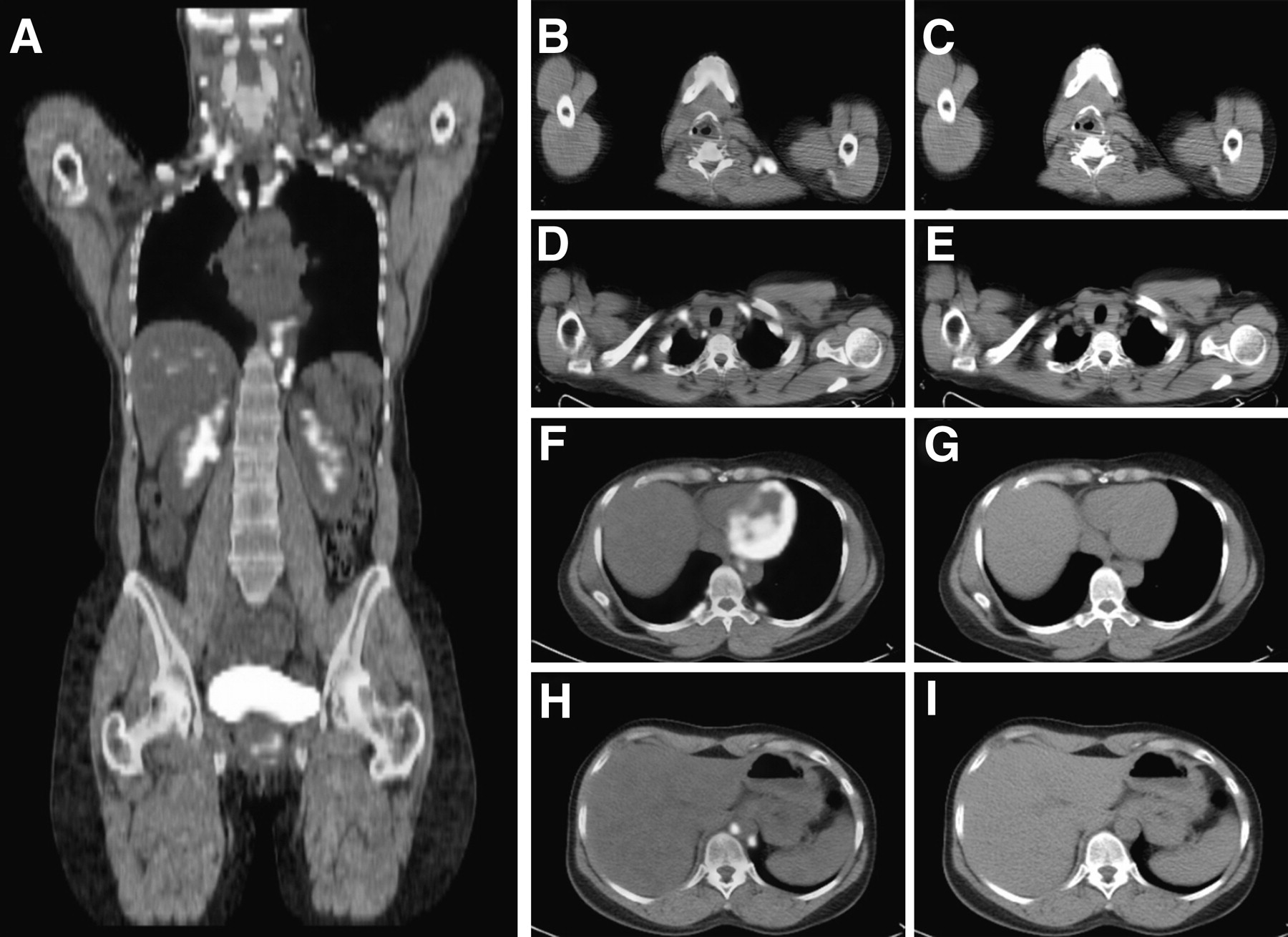
18F-FDG PET/CT showing extended uptake of 18F-FDG in fat-containing spaces of body in gray and white (A). This BAT uptake is seen only on axial fused PET/CT (B, D, F, and H), without abnormalities on CT (C, E, G, and I) in neck (B and C), supraclavicular area (D and E), mediastinal and paravertebral areas (F and G), and paraaortic area (H and I).
Table 2 summarizes the effect of the new protocol. 18F-FDG BAT uptake was diagnosed in 4 of 25 patients (16%) in the group before the intervention started, and in 1 of 28 patients (4%) in the group treated according to the new protocol (Fig. 2). No significant differences were seen between the groups in age and BMI (Table 2). The mean score of 18F-FDG uptake of BAT evaluated with the 4-point system was 0.04 in the group treated according to the new protocol and 0.16 in the group treated according to the old protocol.
Characteristics of the 2 Cohort Groups
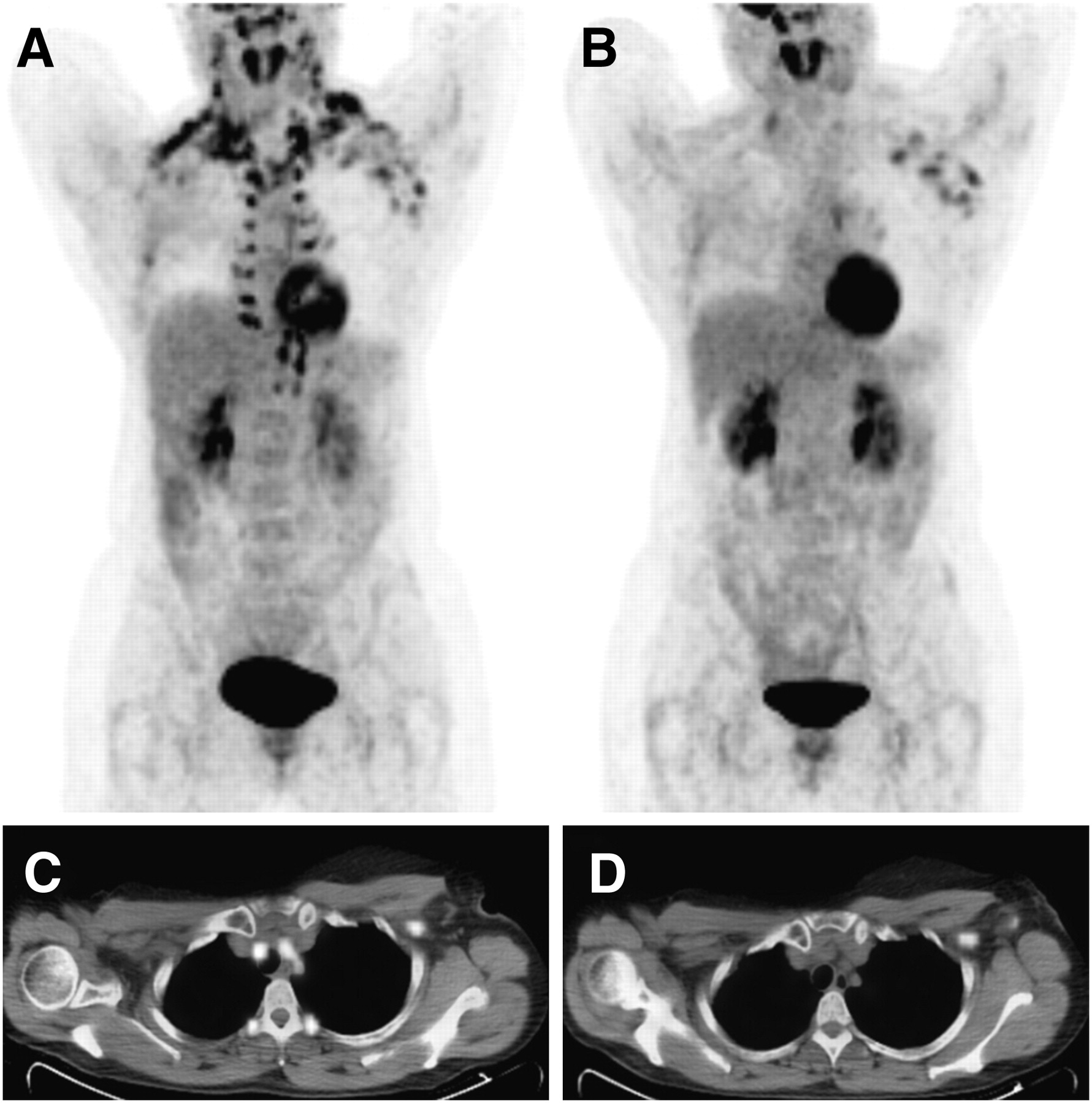
Coronal maximum-intensity-projection PET image (A) shows extended BAT uptake of 18F-FDG in neck, supraclavicular, axillary, mediastinal, paravertebral, and abdominal regions in breast cancer patient with axillary metastases. Second PET study of same patient (B) performed 1 wk later after preparation with diazepam and controlled room temperature shows no BAT uptake of 18F-FDG. Note on PET/CT scan that initial 18F-FDG uptake of BAT disappears (C) and only hypermetabolic lymph metastasis in left axilla is seen (D).
Diazepam was well tolerated by all patients. No serious adverse events were reported. The main side effect was somnolence. Patients were not allowed to drive home after having been given oral diazepam.
DISCUSSION
In this prospective cohort study, patient preparation using a single oral dose of 10 mg of diazepam as premedication and adaptation to a stable indoor temperature reduced the occurrence of 18F-FDG uptake in BAT in breast cancer patients.
Patients for the control group were recruited in the fall in The Netherlands, whereas patients in the intervention group were included during the colder winter. Therefore, our results are not biased by the influence of outdoor temperature on BAT uptake as described by Cohade et al. (11). Recently, Zukotynski et al. (17) and Marken Lichtenbelt et al. (18) demonstrated that maintaining a thermoneutral room temperature significantly decreased BAT uptake, providing more evidence that supports our conclusions. Cypess et al. (19) recently demonstrated that the amount of BAT is inversely correlated with the BMI. Our study was not confounded by this parameter because no significant difference was seen in BMI between the control group and the intervention group.
In our study, we chose to administer diazepam as a pharmacologic intervention, in concordance with the methods described by several other groups (6,14,15). Other studies describe a reduction of 18F-FDG uptake in BAT by a single dose of propranolol (15,20,21)..Williams and Kolodny (22) showed that the use of a high-fat preparation protocol significantly lowered the frequency of uptake of 18F-FDG by BAT. Interestingly, Baba et al. (23) showed that nicotine increased the 18F-FDG BAT uptake significantly. These findings suggest that the activation of BAT is highly multifactorial. On the basis of our results and the current literature (Table 3), we conclude that our combined approach of diazepam and maintained room temperature adequately prevents 18F-FDG BAT uptake in breast cancer patients.
Protocol to Reduce 18F-FDG Uptake in BAT in Breast Cancer Patients Undergoing 18F-FDG PET/CT
To our knowledge, ours is the first study that demonstrates the effect of intervention in the specific group of breast cancer patients who are principally referred for therapy response monitoring. Other studies have described BAT uptake in a heterogeneous group of patients (9,11, 12,17). Rousseau et al. (24) described BAT 18F-FDG uptake in breast cancer patients; however, no intervention was performed.
Our clinical experience, based on more than 6,000 oncologic patients, was that BAT uptake was most frequently seen in patients with breast cancer. This can be partially explained by the sex of most breast cancer patients because BAT uptake is more common in female patients (11,12). However, these results were not sufficient to change the clinical management of all female patients. As a result of our study, we changed the management in our institute specifically for breast cancer patients.
Some limitations of our study should be acknowledged. First, a limited number of 53 patients were included from September 2007 until March 2008, and this resulted in a low number of positive cases (5 patients). A larger number of patients could potentially increase the power of the study. However, including more patients during a longer period would affect our results because the outside temperature would rise, which could potentially bias our results.
Second, this study was not a randomized clinical controlled trial. However, the potential for selection bias was minimal because 18F-FDG BAT uptake is hardly missed, and all PET/CT images were discussed in a meeting of 3 experienced nuclear physicians. This study was a prospective cohort study, which ruled out the potential retrospective biases.
CONCLUSION
In the clinically relevant group of breast cancer patients, 18F-FDG uptake in BAT can be reduced by a single oral administration of diazepam combined with controlled room temperature in resting rooms.
Footnotes
-
COPYRIGHT © 2010 by the Society of Nuclear Medicine, Inc.
References
- Received for publication April 27, 2009.
- Accepted for publication January 14, 2010.


Jump to section
Related Articles
Cited By...
- Physiologic 18F-FDG Uptake in Brown Adipose Tissue and Lactating Breast in a Patient with Hodgkin Lymphoma
- Brown adipose tissue: a potential target in the fight against obesity and the metabolic syndrome
- Intervention to Lower Anxiety of 18F-FDG PET/CT Patients by Use of Audiovisual Imagery During the Uptake Phase Before Imaging