


Article Figures & Data
Figures
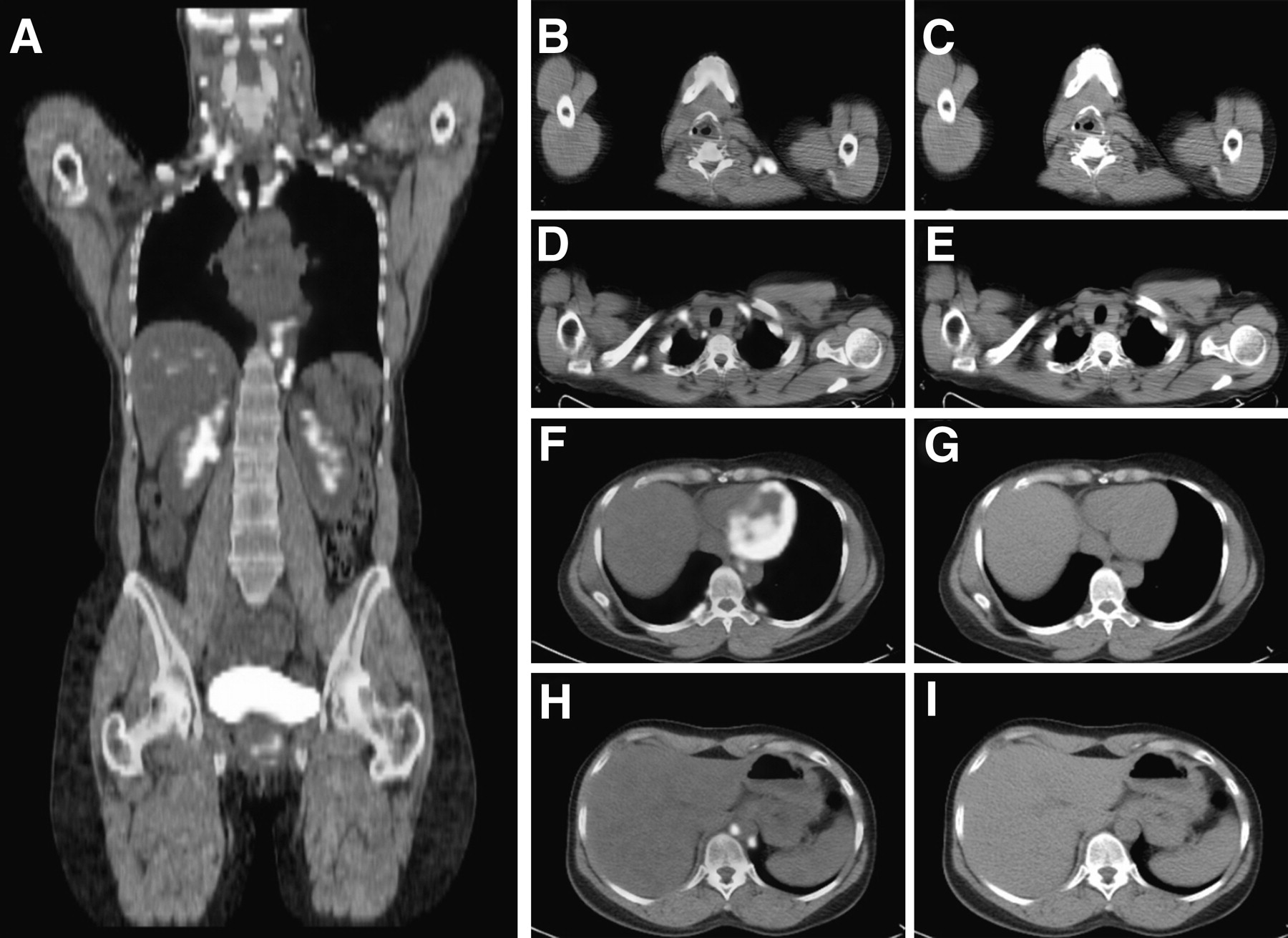
- FIGURE 1.
18F-FDG PET/CT showing extended uptake of 18F-FDG in fat-containing spaces of body in gray and white (A). This BAT uptake is seen only on axial fused PET/CT (B, D, F, and H), without abnormalities on CT (C, E, G, and I) in neck (B and C), supraclavicular area (D and E), mediastinal and paravertebral areas (F and G), and paraaortic area (H and I).
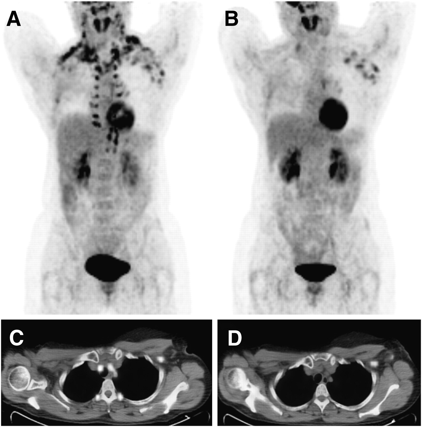
- FIGURE 2.
Coronal maximum-intensity-projection PET image (A) shows extended BAT uptake of 18F-FDG in neck, supraclavicular, axillary, mediastinal, paravertebral, and abdominal regions in breast cancer patient with axillary metastases. Second PET study of same patient (B) performed 1 wk later after preparation with diazepam and controlled room temperature shows no BAT uptake of 18F-FDG. Note on PET/CT scan that initial 18F-FDG uptake of BAT disappears (C) and only hypermetabolic lymph metastasis in left axilla is seen (D).
Tables
BAT location areas Intervention* Visual score† Cervical and supraclavicular No 3 Cervical, supraclavicular, axillary, and mediastinal No 3 Axillary, paravertebral, perirenal, and prehepatic No 3 Cervical, supraclavicular, axillary, paraspinal, and mediastinal No 2 Cervical, supraclavicular, axillary, and mediastinal Yes 2 - TABLE 3
Protocol to Reduce 18F-FDG Uptake in BAT in Breast Cancer Patients Undergoing 18F-FDG PET/CT
Preparation protocol Time of administration in relation to PET/CT acquisition High-fat, very-low-carbohydrate diet (22) Night before and morning of imaging No smoking (23) Day of imaging Constant temperature of 22–24°C in injection room (17,18) 15−30 min before intravenous tracer administration and during 1 h in resting room Oral administration of 20 mg of propranolol (20) or 10 mg of diazepam 15−30 min before intravenous tracer administration


Jump to section
Related Articles
Cited By...
- Physiologic 18F-FDG Uptake in Brown Adipose Tissue and Lactating Breast in a Patient with Hodgkin Lymphoma
- Brown adipose tissue: a potential target in the fight against obesity and the metabolic syndrome
- Intervention to Lower Anxiety of 18F-FDG PET/CT Patients by Use of Audiovisual Imagery During the Uptake Phase Before Imaging