


Article Figures & Data
Figures
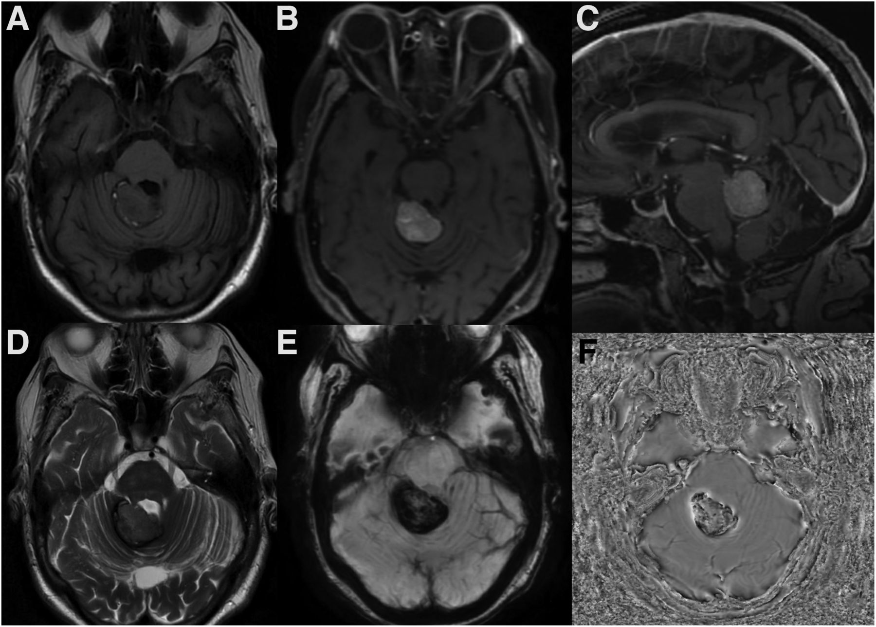
- FIGURE 1.
(A) Axial T1 unenhanced image demonstrates an extra-axial posterior fossa lesion in the right superior cerebellar region with predominantly intermediate T1 signal. (B) Axial and (C) sagittal T1 contrast-enhanced images show avid enhancement within this lesion. (D) Corresponding axial T2 image demonstrates intermediate signal intensity. (E) Susceptibility-weighted image and (F) phase image demonstrate signal loss within the lesion, suggesting intralesional hemorrhage.
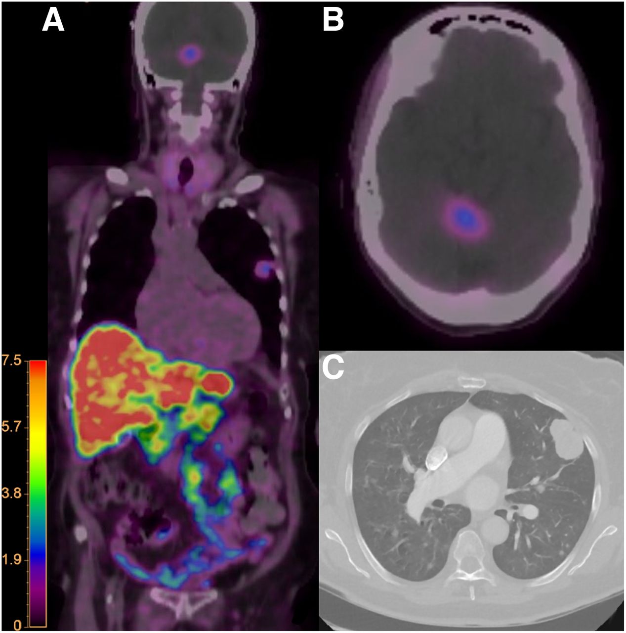
- FIGURE 2.
(A) Whole body coronal color fusion map and (B) axial color fusion map 68Ga-DOTATATE PET/CT demonstrate corresponding uptake within the posterior fossa lesion and left lung mass. (C) Chest CT demonstrates the mass in the left upper lob of the lung. Intensity scale bar in units of SUV.
- FIGURE 3.
18F-FDG PET/CT. (A) Whole-body coronal fusion map maximal-intensity-projection and (C) axial image of the chest color fusion map demonstrate increased uptake in the left upper lobe lung mass. (B) Axial image of the brain demonstrates uptake in the posterior fossa lesion similarly to that in the hypermetabolic brain parenchyma.

In this issue


Jump to section
Related Articles
Cited By...
- No citing articles found.