


Article Figures & Data
Figures
- FIGURE 1.
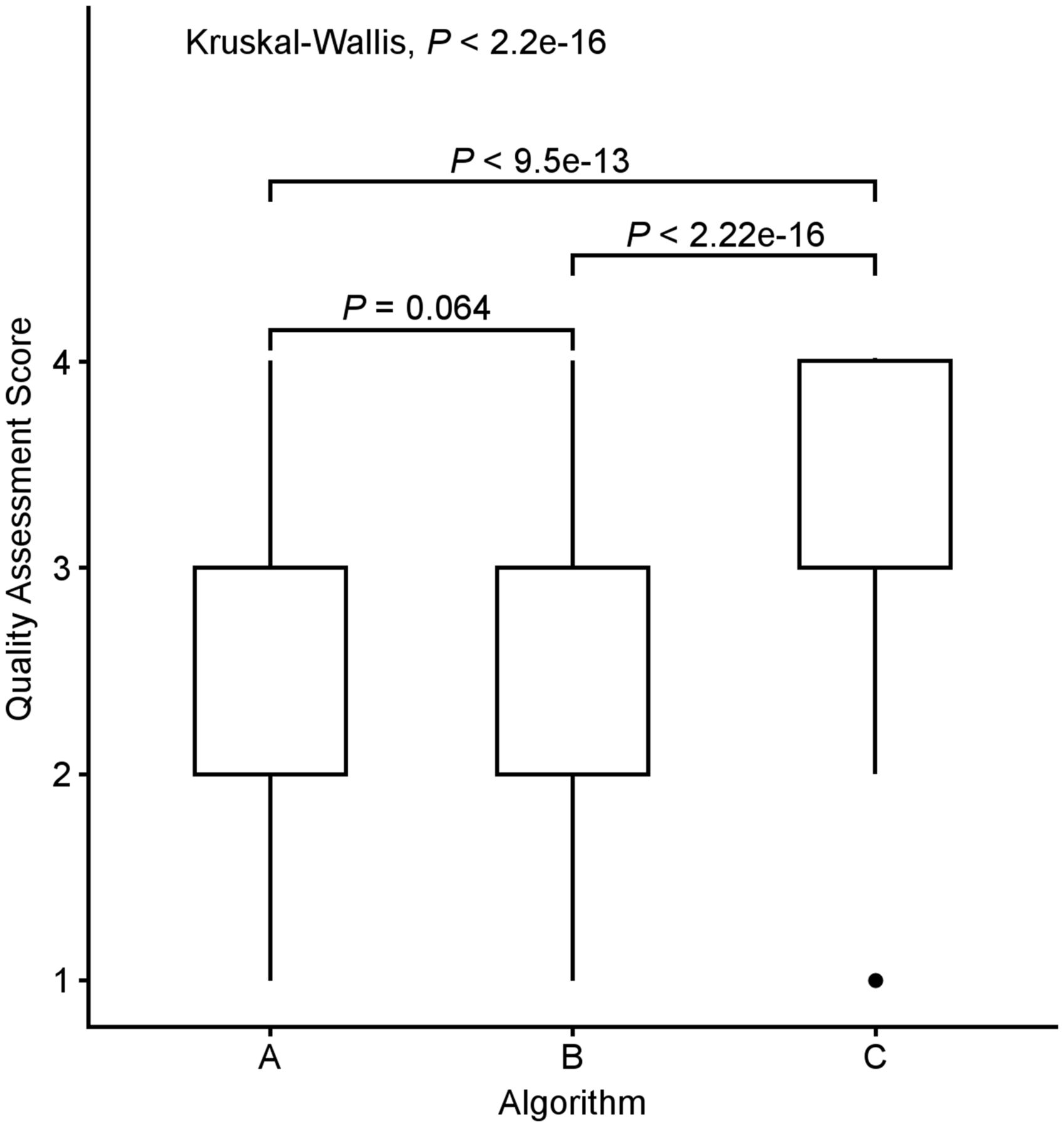
Box plots comparing median quality assessment scores among algorithms. Results from Kruskal–Wallis rank sum test and Wilcoxon rank sum test are included.
- FIGURE 2.
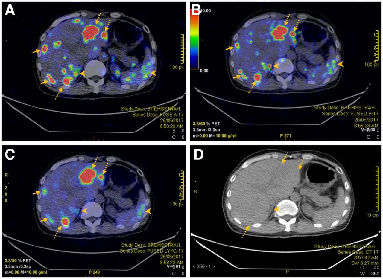
A 52-y-old man with rectal neuroendocrine cancer metastatic to liver underwent 90Y-PRRT therapy. Reconstructed PET/CT using algorithms A (A), B (B), and C (C) managed to detect hepatic metastases (dotted arrows) seen on corresponding CT images (D). However, there was more visible noise within liver for PET using algorithms A and B than for PET using algorithm C (solid arrows). In addition. extrahepatic noise such as that in right adrenal gland and spleen (arrowheads) was less apparent using algorithm C. Right adrenal noise can potentially be mistaken as hepatic metastasis using algorithms A and B (arrowheads).


Tables
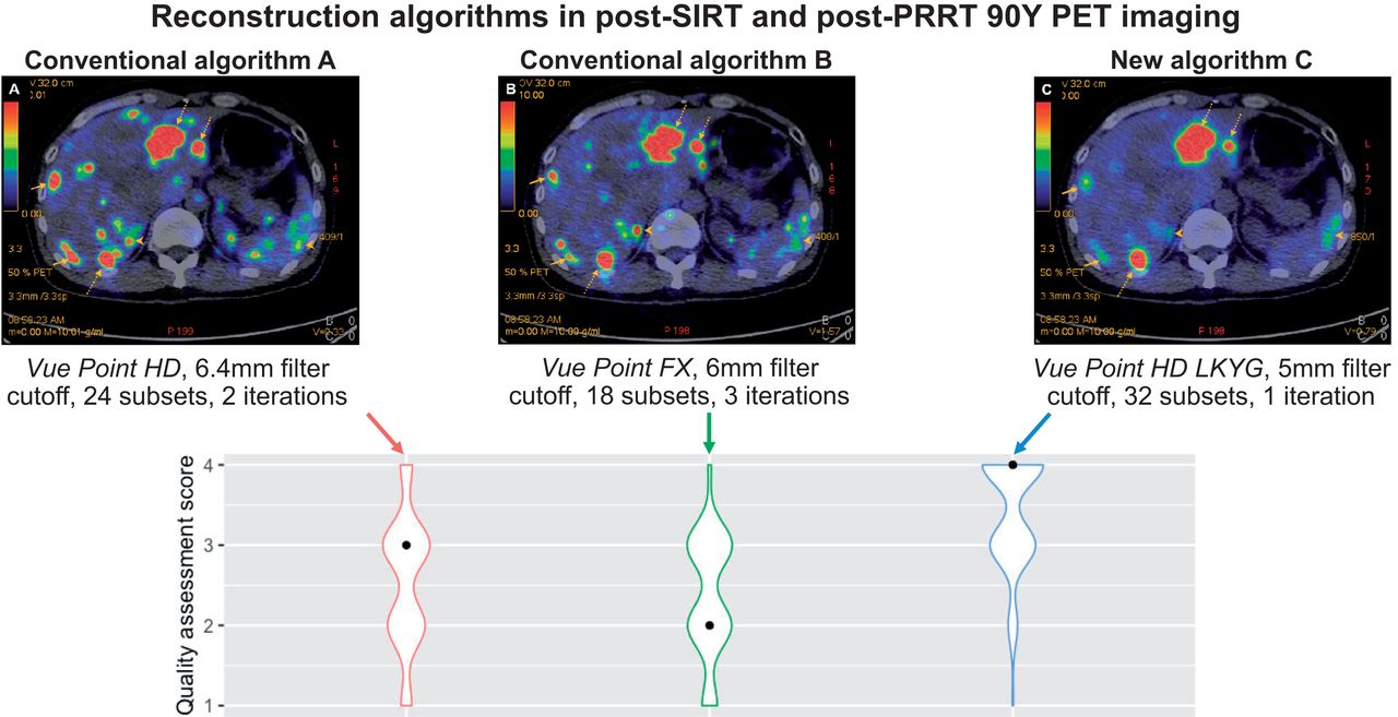
Parameter Algorithm A Algorithm B Algorithm C VUE Point VUE Point HD (OSEM) VUE Point FX (OSEM + TOF) VUE Point HD (OSEM) Gaussian filter cutoff 6.4 mm 6.0 mm 5.0 mm Number of subsets 24 18 32 Sharp IR (point-spread function) On On On z-axis filter Standard Heavy Standard Number of iterations 2 3 1 Matrix 192 × 192 192 × 192 192 × 192 Minutes per bed position 30 30 30 OSEM = ordered-subset expectation maximization.
QS Rating Description 1 Nondiagnostic Excessive noise or artifacts; delineation of tumor and background uptake mostly impossible 2 Barely diagnostic Substantial noise and artifacts; delineation of tumor and background uptake difficult but possible 3 Fairly diagnostic Somewhat noisy and artifacts that interfere with reading; delineation of tumor and background uptake feasible but not satisfactory 4 Diagnostically excellent No interfering noise or artifacts; delineation of tumor and background uptake satisfactory Participant Age (y) Sex BMI (kg/m2) Diagnosis Therapy Radiotracer dose (GBq) 1 52 M 20.6 Rectal NET PRRT 3.70 2 58 M 24.9 Midgut NET PRRT 3.70 3 39 M 19.2 Paraganglioma PRRT 4.22 4 54 F 21.8 Midgut NET metastatic to liver PRRT 3.03 5 41 M 19.7 Pancreatic NET metastatic to liver SIRT 2.97 6 68 M 25.9 HCC SIRT 1.30 7 69 M 26.1 HCC SIRT 0.58 8 59 M 29.9 HCC SIRT 2.50 9 96 M 25.9 HCC SIRT 0.73 10 79 F 21.4 HCC SIRT 3.00 BMI = body mass index; NET = neuroendocrine tumor; HCC = hepatocellular carcinoma.
- TABLE 4.
Number and Percentage of Discrete Scores Rated by 10 Readers on 10 Patients’ Scans Reconstructed Using Algorithms A–C
Algorithm Therapy Score 1 Score 2 Score 3 Score 4 P A SIRT 0 (0.0%) 11 (18.3%) 38 (63.3%) 11 (18.3%) <0.001 PRRT 10 (25.0%) 24 (60.0%) 6 (15.0%) 0 (0.0%) SIRT + PRRT 10 (10.0%) 35 (35.0%) 44 (44.0%) 11 (11.0%) B SIRT 0 (0.0%) 28 (46.7%) 29 (48.3%) 3 (5.0%) <0.001 PRRT 14 (35.0%) 13 (32.5%) 13 (32.5%) 0 (0.0%) SIRT + PRRT 14 (14.0%) 41 (41.0%) 42 (42.0%) 3 (3.0%) C SIRT 0 (0.0%) 0 (0.0%) 15 (25.0%) 45 (75.0%) <0.001 PRRT 1 (2.5%) 8 (20.0%) 24 (60.0%) 7 (17.5%) SIRT + PRRT 1 (1.0%) 8 (8.0%) 39 (39.0%) 52 (52.0%) Variable Multivariable model Adjusted OR 95% CI P Age 0.98 0.95–0.997 0.024 Sex Male Reference — — Female 0.83 0.44–1.58 0.576 Body mass index 0.90 0.81–0.99 0.026 Radioligand PRRT Reference — — SIRT 23.99 11.87–50.35 <0.001 Dose 0.89 0.66–1.19 0.418 Algorithm A Reference — — B 0.46 0.26–0.80 0.007 C 17.4 9.16–34.15 <0.001
In this issue


Jump to section
Related Articles
Cited By...
- No citing articles found.