
Main menu

  • Home
  • Content
    • Current
      • JNMT Supplement
    • Ahead of print
    • Past Issues
    • Continuing Education
    • JNMT Podcast
    • SNMMI Annual Meeting Abstracts
  • Subscriptions
    • Subscribers
    • Rates
    • Journal Claims
    • Institutional and Non-member
  • Authors
    • Submit to JNMT
    • Information for Authors
    • Assignment of Copyright
    • AQARA Requirements
  • Info
    • Reviewers
    • Permissions
    • Advertisers
    • Corporate & Special Sales
  • About
    • About Us
    • Editorial Board
    • Contact Information
  • More
    • Alerts
    • Feedback
    • Help
    • SNMMI Journals
  • SNMMI
    • JNMT
    • JNM
    • SNMMI Journals
    • SNMMI

User menu

  • Subscribe
  • My alerts
  • Log in
  • Log out
  • My Cart

Search

  • Advanced search
Journal of Nuclear Medicine Technology
  • SNMMI
    • JNMT
    • JNM
    • SNMMI Journals
    • SNMMI
  • Subscribe
  • My alerts
  • Log in
  • Log out
  • My Cart
Journal of Nuclear Medicine Technology

Advanced Search

  • Home
  • Content
    • Current
    • Ahead of print
    • Past Issues
    • Continuing Education
    • JNMT Podcast
    • SNMMI Annual Meeting Abstracts
  • Subscriptions
    • Subscribers
    • Rates
    • Journal Claims
    • Institutional and Non-member
  • Authors
    • Submit to JNMT
    • Information for Authors
    • Assignment of Copyright
    • AQARA Requirements
  • Info
    • Reviewers
    • Permissions
    • Advertisers
    • Corporate & Special Sales
  • About
    • About Us
    • Editorial Board
    • Contact Information
  • More
    • Alerts
    • Feedback
    • Help
    • SNMMI Journals
  • Watch or Listen to JNMT Podcast
  • Visit SNMMI on Facebook
  • Join SNMMI on LinkedIn
  • Follow SNMMI on Twitter
  • Subscribe to JNMT RSS feeds
Research ArticlePRACTICAL PROTOCOL TIP

Diuretic Renal Scintigraphy

Mary Beth Farrell, Kevin P. Banks and Justin G. Peacock
Journal of Nuclear Medicine Technology December 2022, 50 (4) 319-321; DOI: https://doi.org/10.2967/jnmt.122.264804
Mary Beth Farrell
  • Find this author on Google Scholar
  • Find this author on PubMed
  • Search for this author on this site
Kevin P. Banks
  • Find this author on Google Scholar
  • Find this author on PubMed
  • Search for this author on this site
Justin G. Peacock
  • Find this author on Google Scholar
  • Find this author on PubMed
  • Search for this author on this site
  • Article
  • Figures & Data
  • Info & Metrics
  • PDF
Loading

RATIONALE/INTRODUCTION

Hydronephrosis can be caused by obstruction or by nonobstructive disorders such as infection, vesicoureteral reflux, or congenital anomalies. Diuretic renal scintigraphy is critical in differentiating between obstructive and nonobstructive hydronephrosis. Under physiologic conditions, urine may pool in the renal pelvis during hydronephrosis because there is an anatomic obstruction or because of the reservoir effect, in which urine remains in the dilated renal pelvis until enough accumulates to spill over into the ureter. During diuretic administration, the resultant high flow condition can overcome the reservoir effect. The presence of obstruction can be assessed by evaluating the washout of radiopharmaceutical from the collecting system and assessing parenchymal function.

INDICATIONS

  • Evaluation of obstructive renal nephropathy.

  • Evaluation of hydronephrosis.

  • Differentiation between obstructive hydronephrosis and nonobstructive collecting system dilation.

CONTRAINDICATIONS/TECHNICAL CONSTRAINTS

  • Pregnancy/breastfeeding (pregnancy must be excluded in accordance with local institutional policy; if the patient is breastfeeding, appropriate radiation safety instructions should be provided).

  • A recent nuclear medicine study (radiopharmaceutical-dependent).

  • Furosemide (contraindicated in anuric or dehydrated patients).

PATIENT PREPARATION/EDUCATION

  • The patient may eat and take medications as necessary before the study.

  • The patient should be well hydrated and instructed to increase fluid intake the day before and morning of the study. Thirty minutes to 1 h before the study, the patient should drink an additional 500 mL (16 oz) of water. Children should receive intravenous hydration with a 10- to 15-mL/kg volume of normal saline over 30 min (5% dextrose for patients younger than 1 y).

  • Diuretics should be discontinued 24 h before the study.

  • A focused history containing the following elements should be obtained:

    • Past medical history, including diabetes, hypertension, kidney disease (urinary tract infections, calculi, renal surgery, stents), and autoimmune disease.

    • Signs and symptoms.

    • Current medications, especially the chronic use of diuretics.

    • The results of clinical laboratory tests, including serum creatinine or estimated glomerular filtration rate.

    • The results of other diagnostic tests, including ultrasonography, MRI, and CT (including contrast administration).

    • Fluid input and output.

RADIOPHARMACEUTICAL IDENTITY, DOSE, AND ROUTE OF ADMINISTRATION

Table 1 provides the radiopharmaceutical identity, dose, and route of administration.

View this table:
  • View inline
  • View popup
TABLE 1.

Radiopharmaceutical Identity, Dose, and Route of Administration

ACQUISITION PARAMETERS: DYNAMIC/STATIC/PLANAR

Table 2 provides the dynamic, static, and planar acquisition parameters

View this table:
  • View inline
  • View popup
TABLE 2.

Acquisition Parameters: Dynamic/Static/Planar

ACQUISITION INSTRUCTIONS

  • Instruct the patient to void immediately before the study to reduce the possibility of patient motion or of the need to terminate the study prematurely. Voiding also minimizes the backpressure effect of a distended bladder, which may slow draining of the upper urinary tracts.

  • Position the patient supine on the imaging table with the camera in the posterior position, ensuring that the kidneys and bladder are in the field of view.

  • Administer the radiopharmaceutical intravenously as a bolus, and begin imaging when the activity is about to enter the abdominal aorta.

  • Acquire immediate blood flow images at 2–4 s/frame for 60–120 s to assess renal arterial flow.

  • Acquire serial sequential images at 1–2 min/frame for 20–30 min to evaluate parenchymal function and urine outflow from the collecting system.

  • On the basis of the Santa Fe consensus, have the patient stand and walk around for a few minutes, followed by voiding of the bladder. The furosemide is then administered over 1–2 min by slow intravenous injection at the start of an additional 20-min acquisition at 1–2 min/frame.

  • Obtain a prevoid and postvoid kidney and bladder 60-s image; an erect postvoid image is optional.

COMMON OPTIONS

If the patient is unable to void, bladder catheterization may be required. Increased bladder pressure may diminish the effect of the diuretic.

PROCESSING INSTRUCTIONS

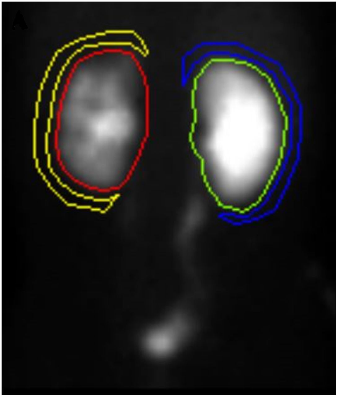
  • Draw regions of interest around the entire kidney and pelvis. Draw additional C-shaped perirenal background regions 2 pixels thick and 1 pixel away from the whole kidney region (Fig. 1).

  • Generate time–activity curves.

  • Calculate the collection system clearance half-time or washout half-time (T½). T½ may be measured from the time the diuretic is given to the time it takes to reach half the original level. Computer quantitative software can more precisely calculate T½ by fitting a curve to the steepest portion of the washout time–activity curve. Normally, a T½ of less than 10 min indicates that no obstruction is present.

  • Use the automatic software to calculate relative function (if available), which is the percentage of the total function of both kidneys. It is calculated by comparing the area under the curve for both kidneys between the 1- and 3-min intervals. Normal values are between 45% and 55%.

  • Sum the dynamic images, and display them for optimal visualization of the area of interest.

  • Scale planar images to accurately visualize areas of normal anatomy and increased uptake.

FIGURE 1.
  • Download figure
  • Open in new tab
  • Download powerpoint
FIGURE 1.

Whole kidney region of interest with perirenal background region. Yellow = left kidney background; red = left kidney; green = right kidney; blue = right kidney background.

PRECAUTIONS

Caution should be used when furosemide is given to patients who have just undergone urologic surgery, because of the increased urine flow rate.

SUGGESTED READING

  1. 1.
    1. Banks K,
    2. Farrell M,
    3. Peacock J
    . Diuretic renal scintigraphy protocol considerations. J Nucl Med Technol. May 24, 2022 [Epub ahead of print].
  2. 2.
    1. Mettler FA,
    2. Guiberteau MJ
    . Essentials of Nuclear Medicine Imaging. 5th ed. Saunders Elsevier; 2006:304–305, 539–540.
  3. 3.
    1. Shackett P
    . Nuclear Medicine Technology and Techniques: Procedures and Techniques. 2nd ed. Lippincott Williams & Wilkins; 2009:249–253.
  4. 4.
    1. Waterstram-Rich KM,
    2. Gilmore D
    1. Sharma A,
    2. Younglove SM
    . Genitourinary system. In: Waterstram-Rich KM, Gilmore D, eds. Nuclear Medicine and PET/CT Technology and Techniques. 8th ed. Mosby Elsevier; 2017:567–569.
  5. 5.
    1. Taylor A,
    2. Schuster DM,
    3. Alazraki N
    . A Clinician’s Guide to Nuclear Medicine. 2nd ed. Society of Nuclear Medicine and Molecular Imaging; 2006:52–53.
  6. 6.
    1. Ziessman HA,
    2. O’Malley JP,
    3. Thrall JH,
    4. Fahey FH
    . The Requisites: Nuclear Medicine. 4th ed. Elsevier Saunders; 2014:168–203.
  7. 7.
    Taylor AT, Brandon DC, De Palma D, et al. SNMMI procedure standard/EANM practice guideline for diuretic renal scintigraphy in adults with suspected upper urinary tract obstruction 1.0. Semin Nucl Med. 2018;48:377–390.
PreviousNext
Back to top

In this issue

Journal of Nuclear Medicine Technology: 50 (4)
Journal of Nuclear Medicine Technology
Vol. 50, Issue 4
December 1, 2022
  • Table of Contents
  • About the Cover
  • Index by author
  • Complete Issue (PDF)
Print
Download PDF
Article Alerts
Sign In to Email Alerts with your Email Address
Email Article

Thank you for your interest in spreading the word on Journal of Nuclear Medicine Technology.

NOTE: We only request your email address so that the person you are recommending the page to knows that you wanted them to see it, and that it is not junk mail. We do not capture any email address.

Enter multiple addresses on separate lines or separate them with commas.
Diuretic Renal Scintigraphy
(Your Name) has sent you a message from Journal of Nuclear Medicine Technology
(Your Name) thought you would like to see the Journal of Nuclear Medicine Technology web site.
Citation Tools
Diuretic Renal Scintigraphy
Mary Beth Farrell, Kevin P. Banks, Justin G. Peacock
Journal of Nuclear Medicine Technology Dec 2022, 50 (4) 319-321; DOI: 10.2967/jnmt.122.264804

Citation Manager Formats

  • BibTeX
  • Bookends
  • EasyBib
  • EndNote (tagged)
  • EndNote 8 (xml)
  • Medlars
  • Mendeley
  • Papers
  • RefWorks Tagged
  • Ref Manager
  • RIS
  • Zotero
Share
Diuretic Renal Scintigraphy
Mary Beth Farrell, Kevin P. Banks, Justin G. Peacock
Journal of Nuclear Medicine Technology Dec 2022, 50 (4) 319-321; DOI: 10.2967/jnmt.122.264804
Twitter logo Facebook logo LinkedIn logo Mendeley logo
  • Tweet Widget
  • Facebook Like
  • Google Plus One
Bookmark this article

Jump to section

  • Article
    • RATIONALE/INTRODUCTION
    • INDICATIONS
    • CONTRAINDICATIONS/TECHNICAL CONSTRAINTS
    • PATIENT PREPARATION/EDUCATION
    • RADIOPHARMACEUTICAL IDENTITY, DOSE, AND ROUTE OF ADMINISTRATION
    • ACQUISITION PARAMETERS: DYNAMIC/STATIC/PLANAR
    • ACQUISITION INSTRUCTIONS
    • COMMON OPTIONS
    • PROCESSING INSTRUCTIONS
    • PRECAUTIONS
    • SUGGESTED READING
  • Figures & Data
  • Info & Metrics
  • PDF

Related Articles

  • No related articles found.
  • Google Scholar

Cited By...

  • Five Years in Review
  • Google Scholar

More in this TOC Section

  • Fatty Meal Hepatobiliary Scintigraphy for Gallbladder Ejection Fraction Determination
  • Gastric Emptying Study: Liquids
  • Ventricular Shunt Patency
Show more Practical Protocol Tip

Similar Articles

SNMMI

© 2025 SNMMI

Powered by HighWire