


Article Figures & Data
Figures
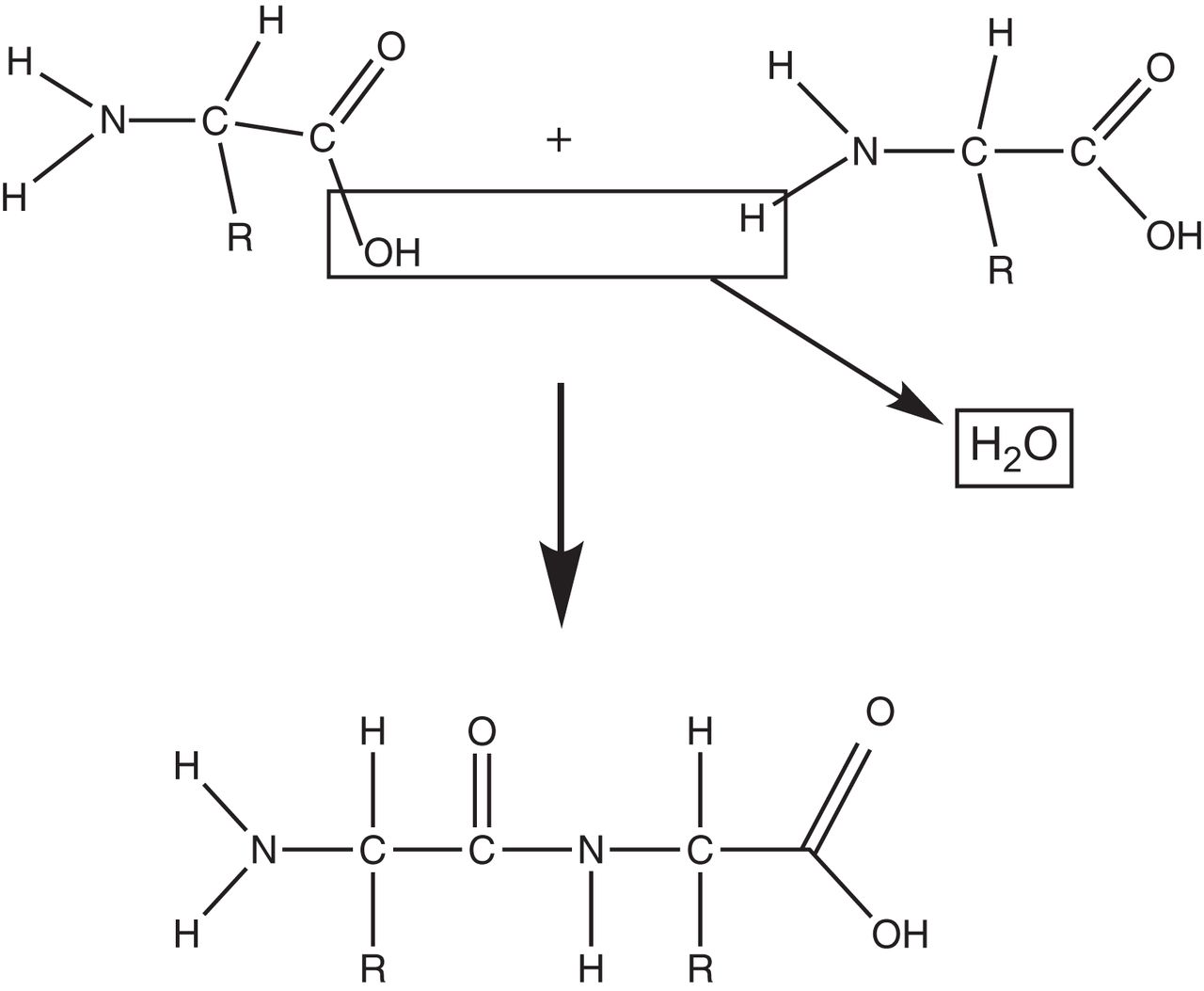
- FIGURE 1.
Condensation reaction. Two amino acids are joined together to form peptide bond with release of water molecule.
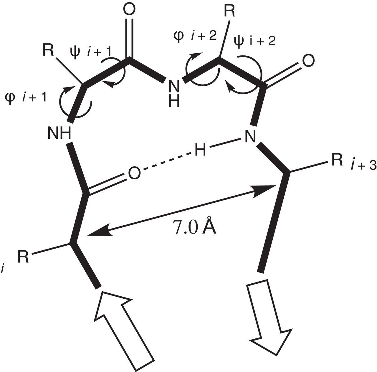
- FIGURE 2.
Protein secondary structure formation. Precise folding of polypeptide chain is achieved by rotational angles (φ, ψ) of backbone bonds flanking central α-carbon atom of each amino acid. These rotational angles are specific for each amino acid and are instrumental in shaping protein structure as prescribed by genetic code.
- FIGURE 3.
Protein tertiary structure: intra- and intermolecular bonds help form and stabilize precise 3-dimensional protein structure into helices and sheets.
- FIGURE 4.
Protein quaternary structure: helices and pleated sheets of different polypeptide chains may further associate together to form quaternary structures as in case of hemoglobin molecule in red blood cells. Each hemoglobin molecule is composed of 4 polypeptide subunits (2 α-chains and 2 β-chains), each stabilized by ion group (heme) in center.
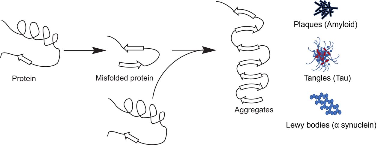
- FIGURE 5.
Protein misfolding can lead to pathology. Correct folding of protein into its proper 3-dimensional structure is important to function correctly. Incorrectly folded proteins either are destroyed by proteasomes or may form insoluble aggregates such as plaques, tangles, and Lewy bodies, which can lead to pathologic conditions as in Alzheimer disease.
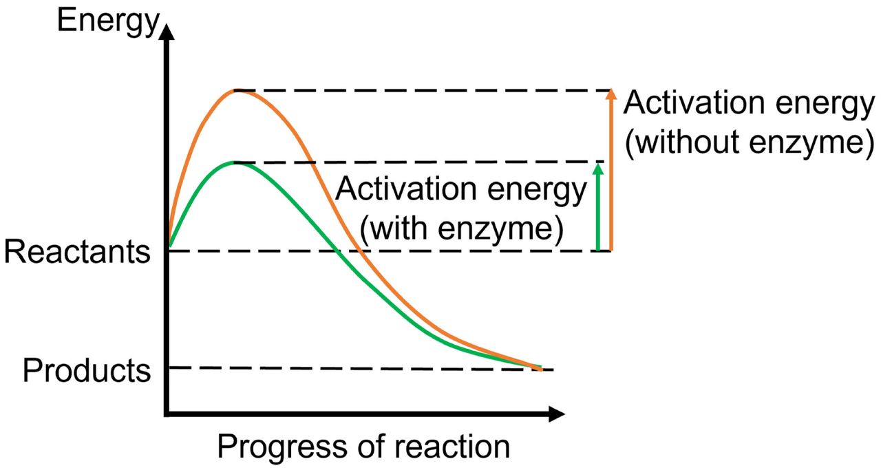
- FIGURE 6.
Enzyme as catalysts. Enzymes catalyze chemical reactions by lowering activation energy required for reactants to progress through steps of chemical reaction. This lowering of energy includes that of the high-energy transition state at the peak of the energy profile, which is lower when enzymes are present, hence making it easier for reaction to progress.


Tables
- TABLE 1.
Clinical Properties of Current Food and Drug Administration–Approved Molecular Tracer (18F-Fluciclovine) Using Amino Acid Metabolism
Property Description Indication Imaging in men with suspected prostate cancer recurrence based on elevated blood level of prostate-specific antigen after treatment Administered dose for adults 370 MBq (10 mCi) Injection route Intravenous Injection-to-imaging time 4–10 min Normal biodistribution Pancreas, liver, bone marrow, muscle - TABLE 2.
Clinical Properties of Current Food and Drug Administration–Approved Molecular Tracers Detecting Protein Folding or Misfolding
Property Description Indication 18F-florbetapir Imaging β-amyloid plaques in suspected AD patients 18F-florbetaben Imaging β-amyloid plaques in suspected AD patients 18F-flutemetamol Imaging β-amyloid plaques in suspected AD patients 18F-flortaucipir Imaging aggregated tau neurofibrillary tangles in suspected AD patients Administered dose for adults 18F-florbetapir 370 MBq (10 mCi) 18F-florbetaben 296 MBq (8 mCi) 18F-flutemetamol 185 MBq (5 mCi) 18F-flortaucipir 370 MBq (10 mCi) Injection route Intravenous Injection-to-imaging time 18F-florbetapir 30–50 min 18F-florbetaben 45–130 min 18F-flutemetamol 80–100 min 18F-flortaucipir 80–100 min Normal biodistribution 18F-florbetapir Inner white matter, from which blood clearance is slower 18F-florbetaben Inner white matter, from which blood clearance is slower 18F-flutemetamol Inner white matter, from which blood clearance is slower 18F-flortaucipir Some normal retention in choroid plexus, striatum, and brain stem nuclei AD = Alzheimer disease.
- TABLE 3.
Clinical Properties of Current Food and Drug Administration–Approved Molecular Tracers Targeting Cell Surface Protein Receptors
Property 18F-fluoroestradiol 99mTc-tilmanocept 68Ga-DOTATATE, 68Ga-DOTATOC, 177Lu-DOTATATE, 64Cu-DOTATATE, and 111In-pentetreotide 90Y-ibritumomab tiuxetan Indication Detecting estrogen receptor–positive lesions as adjunct to biopsy in patients with recurrent or metastatic breast cancer Mapping lymph nodes draining from primary tumor site and guiding sentinel lymph node biopsy with intraoperative γ-probe 68Ga-DOTATATE or 68Ga-DOTATOC: locating SSTR-positive neuroendocrine tumors in adult and pediatric patients; 177Lu/64Cu-DOTATATE: treating SSTR-positive gastroenteropancreatic neuroendocrine tumors, including foregut, midgut, and hindgut neuroendocrine tumors in adults* Evaluating relapsed or refractory, low-grade or follicular B-cell non-Hodgkin lymphoma Administered dose for adults 111–222 MBq (3–6 mCi) 18.5 MBq (0.5 mCi) 68Ga-DOTATATE: 2 MBq/kg (0.054 mCi/kg) up to 200 MBq (5.4 mCi); 68Ga-DOTATOC: adult—148 MBq (4 mCi); pediatric—1.59 MBq/kg (0.043 mCi/kg) with range of 11.1 MBq (0.3 mCi) to 111 MBq (3 mCi); 177Lu-DOTATATE: 7.4 GBq (200 mCi) every 8 wk for total of 4 doses; 64Cu-DOTATATE: 148 MBq (4 mCi); 111In-pentetreotide: 111--222 MBq (3--6 mCi) 14.8 MBq per kg (0.4 mCi/kg); dose adjustment needed if platelet counts are low Injection route Intravenous Subcutaneous, intradermal, subareolar, or peritumoral injection in 1 mL or less Intravenous Intravenous Injection-to-imaging time 80 min 10–15 min 68Ga-DOTATATE: 40–90 min; 68Ga-DOTATOC: 60 min† Imaging not usually done Normal biodistribution Hepatobiliary system (excretion); intestines (excretion); heart, blood, uterus, kidney (excretion); and bladder (excretion) Lymphatic channels draining injection site Pituitary, thyroid, spleen, adrenals, kidney, pancreas, prostate, liver, and salivary glands Significant marrow and splenic distribution without cold antibody pretreatment; pretreatment with rituximab cold anti-CD20 antibody blocks CD20 sites of normal circulating B-cells in spleen and bone marrow by binding to it, thereby allowing the following hot antibodies to reach tumor areas *111In-pentetreotide: detection of neuroendocrine tumors bearing somatostatin receptors.
†177Lu-DOTATATE: not imaged; 64Cu-DOTATATE: not imaged; 111In-pentetreotide: 24--48 h.
- TABLE 4.
Clinical Properties of Current Food and Drug Administration–Approved Molecular Tracers Targeting Cell Surface Protein Transporters
Property 123I-ioflupane 123I-iobenguane and 131I-iobenguane 68Ga-PSMA11 and 18F-piflufolastat Indication Striatal dopamine transporter imaging to assist in evaluation of adults with suspected Parkinsonian syndromes 123I-iobenguane: detection of primary or metastatic pheochromocytoma or neuroblastoma; 131I-iobenguane: treatment of adults and children older than 12 y with iobenguane-positive, unresectable, locally advanced or metastatic pheochromocytoma or paraganglioma PET of PSMA-positive lesions in prostate cancer patients who have suspected metastasis and are candidates for initial definitive therapy or who have suspected recurrence based on elevated serum prostate-specific antigen Administered dose for adults 111–185 MBq (3–5 mCi) 123I-iobenguane: 370 MBq (10 mCi); 131I-iobenguane: 185–222 MBq (5–6 mCi) (dosimetric dose); 18,500 MBq (500 mCi) × 2 doses 90 d apart (therapeutic dose) 68Ga-PSMA11: 111–259 MBq (3–7 mCi); 18F-piflufolastat: 333 MBq (9 mCi) recommended; acceptable range, 8–296 to 370 MBq (10 mCi) Injection route Intravenous Intravenous Intravenous (bolus) Injection-to-imaging time 3–6 h 24 ± 6 h 60 min Normal biodistribution Prominent comma-shaped striatal activity compared with surrounding brain tissue Adrenals (not always visualized, but activity < liver), liver, heart (uptake inversely proportional to catecholamine levels), bowel (large intestine), salivary glands, lung, spleen, urinary bladder, and uterine/neck muscles Kidneys, salivary glands, small intestine, tear glands, and spleen
In this issue


Jump to section
- Article
- Abstract
- MOLECULAR TRACER USING AMINO ACID METABOLISM (18F-FLUCICLOVINE (AXUMIN; BLUE EARTH DIAGNOSTICS) (32))
- MOLECULAR TRACERS DETECTING PROTEIN FOLDING OR MISFOLDING
- MOLECULAR TRACERS TARGETING CELL SURFACE PROTEIN RECEPTORS
- MOLECULAR IMAGING TRACERS TARGETING CELL SURFACE PROTEIN TRANSPORTERS
- CONCLUSION
- DISCLOSURE
- Footnotes
- REFERENCES
- Figures & Data
- Info & Metrics