Abstract
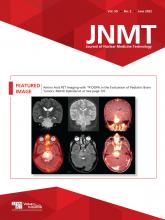
Although MRI is the workhorse of initial evaluation and follow-up in brain tumors, there are growing data recommending incorporation of amino acid PET imaging at different stages in the management of these patients. Recent recommendations on nuclear medicine and neurooncology clinical practice support the use of amino acid imaging for brain tumors. Methods: We present 4 pediatric brain tumor cases imaged with 3,4-dihydroxy-6-18F-fluoro-l-phenylalanine (18F-DOPA), and we review the literature. Results: 18F-DOPA showed low level/no uptake in benign conditions and in inflammatory postoperative changes. It showed better definition of viable tumor boundaries than MRI. Conclusion: Considering that 18F-DOPA is Food and Drug Administration–approved for the evaluation of parkinsonian syndromes, this tracer could be used clinically for other valuable clinical indications, such as brain tumor evaluations. The value of this use seems to be well established in adults, and evidence of its value in pediatrics has been growing as well.
PET has been used in the assessment of brain tumors for 40 y. State-of-the-art MRI has become the gold standard in brain tumor imaging during the various steps of clinical management. 18F-FDG was initially used with some success but faced limitations due to the high background uptake from normal metabolism of glucose by the brain. Amino acid PET has proven superior to 18F-FDG PET and adds incremental value to MRI in imaging of brain tumors. 11C-methyl-l-ethionine (11C-MET), O-(2-18F-fluoroethyl)-l-tyrosine (18F-FET), and 3,4-dihydroxy-6-18F-fluoro-l-phenylalanine (18F-DOPA) have shown great clinical value. Considering that 18F-DOPA is Food and Drug Administration–approved for the evaluation of Parkinsonian syndromes, it can be used clinically for other valuable clinical indications as well. 18F-fluciclovine (Axumin; Blue Earth Diagnostics) is also Food and Drug Administration–approved for prostate cancer imaging and is being investigated for brain tumor imaging in adults. Bearing in mind that radiation exposure (albeit minimal) is a matter of concern in pediatrics, amino acid imaging can judiciously be used in the highest-yield case scenarios.
MATERIALS AND METHODS
We highlight 4 cases of pediatric brain tumor in which 18F-DOPA imaging was used. We discuss its main value, and we review use of amino acid PET imaging in the pediatric population. Scans were acquired on a 3-T MRI scanner and a GE Healthcare Discovery 690 PET/CT scanner at Sidra Medicine. The radiopharmaceutical activities ranged from 99.9 to 118.4 MBq (2.7–3.2 mCi).
RESULTS
Case 1
A 7-y-old boy was referred to our hospital because of visual hallucinations, abdominal pain, headache, and seizures. He was diagnosed with temporal lobe epilepsy and had a further work-up including CT and MRI scans of his brain. A well-defined calcified lesion was identified in the right temporal lobe, and surgical excision was recommended. His parents were hesitant to consent to surgery. Amino acid PET imaging using 18F-DOPA was performed to gain additional prognostic information. The lack of 18F-DOPA uptake by the lesion (Fig. 1) was indicative of a more benign or a lower-grade process, and the parents elected for watchful waiting. A few months later, the parents agreed to surgical excision of the lesion, and a right temporal lobe lesionectomy was performed. Histopathologic examination of this lesion revealed meningioangiomatosis, which is a rare, benign disease of the brain. Lack of 18F-DOPA uptake predicted a benign nature for the lesion and provided additional confidence in the management and reassurance to the family.
No 18F-DOPA uptake in calcified benign right temporal lesion (meningioangiomatosis) (arrows). (A) From left to right: axial T2-weighted, axial T1-weighted, axial fluid-attenuated inversion recovery, and coronal T2-weighted MR images. (B) From left to right: axial 18F-DOPA PET/MR, axial 18F-DOPA PET/CT, coronal 18F-DOPA PET/CT, and axial 18F-DOPA PET images.
Case 2
A 3-y-old boy was evaluated for gait issues and lower-extremity weakness. He was then diagnosed with diffuse intrinsic pontine glioma. He received standard focal conformal radiotherapy, with a total dose of 54 Gy. Eight months after finishing treatment, he presented with mild left-sided weakness and limp. He was reevaluated with an MRI scan, which showed that his tumor was overall smaller than on the scan at diagnosis. However, there were areas of signal change, which created concern about disease progression. An 18F-DOPA scan demonstrated uptake in the area showing contrast enhancement on the MRI scan. The 18F-DOPA scan was also able to delineate the tumor margins with higher accuracy than the MRI scan and provided an additional negative prognostic indicator with regard to the intensity of 18F-DOPA uptake (Fig. 2).
Intense 18F-DOPA uptake in recurrent diffuse intrinsic pontine glioma showing better tumor delineation of true tumor boundaries than on the MRI alone and showing that intensity of uptake is likely a prognostic parameter. (A) From left to right: coronal T2-weighted fat-saturated initial, coronal T2-weighted fat-saturated 9-mo follow-up, and coronal 18F-DOPA 9-mo follow-up PET/MR images. (B) From left to right: axial 18F-DOPA PET fused to initial MR, axial T2-weighted fat-saturated 9-mo follow-up MR, and axial 18F-DOPA 9-mo follow-up PET/MR images.
Case 3
A 6-y-old boy presented with an approximately 2-mo history of an abnormally wide gait and leg weakness. He then had increased somnolence at school and an increasing number of falls for 3 wk before the initial diagnosis. He underwent brain MRI. It demonstrated a midline pontine tumor (diffuse intrinsic pontine glioma), which was treated with focal radiotherapy to a total dose of 54 Gy. Six months after completing treatment, he presented to the clinic with recurrent gait abnormalities. An MRI scan showed an alteration in the internal architectural appearance of the brain stem glioma, with marginal dimensional reduction (the glioma was smaller than on the previous scan). There were also changes in diffusion-weighted imaging patterns between scans that could not clearly be characterized. An 18F-DOPA scan was then performed a few days later and demonstrated tumor viability and recurrence (Fig. 3).
No clear change in size of initial diffuse intrinsic pontine glioma tumor 9 mo after initial MRI. Changes in appearance on different sequences showed T2-weighted and diffusion-weighted MRI signal distribution changes. 18F-DOPA scan fused with follow-up MRI clearly delineates tumor viability and recurrence. (A) From left to right: initial axial T1-weighted contrast-enhanced, initial axial T2-weighted, and initial axial diffusion-weighted MR images. (B) From left to right: axial 18F-DOPA T2-weighted 9-mo follow-up PET/MR, axial T2-weighted 9-mo follow-up MR, and axial diffusion-weighted 9-mo follow-up MR images. (C) From left to right: axial 18F-DOPA, coronal 18F-DOPA, and sagittal 18F-DOPA PET images.
Case 4
A 3-y-old girl presented with a 2-mo history of gait disturbance followed by an inability to walk for 3 d before her admission to the hospital. MRI revealed a large tumor in the posterior fossa, prompting neurosurgical intervention and resection. Histopathologic examination of the excised specimen revealed anaplastic ependymoma. Immediate postoperative MRI showed a small, 8-mm, nodule in the resection bed suggestive of residual tumor. Correlative 18F-DOPA PET and MRI performed 3 wk after surgery revealed no uptake in a much smaller nodular density, indicating a favorable postoperative change. This finding obviated second-look surgery (Fig. 4).
A 3-y-old girl after resection of posterior fossa tumor (anaplastic ependymoma: World Health Organization grade III). Three weeks after surgery, 8-mm left-sided nodule (blue arrows) in resection cavity showed no abnormal 18F-DOPA uptake. Follow-up MRI on same day as 18F-DOPA scan showed interval reduction in size and near-complete resolution of this nodule (white arrows), most consistent with initial postoperative changes. (A) From left to right: axial T2-weighted preoperative MR, axial immediately postoperative T1-weighted contrast-enhanced MR, and axial immediately postoperative 18F-DOPA PET/MR images (B) From left to right: axial follow-up postoperative T1-weighted MR (3 wk), axial follow-up 18F-DOPA PET/MR, and axial 18F-DOPA PET images.
DISCUSSION
Amino acid PET imaging has been used successfully for about 15 y and has gained traction in the clinical management of brain tumors as several fluorine-labeled compounds have become more readily available and as data have mounted on a major impact on clinical management (1–4). 18F-DOPA and 18F-FET seem to be the agents with the highest accuracy, although only 18F-DOPA is Food and Drug Administration–approved (1,5,6). 18F-DOPA can offer complementary and additive valuable information at different stages of brain tumor management (Fig. 5).
Summary diagram. EANM = European Association of Nuclear Medicine; EANO = European Association of Neuro-Oncology; RANO = Response Assessment in Neuro-Oncology; RAPNO = Response Assessment in Pediatric Neuro-Oncology; SIOPE = European Society for Paediatric Oncology; SNMMI = Society of Nuclear Medicine and Molecular Imaging.
Historically, 18F-FDG and 11C-MET have been used in brain tumor evaluation. Higher 18F-FDG uptake has been correlated with higher grade and worse prognosis but is no longer routinely used in the clinic because of its low sensitivity (2,6). 11C-MET is limited by its short half-life of 20 min and is used only in centers with an on-site cyclotron. When available, it can provide useful diagnostic and prognostic information (2,3). 18F-FET and 18F-DOPA radiotracers are newer and significantly superior and have largely taken over brain tumor imaging with PET (3,4). Most recently, the European Association of Nuclear Medicine, Society of Nuclear Medicine and Nuclear Imaging, European Association of Neuro-Oncology, and Response Assessment in Neuro-Oncology Group published practice guidelines and procedure standards for imaging of gliomas using PET with radiolabeled amino acids and 18F-FDG (6). These guidelines have consolidated the benefit of PET imaging in the different stages of brain tumor management as described earlier by the Response Assessment in Neuro-Oncology Group (5).
Pirotte’s group has extensively explored amino acid imaging in pediatrics, described their experience over 10 y with 18F-FDG and 11C-MET, and outlined the value of PET in the presurgical, surgical, and postsurgical management of 126 pediatric cases (7). In another study, Pirotte et al. showed in 85 pediatric brain tumors that 18F-FDG PET or 11C-MET PET guided treatment when MRI was unable to assist in selecting accurate biopsy targets (35 patients) or to delineate tumors for maximal resection (50 patients) (8). They also showed, in 55 children, that absence of 11C-MET uptake had a high accuracy in excluding high-grade tumors and was able to guide conservative management; increased uptake was also seen in all patients with high-grade tumors (9). Pirotte et al. also concluded, in their evaluation of 9 pediatric cases of infiltrative brain tumor, that PET and MR coguidance for accurate stereotactic biopsy of these lesions improved the diagnostic yield, made it possible to reduce the sampling in high-risk or functional areas, and improved the overall quality of therapeutic management (10). The latter strategy is clinically relevant because of the histologic heterogeneity of brain tumors (11–13). This additional value of PET imaging, when compared with MRI alone, was also highlighted in another series of 103 pediatric cases (14).
Furthermore, Morana’s group has suggested that 18F-DOPA PET, when added to the MRI work-up in cases of infiltrative gliomas, may offer additional value in diagnosis, prognosis, and therapy assessment (15–17). Other groups have also pointed to the impact of a positive 18F-DOPA PET result on overall survival, progression-free survival, and overall management in adults with low-grade primary brain tumors and brain tumor recurrence (18–21). On the other hand, Morana’s group has also highlighted that although 18F-DOPA can aid in differentiating high- and low-grade brain tumors, one should be cautious because developmental venous anomalies may represent a false-positive finding (22). The group recommended that all 18F-DOPA PET interpretations be performed by expert readers and in correlation with the patient’s MR images (22). This recommendation is of greater importance, considering the increased prevalence of developmental anomalies in children with intracranial neoplasms (23). These developmental anomalies were associated with neuronal dysfunction in adjacent brain areas as depicted on 18F-FDG PET (24). Morana et al. also reported on the possibility of seeing 18F-DOPA changes in the basal ganglia secondary to network changes due to cortical resection—a possibility that needs to be considered when interpreting these PET scans (25). 18F-DOPA PET was also highlighted by Bund et al. to be helpful in the evaluation of 53 nonenhancing brain tumors, although this evaluation was in adults (mean age, 39 y) (26). 18F-DOPA PET was able to discriminate between dysembryoplastic neuroepithelial tumor and grade II oligodendroglioma and between low- and high-grade gliomas with no contrast enhancement on MRI (26).
In addition, as the management and classification of brain tumors has been shifting from the plain high- or low-grade spectrum and incorporating molecular markers that define distinct biologic subtypes with different clinical courses, it seems natural that metabolic imaging may add a layer of subcategorizing tumors, stratifying management, and altering the course of disease. Suchorska et al. briefly summarized this paradigm in the context of the new, 2016, World Health Organization classification for brain tumors (27). At this moment, it may be difficult to differentiate subtypes on the basis of amino acid PET imaging (27); however, higher 18F-DOPA uptake has been associated with IDH mutation in diffuse grade II and grade III gliomas (28). Although most dynamic data stem from 18F-FET, it seems that current dynamic 18F-DOPA data are a predictor of progression/recurrence and progression-free survival. However, dynamic 18F-DOPA data do not seem to offer any additional value over static parameters such as mean and maximum tumor–to–normal-brain ratios, tumor-to-striatum ratios, and metabolic tumor volume (29). Ponisio et al., on the other hand, did not uncover any value to dynamic analysis (30). Furthermore, Ginet et al. demonstrated that dynamic 18F-DOPA PET was able to differentiate the molecular features of newly diagnosed gliomas (i.e., presence or absence of isocitrate dehydrogenase mutation), but static uptake parameters were not (31). Lastly, Piccardo et al. compared advanced MRI features and metabolic information obtained by 18F-DOPA PET in 22 pediatric midline gliomas. They showed that in comparison to advanced MRI techniques such as ADC maps, arterial spin labeling perfusion maps, and MR spectroscopy, 18F-DOPA PET tumor-to-striatum ratio was the only parameter able to discriminate H3K27 M-mutant from wild-type diffuse midline gliomas independently from histology (32).
CONCLUSION
Amino acid imaging with 18F-DOPA is a powerful clinical tool and should be used in agreement with the recent joint guidelines from the European Association of Nuclear Medicine, the European Association of Neuro-Oncology, the Response Assessment in Neuro-Oncology Group (2019), and the Neuro-Oncology Working Group (2016). Wider use and pooling of patient data from multicenter registries would provide even more insight.
DISCLOSURE
Cyclomedical (Hamad Medical Corporation) facilitated provision of 18F-DOPA. No other potential conflict of interest relevant to this article was reported.
KEY POINTS
QUESTION: Is amino acid PET imaging with 18F-DOPA of significant value in pediatric brain tumors?
PERTINENT FINDINGS: Amino acid PET imaging with 18F-DOPA offers valuable clinical information in primary diagnosis, treatment response assessment, pseudoprogression, and recurrence applications in pediatric brain tumors.
IMPLICATIONS FOR PATIENT CARE: Amino acid PET imaging with 18F-DOPA in pediatric brain tumors may change clinical management and improve outcomes.
Footnotes
Published online Apr. 19, 2022.
REFERENCES
- Received for publication August 13, 2021.
- Revision received December 28, 2021.


