
Main menu

  • Home
  • Content
    • Current
      • JNMT Supplement
    • Ahead of print
    • Past Issues
    • Continuing Education
    • JNMT Podcast
    • SNMMI Annual Meeting Abstracts
  • Subscriptions
    • Subscribers
    • Rates
    • Journal Claims
    • Institutional and Non-member
  • Authors
    • Submit to JNMT
    • Information for Authors
    • Assignment of Copyright
    • AQARA Requirements
  • Info
    • Reviewers
    • Permissions
    • Advertisers
    • Corporate & Special Sales
  • About
    • About Us
    • Editorial Board
    • Contact Information
  • More
    • Alerts
    • Feedback
    • Help
    • SNMMI Journals
  • SNMMI
    • JNMT
    • JNM
    • SNMMI Journals
    • SNMMI

User menu

  • Subscribe
  • My alerts
  • Log in
  • Log out
  • My Cart

Search

  • Advanced search
Journal of Nuclear Medicine Technology
  • SNMMI
    • JNMT
    • JNM
    • SNMMI Journals
    • SNMMI
  • Subscribe
  • My alerts
  • Log in
  • Log out
  • My Cart
Journal of Nuclear Medicine Technology

Advanced Search

  • Home
  • Content
    • Current
    • Ahead of print
    • Past Issues
    • Continuing Education
    • JNMT Podcast
    • SNMMI Annual Meeting Abstracts
  • Subscriptions
    • Subscribers
    • Rates
    • Journal Claims
    • Institutional and Non-member
  • Authors
    • Submit to JNMT
    • Information for Authors
    • Assignment of Copyright
    • AQARA Requirements
  • Info
    • Reviewers
    • Permissions
    • Advertisers
    • Corporate & Special Sales
  • About
    • About Us
    • Editorial Board
    • Contact Information
  • More
    • Alerts
    • Feedback
    • Help
    • SNMMI Journals
  • Watch or Listen to JNMT Podcast
  • Visit SNMMI on Facebook
  • Join SNMMI on LinkedIn
  • Follow SNMMI on Twitter
  • Subscribe to JNMT RSS feeds
Research ArticleTeaching Case Study

Parathyroid Scintigraphy with an Incidental Indirect Finding of Pancreatic Cancer

Ba D. Nguyen
Journal of Nuclear Medicine Technology June 2021, 49 (2) 188-189; DOI: https://doi.org/10.2967/jnmt.120.248831
Ba D. Nguyen
Department of Radiology, Mayo Clinic, Scottsdale, Arizona
  • Find this author on Google Scholar
  • Find this author on PubMed
  • Search for this author on this site
  • Article
  • Figures & Data
  • Info & Metrics
  • PDF
Loading

Abstract

Modifications of the biodistribution of 99mTc-sestamibi seen during myocardial perfusion and parathyroid imaging may be secondary to benign or malignant processes in visualized anatomic structures not related to the target organs of these imaging procedures. This article presents a case of pancreatic adenocarcinoma indirectly depicted on parathyroid scintigraphy.

  • 99mTc-sestamibi
  • parathyroid scintigraphy
  • biliary ducts
  • pancreas cancer
  • SPECT/CT

99mThe radiopharmaceutical 99mTc-sestamibi has physiologic biliary excretion during scintigraphic evaluation of the parathyroid and the myocardium. Biliary and gallbladder incidental findings have been reported during these 2 main imaging applications of this radiotracer. This article presents a case of an indirect incidental demonstration of pancreatic cancer with the presence of distended intra- and extrabiliary ducts on parathyroid scintigraphy.

CASE REPORT

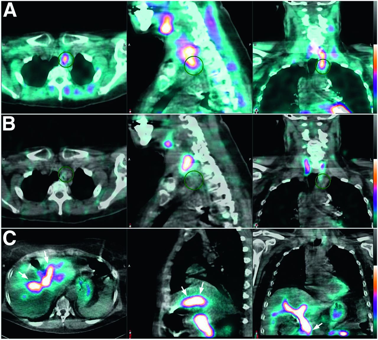
A 67-y-old woman consulted our institution because of primary hyperparathyroidism. Dual-radiotracer parathyroid scintigraphy was performed with 9.25 MBq (250 μCi) of 123I-sodium iodide, given orally as a capsule, and 925 MBq (25 mCi) of 99mTc-sestamibi intravenously. Double-isotope pinhole imaging with subtraction and SPECT/CT were performed. The SPECT/CT was done about 40–60 min after the tracer injection, using a Philips Brightview XCT camera with a low-energy high-resolution collimator, a 128 × 128 matrix, and 128 angles of rotation with 20 s per angle. The images showed a parathyroid adenoma at the left superior mediastinum near the thoracic inlet (Figs. 1A and 1B). In addition, the exam showed, at the right upper abdomen, prominent activity in the biliary ducts, with a cutoff sign at the distal common bile duct (Fig. 1C). The gallbladder was not seen because the patient had previously undergone cholecystectomy.

FIGURE 1.
  • Download figure
  • Open in new tab
  • Download powerpoint
FIGURE 1.

Axial, sagittal, and coronal SPECT/CT images of parathyroid scintigraphy showed parathyroid adenoma in left superior mediastinum positive for 99mTc-sestamibi uptake (A, circles) and negative for 123I-sodium iodide uptake (B, circles). SPECT/CT also showed prominent tracer activity in intra- and extrahepatic biliary ducts, with cutoff sign at distal common bile duct suggestive of obstruction (C, arrows).

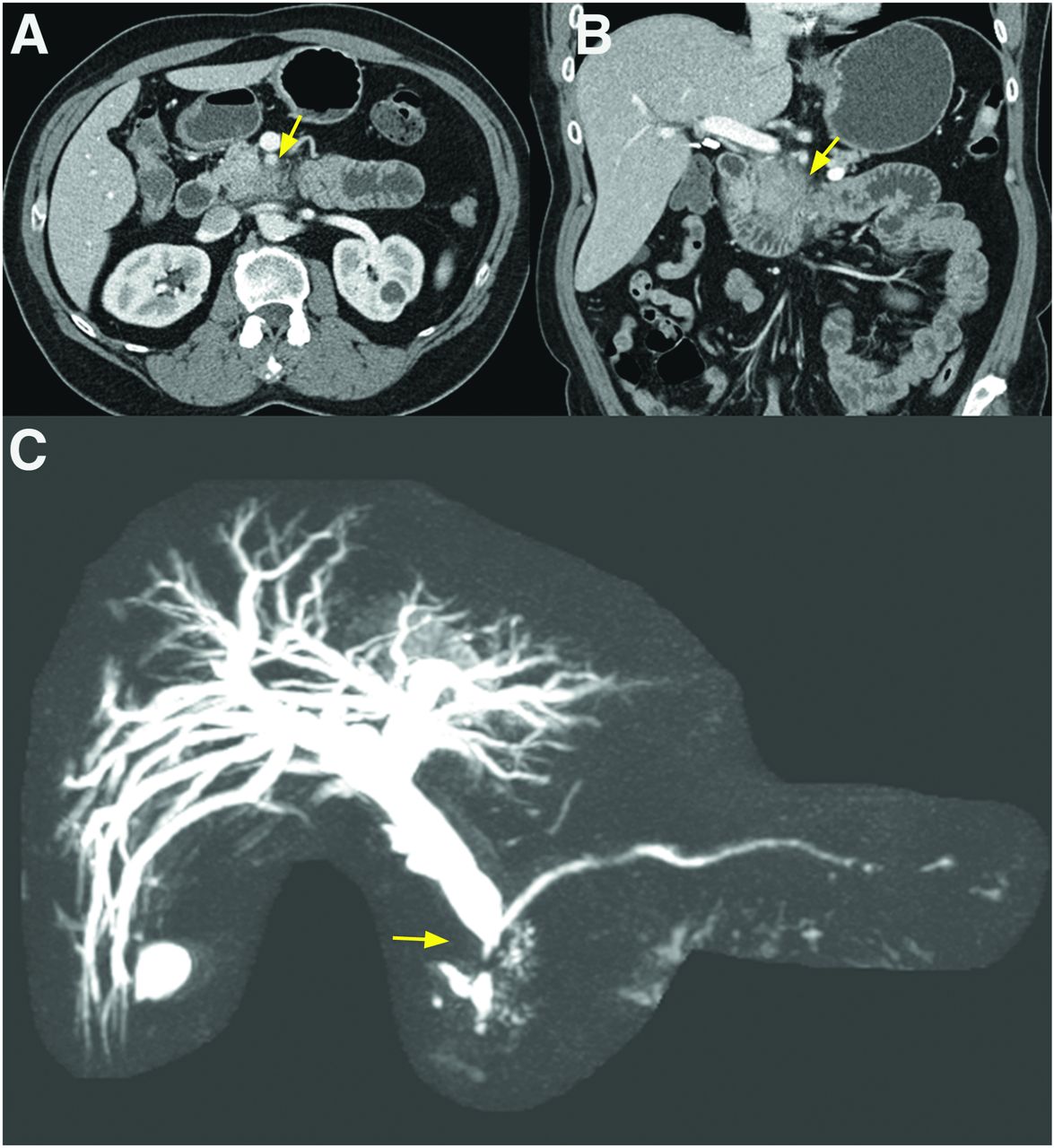
Review of the clinical notes revealed a recent history of progressively worsening pain in the back and abdomen, for which she had undergone abdominal CT, with normal findings, at an outside institution 2 mo before the present consultation. On the basis of the scintigraphic biliary findings, that previous CT scan was obtained, reviewed again, and found to show a soft-tissue mass in the region of the head and uncinate process of the pancreas (Figs. 2A and 2B). At the time of that CT exam, the patient had a vague abdominal pain but no laboratory or CT findings of biliary or pancreatic ductal obstruction.

FIGURE 2.
  • Download figure
  • Open in new tab
  • Download powerpoint
FIGURE 2.

Review of prior CT exam, performed 2 mo before parathyroid scan, showed soft-tissue fullness of head and uncinate process of pancreas suggestive of malignancy (A and B, arrows). MR cholangiography, after parathyroid scan, confirmed dilated biliary ducts from progressing unresectable adenocarcinoma at head of pancreas, with cutoff sign at distal common bile duct (C, arrow).

Subsequent MRI and MR cholangiography showed an unresectable pancreatic mass with compression of the distal common bile duct and tumor encasement of the superior mesenteric artery, the replaced right hepatic artery, and the superior mesenteric vein (Fig. 2C). An MRI exam followed by an endosonographic evaluation staged the pancreatic cancer as T4 Nx Mx. Endoscopic tissue sampling confirmed the diagnosis to be pancreatic adenocarcinoma.

Because of the aggressiveness of the pancreatic adenocarcinoma and the patient’s rapid clinical deterioration, parathyroidectomy was postponed. Twenty-two months after the initial CT exam, the patient died from complicated biliary and gastrointestinal tract obstruction along with widespread bilateral pulmonary metastasis.

DISCUSSION

99mTc-sestamibi is the workhorse radiopharmaceutical for myocardial perfusion imaging and for scintigraphic evaluation of hyperparathyroidism. These 2 procedures include planar, SPECT, or SPECT/CT imaging with a field of view that includes part of the chest and upper abdomen. The radiotracer biodistribution in thoracic and abdominal organs visualized during these procedures may provide important information about coexisting diseases potentially impacting patient care (1–3). 99mTc-sestamibi physiologic uptake is usually homogeneously diffuse in the hepatic parenchyma, often with prominent activity in the gallbladder (4). In our case, the unusually conspicuous intra- and extrabiliary ducts on scintigraphy, with distal common bile duct interruption, led to retrospective detection of an overlooked pancreatic lesion and subsequent diagnosis of an unresectable pancreatic adenocarcinoma.

CONCLUSION

Thorough evaluation of planar, SPECT, and SPECT/CT images of myocardial perfusion and the parathyroid is useful in detecting extracardiac and extracervical pathologic processes.

DISCLOSURE

No potential conflict of interest relevant to this article was reported.

Footnotes

  • Published online Dec. 24, 2020.

REFERENCES

  1. 1.↵
    1. Gedik GK,
    2. Ergün EL,
    3. Aslan M,
    4. Caner B
    . Unusual extracardiac findings detected on myocardial perfusion single photon emission computed tomography studies with Tc-99m sestamibi. Clin Nucl Med. 2007;32:920–926.
    OpenUrlPubMed
  2. 2.
    1. Jones SE,
    2. Aziz K,
    3. Yasuda T,
    4. Gewirtz H,
    5. Scott JA
    . Importance of systematic review of rotating projection images from Tc99m-sestamibi cardiac perfusion imaging for noncardiac findings. Nucl Med Commun. 2008;29:607–613.
    OpenUrlCrossRefPubMed
  3. 3.↵
    1. Chamarthy M,
    2. Travin MI
    . Altered biodistribution and incidental findings on myocardial perfusion imaging. Semin Nucl Med. 2010;40:257–270.
    OpenUrlPubMed
  4. 4.↵
    1. Wackers FJ,
    2. Berman DS,
    3. Maddahi J,
    4. et al
    . Technetium-99m hexakis 2-methoxyisobutyl isonitrile: human biodistribution, dosimetry, safety, and preliminary comparison to thaffium-201 for myocardial perfusion imaging. J Nucl Med. 1989;30:301–311.
    OpenUrlAbstract/FREE Full Text
  • Received for publication May 1, 2020.
  • Accepted for publication September 18, 2020.
PreviousNext
Back to top

In this issue

Journal of Nuclear Medicine Technology: 49 (2)
Journal of Nuclear Medicine Technology
Vol. 49, Issue 2
June 1, 2021
  • Table of Contents
  • About the Cover
  • Index by author
Print
Download PDF
Article Alerts
Sign In to Email Alerts with your Email Address
Email Article

Thank you for your interest in spreading the word on Journal of Nuclear Medicine Technology.

NOTE: We only request your email address so that the person you are recommending the page to knows that you wanted them to see it, and that it is not junk mail. We do not capture any email address.

Enter multiple addresses on separate lines or separate them with commas.
Parathyroid Scintigraphy with an Incidental Indirect Finding of Pancreatic Cancer
(Your Name) has sent you a message from Journal of Nuclear Medicine Technology
(Your Name) thought you would like to see the Journal of Nuclear Medicine Technology web site.
Citation Tools
Parathyroid Scintigraphy with an Incidental Indirect Finding of Pancreatic Cancer
Ba D. Nguyen
Journal of Nuclear Medicine Technology Jun 2021, 49 (2) 188-189; DOI: 10.2967/jnmt.120.248831

Citation Manager Formats

  • BibTeX
  • Bookends
  • EasyBib
  • EndNote (tagged)
  • EndNote 8 (xml)
  • Medlars
  • Mendeley
  • Papers
  • RefWorks Tagged
  • Ref Manager
  • RIS
  • Zotero
Share
Parathyroid Scintigraphy with an Incidental Indirect Finding of Pancreatic Cancer
Ba D. Nguyen
Journal of Nuclear Medicine Technology Jun 2021, 49 (2) 188-189; DOI: 10.2967/jnmt.120.248831
Twitter logo Facebook logo LinkedIn logo Mendeley logo
  • Tweet Widget
  • Facebook Like
  • Google Plus One
Bookmark this article

Jump to section

  • Article
    • Abstract
    • CASE REPORT
    • DISCUSSION
    • CONCLUSION
    • DISCLOSURE
    • Footnotes
    • REFERENCES
  • Figures & Data
  • Info & Metrics
  • PDF

Related Articles

  • No related articles found.
  • PubMed
  • Google Scholar

Cited By...

  • No citing articles found.
  • Google Scholar

More in this TOC Section

  • Crafting a Compelling Abstract That Gets Accepted
  • Cervical Ganglioneuroblastoma Diagnosed by 68Ga-DOTATOC PET/CT in a Child with Opsoclonus Myoclonus Syndrome
  • Utility of 18F-FDG PET/CT in Assessment of Disease Extent and Response to Treatment in Xanthoma Disseminatum
Show more Teaching Case Study

Similar Articles

Keywords

  • 99mTc-sestamibi
  • parathyroid scintigraphy
  • biliary ducts
  • pancreas cancer
  • SPECT/CT
SNMMI

© 2025 SNMMI

Powered by HighWire