


Article Figures & Data
Figures
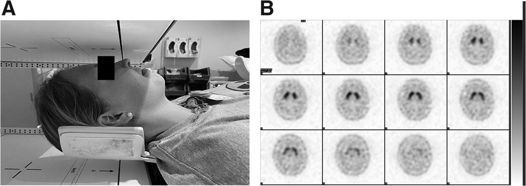
- FIGURE 1.
(Left) Correct positioning for brain SPECT scan, with head resting in head-and-neck holder and chin in neutral position. (Right) Axial brain SPECT images showing normal findings and no head tilt.
- FIGURE 2.
Axial brain SPECT images of 87-y-old woman with parkinsonian features, left-sided weakness, dysarthria, and generalized weakness. Images show head tilt, normal comma shape on left, decreased uptake in right putamen, and dilated ventricles.
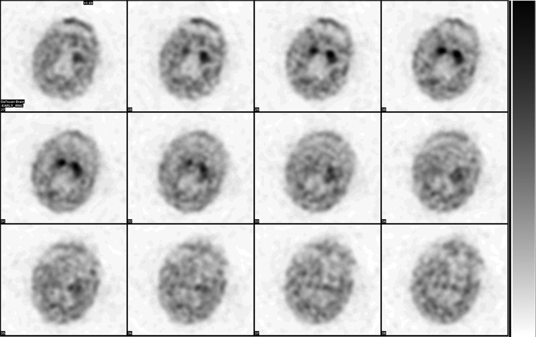
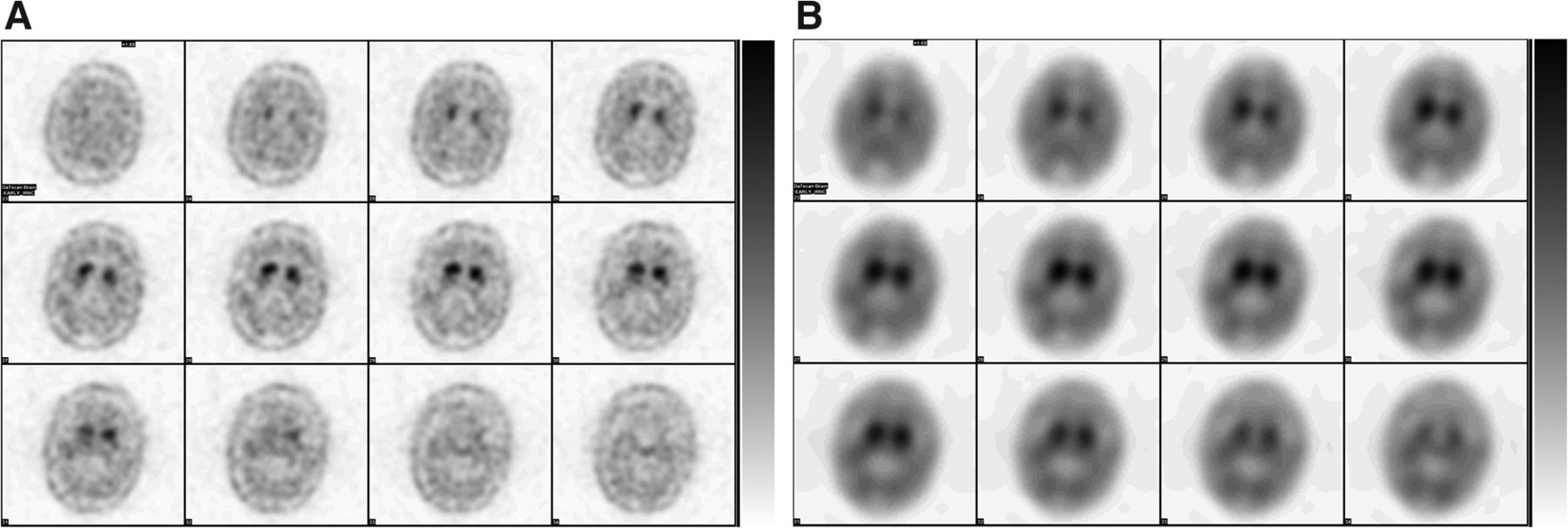
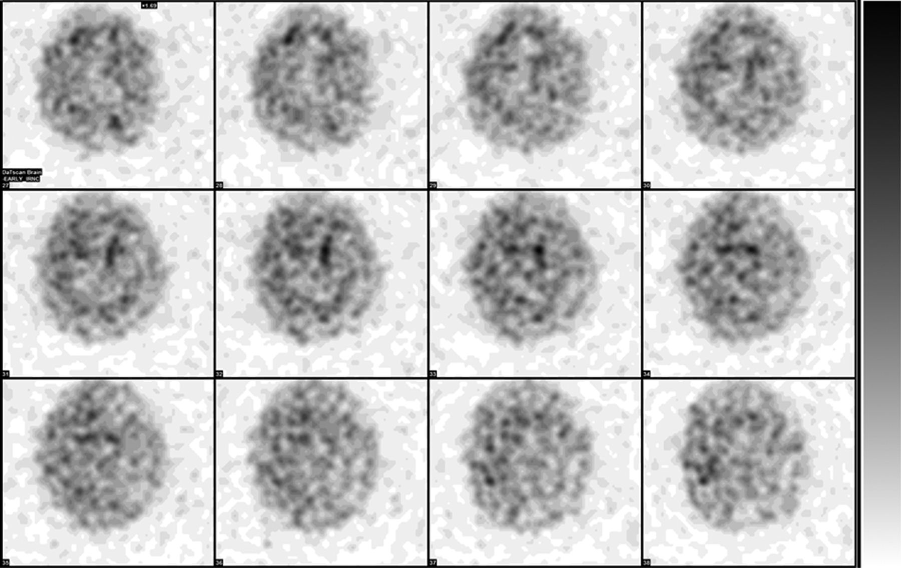
- FIGURE 3.
Axial brain SPECT images of 67-y-old woman who presented for evaluation of progressive gait instability with falls, micrographia, depth-perception problems, and vertical gaze palsy. Study was nondiagnostic because of increased background uptake from patient’s medications, citalopram and bupropion.
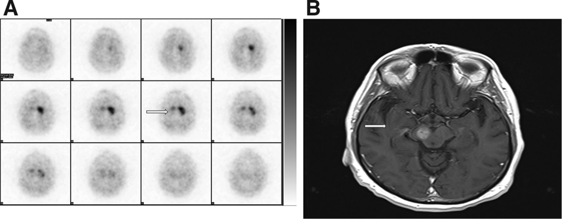
- FIGURE 4.
Images of 65-y-old man who had history of low-grade glioma in right midbrain and was being evaluated for right-arm rigidity and tremor. (Left) Axial brain SPECT image showing decreased uptake in right caudate nucleus and no uptake (arrow) in right putamen because of right midbrain glioma. Normal comma-shaped activity is seen in left caudate nucleus and putamen. (Right) Contrast-enhanced T1-weighted brain MR image showing enhancing lesion (arrow) in right midbrain consistent with history of low-grade glioma.
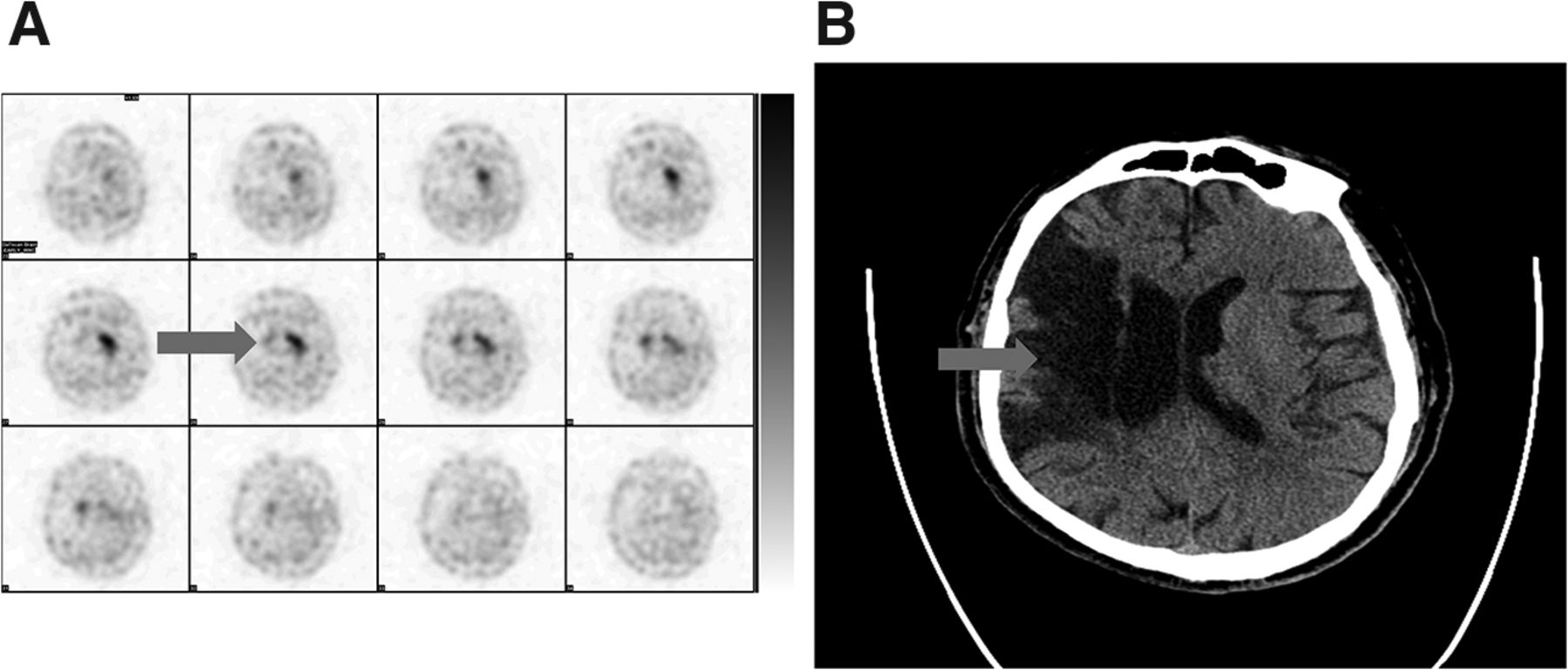
- FIGURE 5.
Images of 61-y-old man who had history of infarct in territory of right middle cerebral artery and was being evaluated for right-upper-extremity tremors of 2- to 4-mo duration. (Left) Axial brain SPECT image showing decreased to absent uptake (arrow) in right caudate nucleus and putamen because of prior infarct. (Right) Unenhanced axial CT scan of head showing infarct (arrow).
- FIGURE 6.
Axial brain SPECT images of 80-y-old man on carbidopa–levodopa without improvement of tremors, shuffling gait, and bradykinesia for 2–3 mo. (Left) Images showing decreased uptake bilaterally in putamen and loss of normal comma shape. These findings are scintigraphic evidence of presynaptic-deficit parkinsonian syndrome with probable age-associated changes due to no improvement with medication. (Right) Images showing oversmoothing artifact.
- FIGURE 7.
Axial brain SPECT images of 76-y-old man on primidone with history of resting and action tremors and cogwheel rigidity for 5 y. Images show decreased uptake bilaterally in putamen and right caudate nucleus, with loss of normal comma shape on right and scintigraphic evidence of probable presynaptic-deficit parkinsonian syndrome. Study was limited because of patient’s body habitus and inability to perform SPECT rotation closer to patient's head with required diameter of 11–15 cm.


Tables
Pitfall type Description Biologic Dopamine transporter density Age Sex Body habitus Ethnicity Allelic variants Medications competing with dopamine transporter Striatal infarct Brain tumors Trauma Surgery Technical Patient motion Patient position Patient orientation Equipment resolution Collimator Dose extravasation Time after injection Photopeak Oversmoothing on filtration Attenuation correction Size and placement of regions of interest Adapted from Morbelli et al. (13).
Effect type Drug Possible increase in binding Opioid: fentanyl Eugeroic: modafinil Antidepressants: bupropion, mazindol, radafaxine Anticholinergic: benztropine Anesthetics: isoflurane, ketamine, phencyclidine Central nervous system stimulant: cocaine Possible increase or decrease in binding Adrenergic agonists: norepinephrine, phenylephrine Amphetamines: d-amphetamine, methamphetamine, methylphenidate Central nervous system stimulants: ephedrine, phentermine No effect Dopamine agonists N-methyl-d-aspartate receptor blockers Monoamine oxidase B inhibitors Catechol-O-methyltransferase inhibitors Adapted from Djang et al. (3).
Drug Withdrawal time (d) Amphetamine 7 Benztropine 5 Bupropion 8 Cocaine 2 Dexamphetamine 7 Mazindol 3 Methylamphetamine 3 Methylphenidate 1–2 Modafinil 3 Phentermine 14 Adapted from Kägi et al. (6).


