


Article Figures & Data
Figures
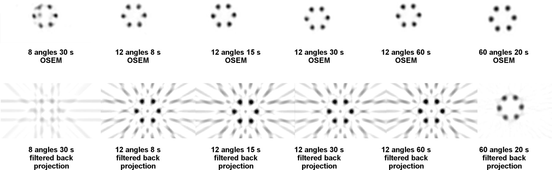
- FIGURE 1.
SPECT images of phantom with symmetrically distributed cylinders acquired with various image angles and times. SPECT acquisitions in top row are processed with OSEM, and same SPECT images in bottom row are processed with filtered backprojection. Images are labeled with number of images and times for acquisitions. Standard SPECT protocol is 60 angles and 20 s per image.
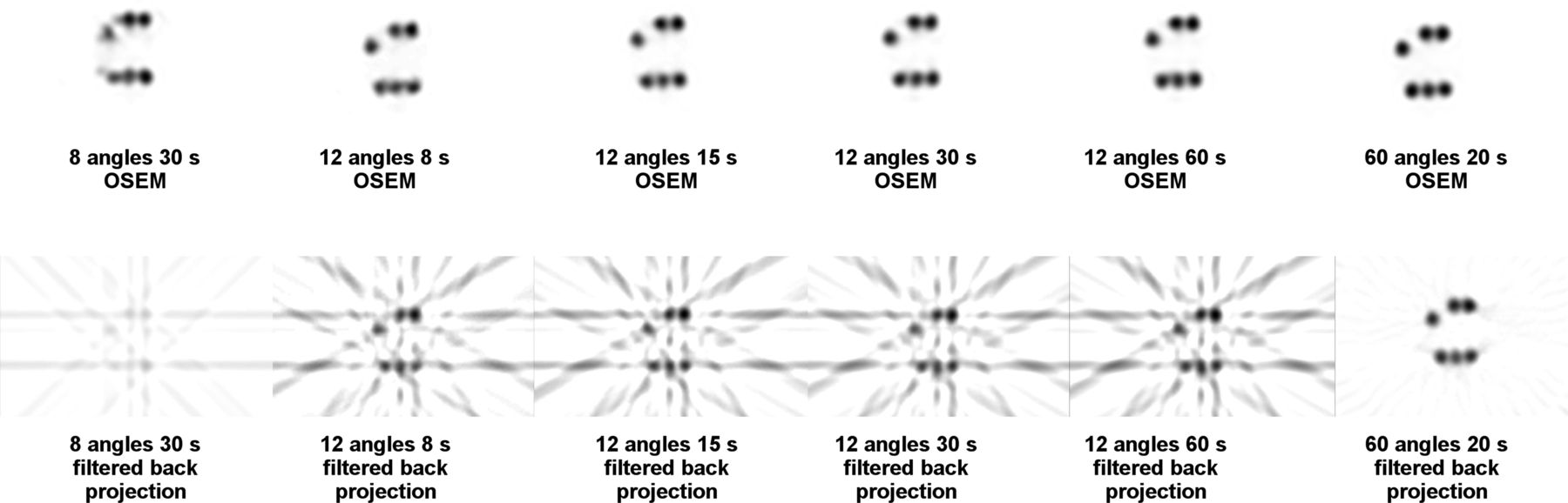
- FIGURE 2.
SPECT images of phantom with randomly placed activity-containing glass vial cylinders. Image parameters are same as in Figure 1.
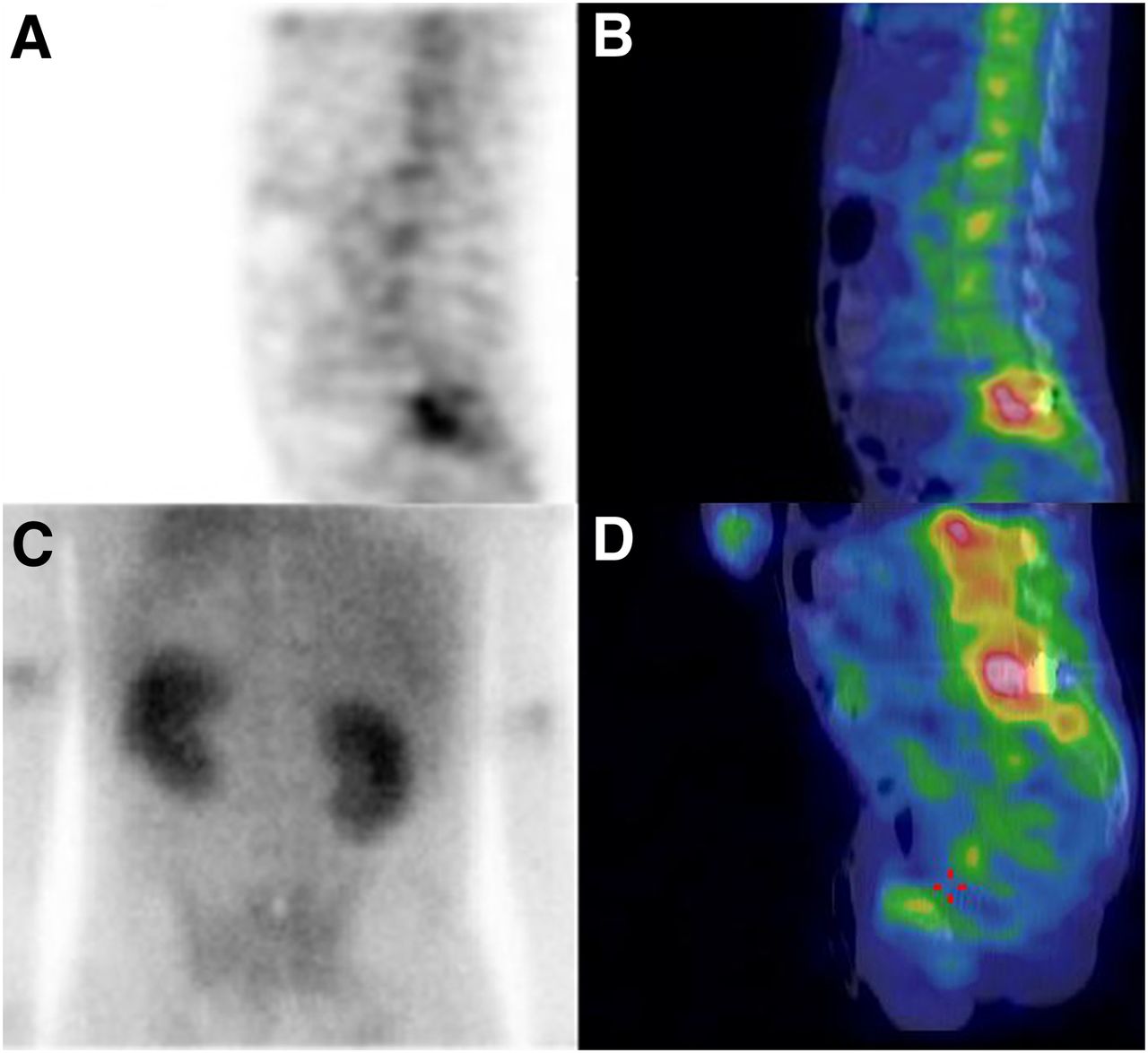
- FIGURE 3.
Imaging of spine: FASpecT blood pool sagittal image (A), FASpecT/CT blood pool sagittal image (B), posterior planar blood pool image (C), and 67Ga citrate SPECT/CT sagittal image (D). FASpecT/CT blood pool images demonstrate intense focally increased uptake at L5–S1 vertebrae that is poorly localized on planar image. Subse-quent 67Ga citrate image demonstrates infection corresponding to region of increased blood pool uptake on FASpecT image.
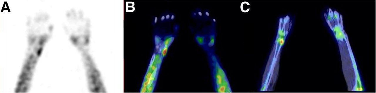
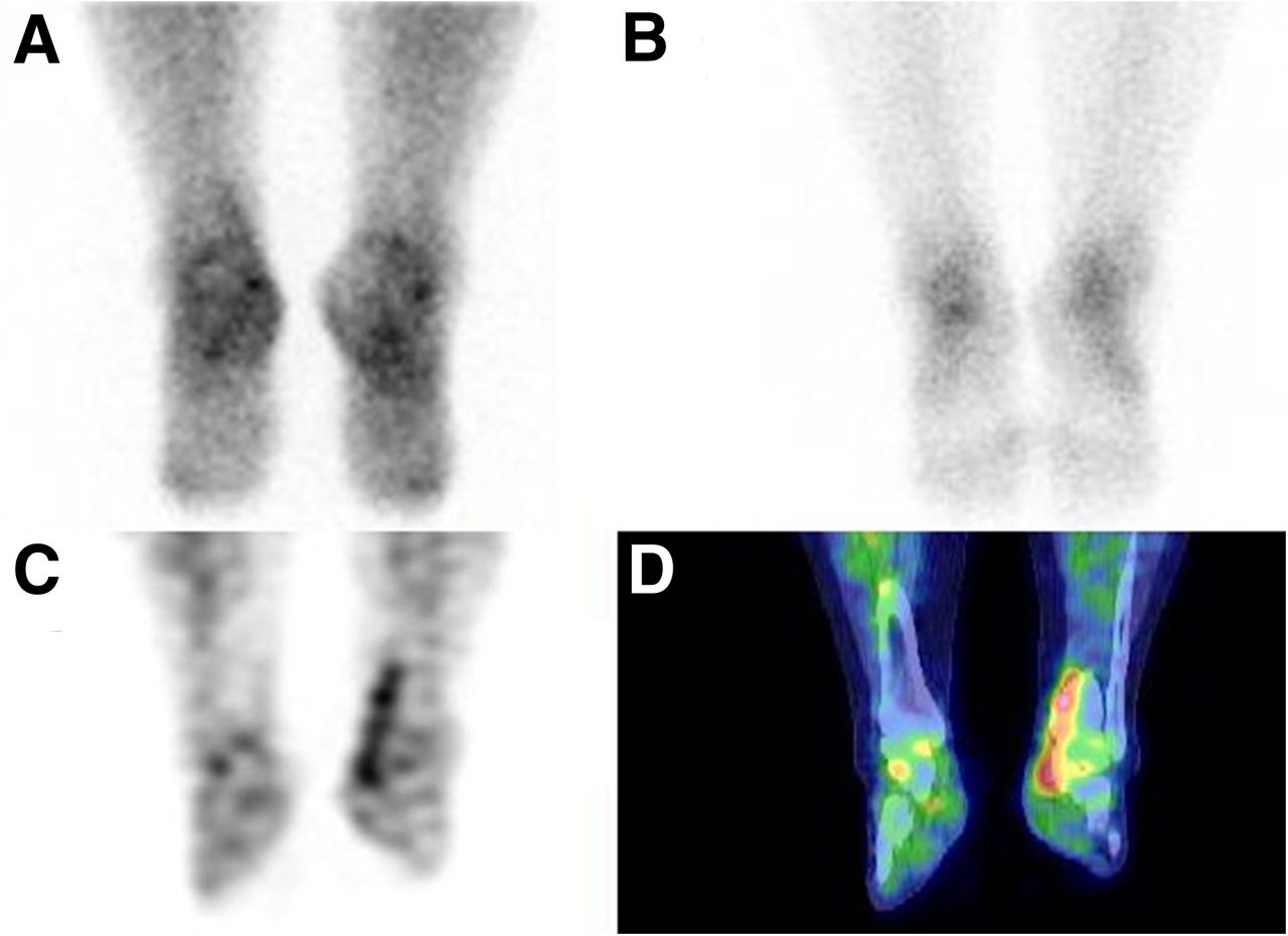
- FIGURE 4.
Imaging of feet: anterior planar blood pool image (A), anterior planar delayed bone scan (B), FASpecT/CT blood pool sagittal image of left foot (C), and 111In-white blood cell SPECT/CT sagittal image of left foot (D). Planar blood pool image demonstrates diffuse increased blood pool activity that cannot be precisely localized. Delayed planar bone scan shows focal bone uptake consistent with 3-phase positivity of region of left distal second metatarsal. However, FASpecT/CT blood pool image demonstrates focal soft-tissue blood pool activity of left foot without apparent bone involvement. Subsequent 111In-white blood cell SPECT/CT image shows focal soft-tissue activity without bone activity, correlating with FASpecT/CT blood pool imaging.
- FIGURE 5.
Imaging of hands: FASpecT blood pool image (A), FASpecT/CT blood pool image (B), and delayed SPECT/CT bone image (C). FASpecT/CT blood pool images show increased linear blood pool activity along distribution of right extensor pollicis brevis tendon characteristic of De Quervain tenosynovitis. Delayed bone scan shows focally increased uptake only in distal radius at origin of extensor pollicis brevis tendon, most likely because of increased regional blood flow. Planar blood pool image (not shown) showed minimal asymmetric increased blood pool uptake in right wrist that could not be localized.
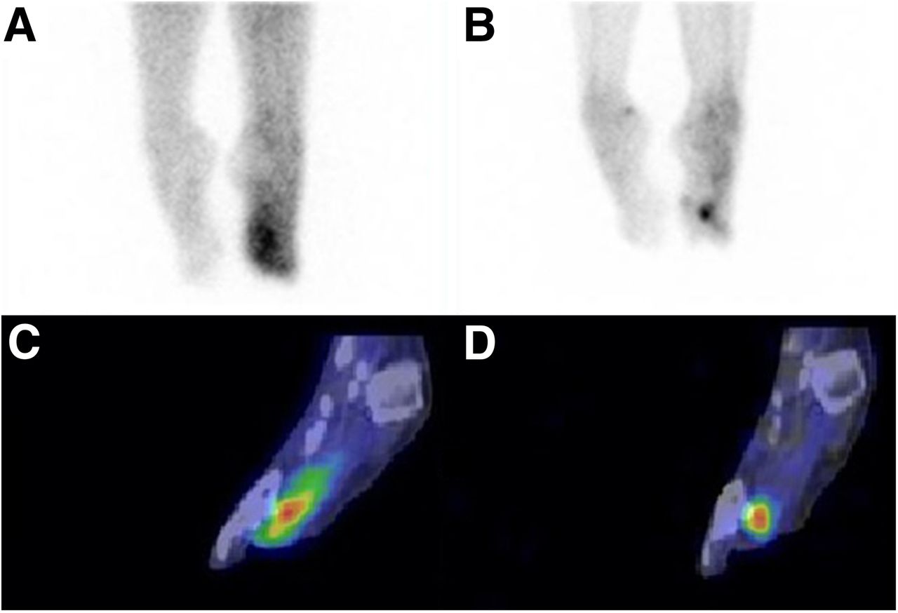
- FIGURE 6.
Imaging of feet and ankles: posterior planar blood pool image (A), posterior planar delayed bone scan (B), FASpecT blood pool coronal image (C), and coronal SPECT/CT blood pool coronal image (D). FASpecT/CT blood pool images demonstrate focally increased uptake along posterior tibial tendon consistent with tendonitis. Findings on delayed bone scan are essentially normal, and posterior planar blood pool scan shows asymmetry that cannot be localized.

Tables
- TABLE 1
Comparison of Acquisition and Processing Parameters Between FASpecT and Standard SPECT for Blood Pool Imaging
Parameter FASpecT/CT protocol Standard SPECT/CT protocol SPECT acquisition 30° steps and total angular range of 360°with dual-head SPECT/CT camera 3°–6° steps and total angular range of 360° with dual-head SPECT/CT camera Acquisition time per projection 30 s 20–30 s Total imaging time required 3 min 20 s 22–65 min Number of steps 6 60 Total images acquired 12 60–120 Matrix size 128 × 128 64 × 64 or 128 × 128 Reconstruction OSEM (2 iterations,10 subsets) Filtered backprojection or OSEM Postreconstruction filter Butterworth: critical frequency, 0.48; power, 10 Butterworth with varying parameters


Jump to section
Related Articles
Cited By...
- No citing articles found.