
Main menu

  • Home
  • Content
    • Current
      • JNMT Supplement
    • Ahead of print
    • Past Issues
    • Continuing Education
    • JNMT Podcast
    • SNMMI Annual Meeting Abstracts
  • Subscriptions
    • Subscribers
    • Rates
    • Journal Claims
    • Institutional and Non-member
  • Authors
    • Submit to JNMT
    • Information for Authors
    • Assignment of Copyright
    • AQARA Requirements
  • Info
    • Reviewers
    • Permissions
    • Advertisers
    • Corporate & Special Sales
  • About
    • About Us
    • Editorial Board
    • Contact Information
  • More
    • Alerts
    • Feedback
    • Help
    • SNMMI Journals
  • SNMMI
    • JNMT
    • JNM
    • SNMMI Journals
    • SNMMI

User menu

  • Subscribe
  • My alerts
  • Log in
  • My Cart

Search

  • Advanced search
Journal of Nuclear Medicine Technology
  • SNMMI
    • JNMT
    • JNM
    • SNMMI Journals
    • SNMMI
  • Subscribe
  • My alerts
  • Log in
  • My Cart
Journal of Nuclear Medicine Technology

Advanced Search

  • Home
  • Content
    • Current
    • Ahead of print
    • Past Issues
    • Continuing Education
    • JNMT Podcast
    • SNMMI Annual Meeting Abstracts
  • Subscriptions
    • Subscribers
    • Rates
    • Journal Claims
    • Institutional and Non-member
  • Authors
    • Submit to JNMT
    • Information for Authors
    • Assignment of Copyright
    • AQARA Requirements
  • Info
    • Reviewers
    • Permissions
    • Advertisers
    • Corporate & Special Sales
  • About
    • About Us
    • Editorial Board
    • Contact Information
  • More
    • Alerts
    • Feedback
    • Help
    • SNMMI Journals
  • Watch or Listen to JNMT Podcast
  • Visit SNMMI on Facebook
  • Join SNMMI on LinkedIn
  • Follow SNMMI on Twitter
  • Subscribe to JNMT RSS feeds
Research ArticleImaging

Added Value of CT Attenuation Correction and Prone Positioning in Improving Breast and Subdiaphragmatic Attenuation in Myocardial Perfusion Imaging

Ahmed E. Tawakol, Hazem M. Tantawy, Rana E. Elashmawy, Yasser G. Abdelhafez and Yasser M. Elsayed
Journal of Nuclear Medicine Technology March 2021, 49 (1) 23-29; DOI: https://doi.org/10.2967/jnmt.120.255943
Ahmed E. Tawakol
1Department of Clinical Oncology and Nuclear Medicine, Faculty of Medicine, Cairo University, Cairo, Egypt
  • Find this author on Google Scholar
  • Find this author on PubMed
  • Search for this author on this site
Hazem M. Tantawy
2Department of Nuclear Medicine Technology, Inaya Medical College, Riyadh, Saudi Arabia; and
  • Find this author on Google Scholar
  • Find this author on PubMed
  • Search for this author on this site
Rana E. Elashmawy
1Department of Clinical Oncology and Nuclear Medicine, Faculty of Medicine, Cairo University, Cairo, Egypt
  • Find this author on Google Scholar
  • Find this author on PubMed
  • Search for this author on this site
Yasser G. Abdelhafez
3Nuclear Medicine Unit, South Egypt Cancer Institute, Assiut University, Assiut, Egypt
  • Find this author on Google Scholar
  • Find this author on PubMed
  • Search for this author on this site
Yasser M. Elsayed
1Department of Clinical Oncology and Nuclear Medicine, Faculty of Medicine, Cairo University, Cairo, Egypt
  • Find this author on Google Scholar
  • Find this author on PubMed
  • Search for this author on this site
  • Article
  • Figures & Data
  • Info & Metrics
  • PDF
Loading

Article Figures & Data

Figures

  • Tables
  • FIGURE 1.
    • Download figure
    • Open in new tab
    • Download powerpoint
    FIGURE 1.

    Corrected and uncorrected artifacts using CT AC and prone imaging.
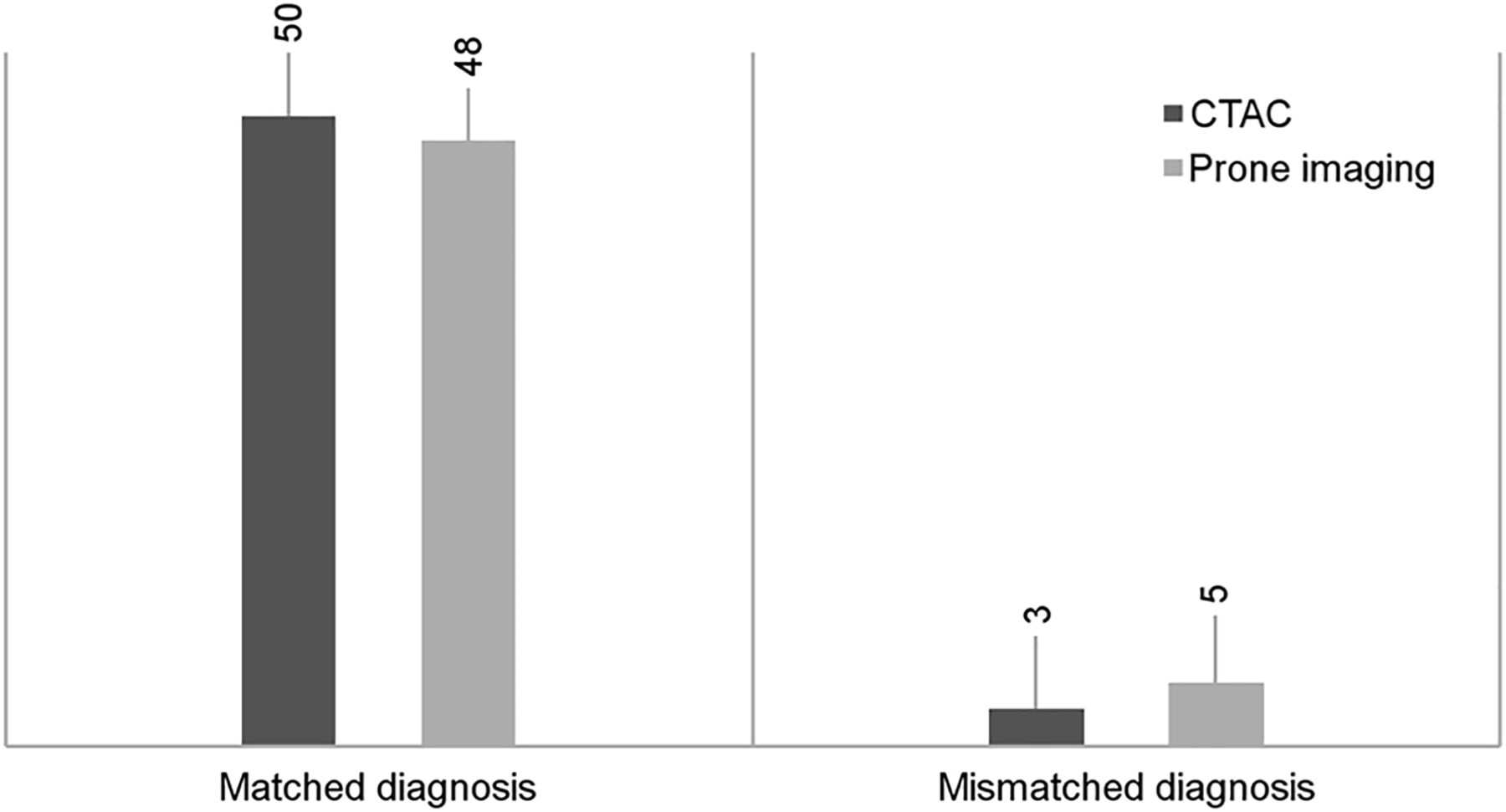
  • FIGURE 2.
    • Download figure
    • Open in new tab
    • Download powerpoint
    FIGURE 2.

    Matching between correction technique and final diagnosis.
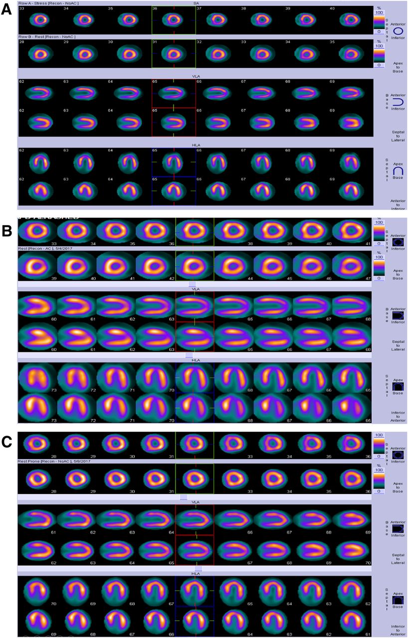
  • FIGURE 3.
    • Download figure
    • Open in new tab
    • Download powerpoint
    FIGURE 3.

    (A) No-AC images showing mild reversible inferior wall hypoperfusion defect. (B) Stress/rest CT AC images showing normalization of inferior wall hypoperfusion defect. (C) Stress/rest prone images showing normalization of inferior wall hypoperfusion defect.
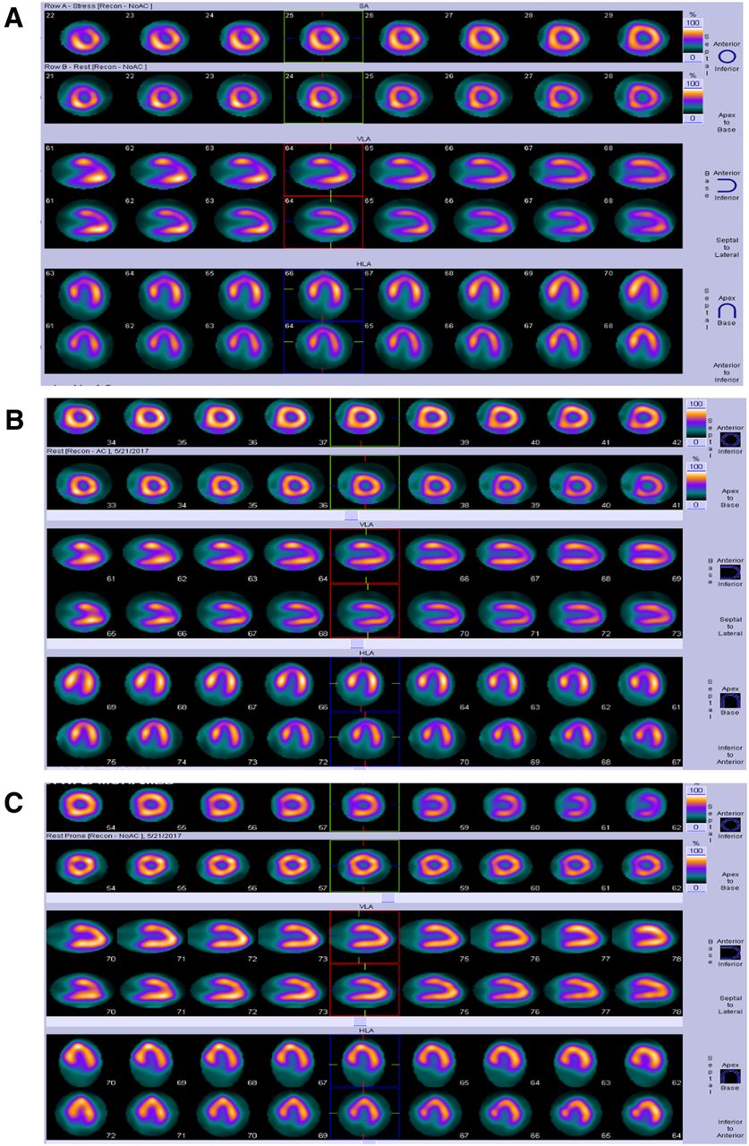
  • FIGURE 4.
    • Download figure
    • Open in new tab
    • Download powerpoint
    FIGURE 4.

    (A) No-AC images showing moderate reversible hypoperfusion defect in anteroseptal wall. (B) Stress/rest CT AC images showing partial improvement of anteroseptal wall defect. (C) Stress/rest prone images showing complete resolution of anteroseptal wall defect (C).
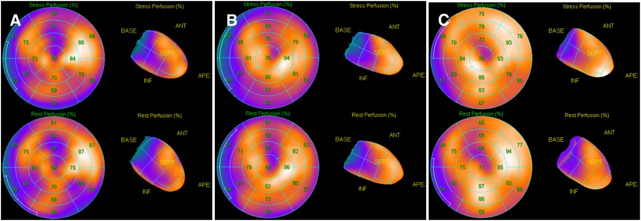
  • FIGURE 5.
    • Download figure
    • Open in new tab
    • Download powerpoint
    FIGURE 5.

    (A) No-AC images showing hypoperfusion defect in inferior wall. (B) Stress/rest CT AC images showing partial improvement of inferior wall defect. (C) Stress/rest prone images showing complete resolution of inferior wall defect.

Tables

  • Figures
    • View popup
    TABLE 1

    Presence or Absence of Myocardial Ischemia in the 14 Excluded Patients

    ParameternNon-ACCT ACProne imaging
    Negative for myocardial ischemia4No perfusion defectNo perfusion defectNo perfusion defect
    Multivessel disease10Perfusion defectPerfusion defectPerfusion defect
    • View popup
    TABLE 2

    Age and Sex Distribution of the 30 Patients

    SexnPercentageAge (mean ± SD)
    Male2067%54.7 ± 12.5
    Female1033%49.8 ± 11
    Total30100%53 ± 12
    • View popup
    TABLE 3

    Agreement Between Correction Technique and Uncorrected Images in All Wall Defects

    Final diagnosisCT AC*Prone imaging*
    True defect (n = 22)2222
    Attenuation artifact (n = 31)
     Corrected2826
     Uncorrected35
    Total (n = 53)53
    • ↵* P < 0.001.

    • View popup
    TABLE 4

    Matching Between Correction Technique and Final Diagnosis

    Correction techniqueFinal diagnosisTotal
    MatchedMismatched
    CT AC50 (94%)3 (6%)53 (100%)
    Prone imaging48 (91%)5 (9%)53 (100%)
    • View popup
    TABLE 5

    Correlation Between Wall Defects and Final Diagnosis

    ParameterWall defects in CT ACWall defects in prone imagingTrue defectsTotal
    Corrected28 (90.32%)26 (83.8%)None26 (49.1%)
    Uncorrected3 (9.68%)5 (16.2%)22 (100%)27 (50.9%)
    Total31 (100%)22 (100%)53 (100%)
    • View popup
    TABLE 6

    Specificity and Sensitivity

    ParameterSpecificitySensitivity
    CT AC90.3%100%
    Prone imaging83.8%100%
PreviousNext
Back to top

In this issue

Journal of Nuclear Medicine Technology: 49 (1)
Journal of Nuclear Medicine Technology
Vol. 49, Issue 1
March 1, 2021
  • Table of Contents
  • About the Cover
  • Index by author
Print
Download PDF
Article Alerts
Sign In to Email Alerts with your Email Address
Email Article

Thank you for your interest in spreading the word on Journal of Nuclear Medicine Technology.

NOTE: We only request your email address so that the person you are recommending the page to knows that you wanted them to see it, and that it is not junk mail. We do not capture any email address.

Enter multiple addresses on separate lines or separate them with commas.
Added Value of CT Attenuation Correction and Prone Positioning in Improving Breast and Subdiaphragmatic Attenuation in Myocardial Perfusion Imaging
(Your Name) has sent you a message from Journal of Nuclear Medicine Technology
(Your Name) thought you would like to see the Journal of Nuclear Medicine Technology web site.
Citation Tools
Added Value of CT Attenuation Correction and Prone Positioning in Improving Breast and Subdiaphragmatic Attenuation in Myocardial Perfusion Imaging
Ahmed E. Tawakol, Hazem M. Tantawy, Rana E. Elashmawy, Yasser G. Abdelhafez, Yasser M. Elsayed
Journal of Nuclear Medicine Technology Mar 2021, 49 (1) 23-29; DOI: 10.2967/jnmt.120.255943

Citation Manager Formats

  • BibTeX
  • Bookends
  • EasyBib
  • EndNote (tagged)
  • EndNote 8 (xml)
  • Medlars
  • Mendeley
  • Papers
  • RefWorks Tagged
  • Ref Manager
  • RIS
  • Zotero
Share
Added Value of CT Attenuation Correction and Prone Positioning in Improving Breast and Subdiaphragmatic Attenuation in Myocardial Perfusion Imaging
Ahmed E. Tawakol, Hazem M. Tantawy, Rana E. Elashmawy, Yasser G. Abdelhafez, Yasser M. Elsayed
Journal of Nuclear Medicine Technology Mar 2021, 49 (1) 23-29; DOI: 10.2967/jnmt.120.255943
Twitter logo Facebook logo LinkedIn logo Mendeley logo
  • Tweet Widget
  • Facebook Like
  • Google Plus One
Bookmark this article

Jump to section

  • Article
    • Abstract
    • MATERIALS AND METHODS
    • RESULTS
    • DISCUSSION
    • CONCLUSION
    • DISCLOSURE
    • Footnotes
    • REFERENCES
  • Figures & Data
  • Info & Metrics
  • PDF

Related Articles

  • No related articles found.
  • PubMed
  • Google Scholar

Cited By...

  • No citing articles found.
  • Google Scholar

More in this TOC Section

  • Early 10-Minute Postinjection [18F]F-FAPI-42 uEXPLORER Total-Body PET/CT Scanning Protocol for Staging Lung Cancer Using HYPER Iterative Reconstruction
  • Single- Versus Dual-Time-Point Imaging for Transthyretin Cardiac Amyloid Using 99mTc-Pyrophosphate
  • Effectiveness of Data-Driven Gating FDG PET/CT for Abdominal Region
Show more Imaging

Similar Articles

Keywords

  • SPECT/CT
  • attenuation correction
  • prone imaging
  • MPI
SNMMI

© 2025 SNMMI

Powered by HighWire