


Article Figures & Data
Figures
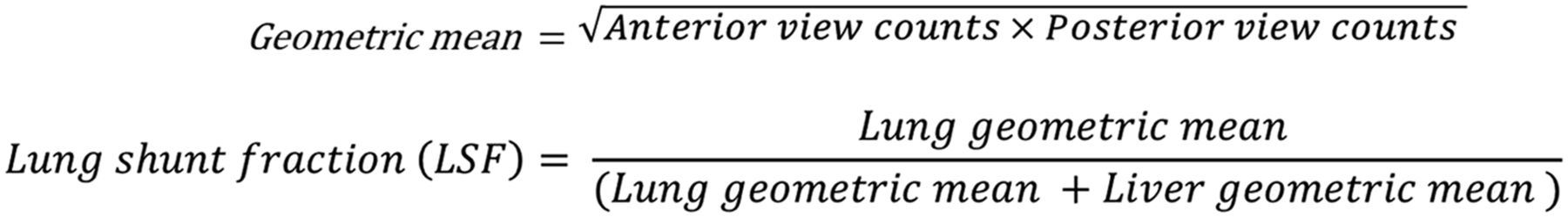
- FIGURE 1.
Formulas used for calculating geometric mean (for designated ROI) and LSF.
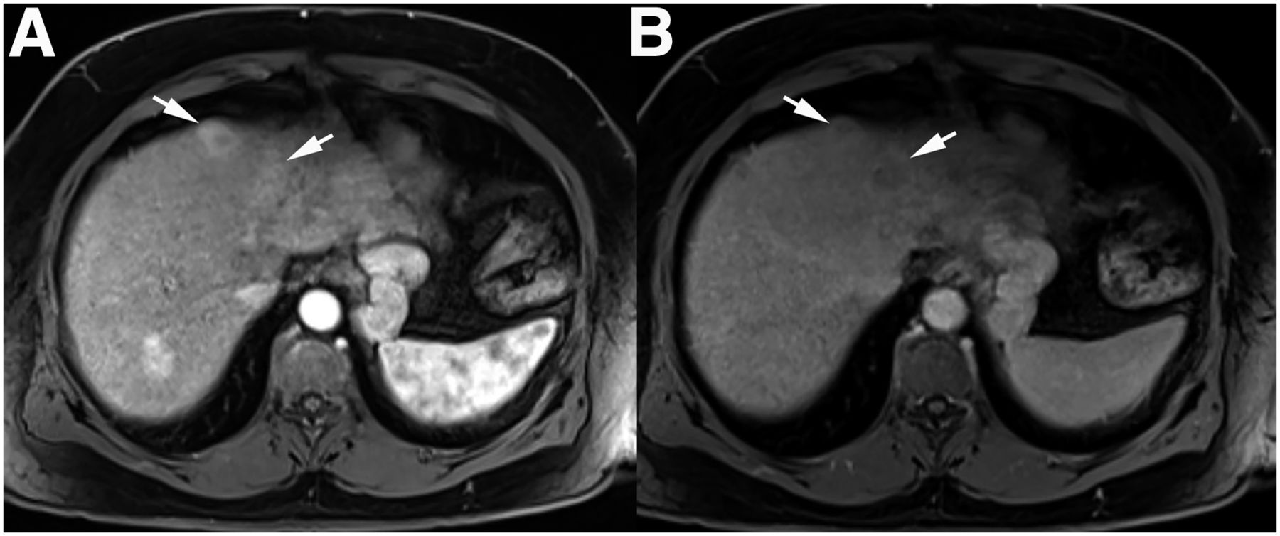
- FIGURE 2.
Contrast-enhanced axial T1-weighted images during arterial (A) and delayed (B) phases demonstrate washout within 2 adjacent hepatocellular carcinomas (arrows) located anteriorly within segment 4A of liver.
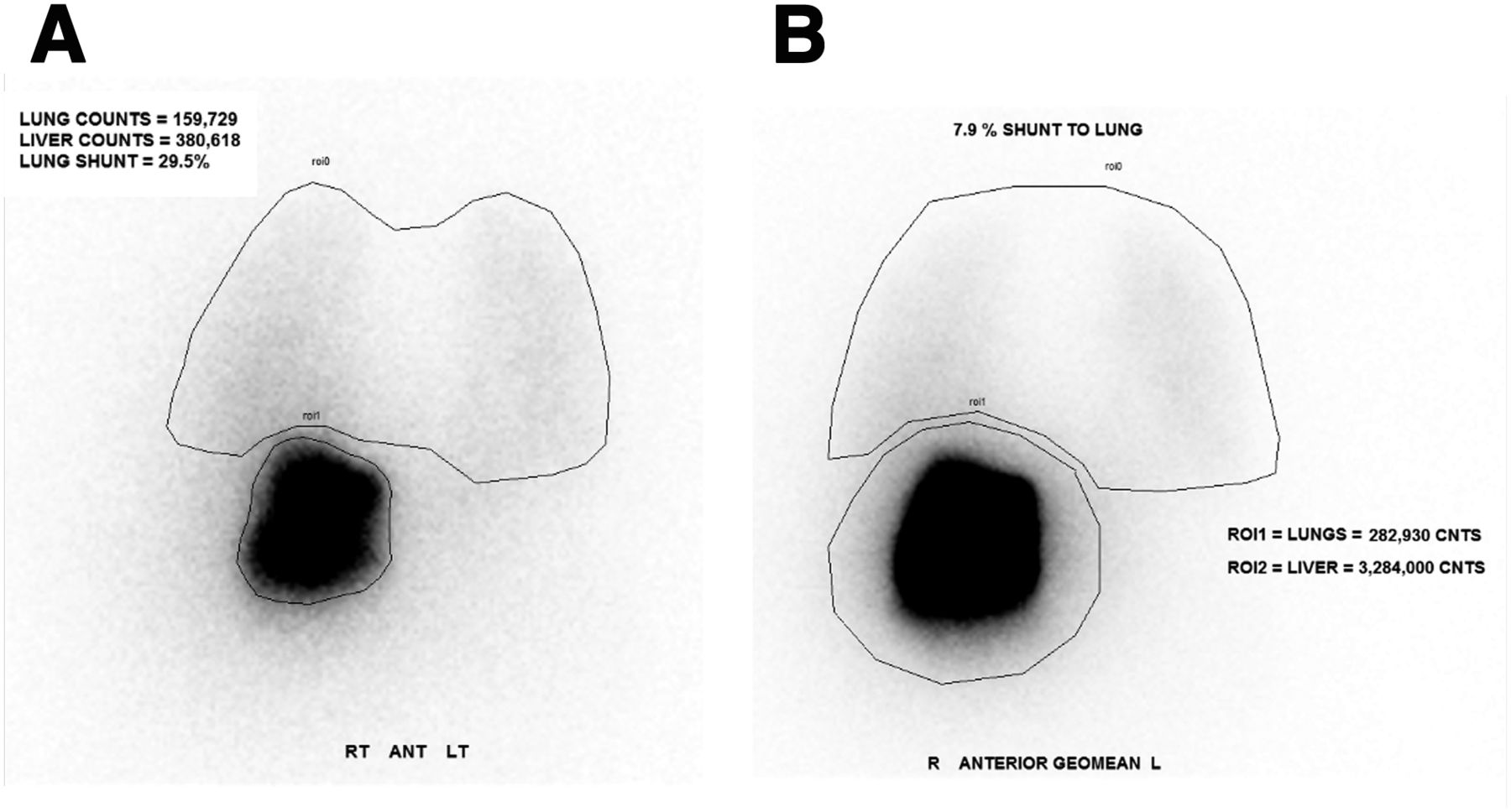
- FIGURE 3.
(A) Initial 99mTc-MAA (flipped) posterior planar image with lung and liver ROIs. Incorrect LSF calculated was 29.5%. (B) 99mTc-MAA geometric mean image with lung and liver ROIs. Correct LSF calculated was 7.9%.
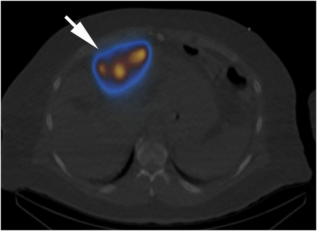
- FIGURE 4.
Selected axial 99mTc-MAA SPECT/CT image confirming radiotracer delivery to anteriorly positioned hepatocellular carcinomas (arrow).
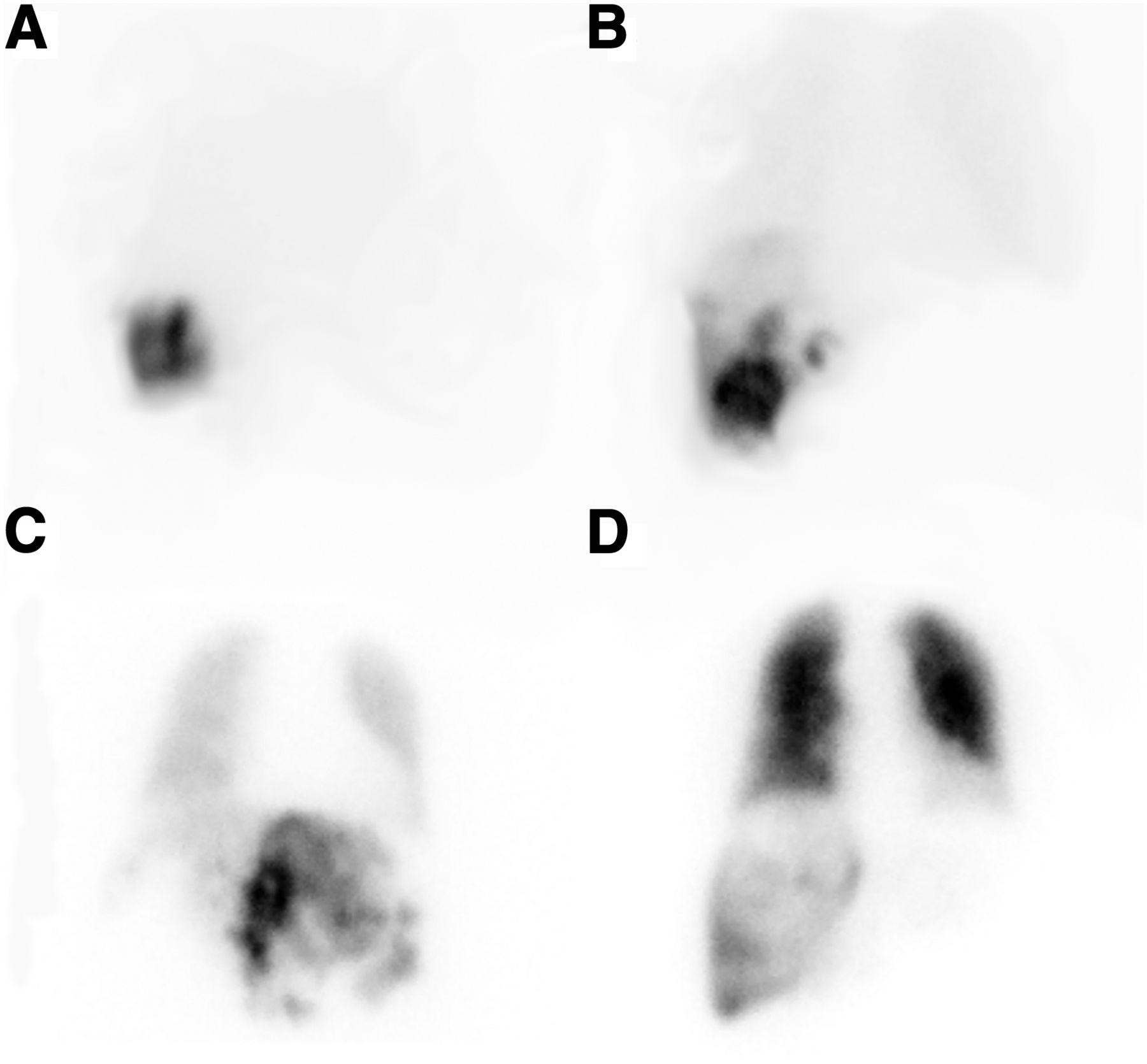
- FIGURE 5.
Lung shunt fraction visual reference for LSFs of 3% (A), 15% (B), 37% (C), and 79% (D).


