


Abstract
Textitis is a new term used to refer to the degenerative-strain osteoarthritis that comes from excessive use of a smart phone. 18F-NaF is increasingly used in diagnosing skeletal pain that is not identified on radiographs. We report a case of a 26-y-old woman with left breast cancer referred for 18F-NaF PET/CT, who was complaining of right thumb and wrist pain. Findings were negative for bone secondaries. Dedicated hands views were acquired on a positron emission mammography scanner and showed focal uptake at the first carpometacarpal and second metacarpophalangeal joints. On the basis of the strong history, the findings were likely due to active arthritic changes caused by repetitive strain injury from excessive text messaging.
Textitis is a new term used to describe a kind of osteoarthritis, or degenerative strain disease, that arises from excessive use of a smart phone. Smart-phone users are at risk of developing various repetitive strain injuries to the soft tissues because of repetitive use of the phone for text messaging (1). Although trauma, osteoarthritis, and rheumatoid arthritis can also present similarly, in textitis patients the symptoms of pain, tenderness, throbbing, tingling, or numbness and weakness are strongly associated with extensive texting (2). Thumb pain from texting can be due to strain in the tendon caused by constant holding of a cell phone or stretching of the thumb to type text messages. Pain can also be due to arthritis at the carpometacarpal joint, where the wrist and thumb are joined (3).
Usually, no radiologic tests are advised for mild symptoms, but for moderate to severe pain a radiograph is usually recommended to rule out any underlying condition. Treatment of repetitive strain injuries includes activity modification, painkillers, cock-up wrist splints, and limitation of texting. But without treatment, the symptoms become constant and cause swelling in the affected area.
CASE REPORT
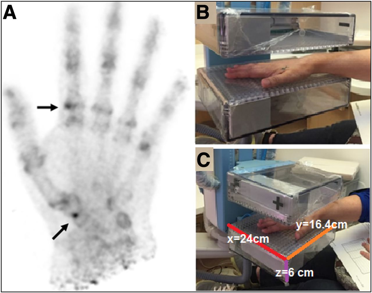
We here report a case in which textitis was found incidentally in a 26-y-old woman with known left breast cancer referred for 18F-NaF PET/CT for staging. A 144.3-MBq (3.9-mCi) dose of 18F-NaF (2.22 MBq [0.06 mCi]/kg) was injected, followed 60 min later by acquisition of whole-body images. Because the patient was complaining of pain at the right thumb and radial aspect of the wrist, dedicated hand images for 10 min were also acquired a positron emission mammography (PEM) scanner. The imaging parameters and reconstruction were according to local protocols (Table 1). The whole-body images were negative for bone secondaries. The dedicated hand views showed focal uptake (Fig. 1A) at the first carpometacarpal, trapezium-scaphoid, and second metacarpophalangeal joints. When correlated with the history of excessive smart-phone use for text messaging, these findings favored active arthritic changes due to repetitive strain injury. The institutional review board approved this study, and the patient gave written informed consent to publication of her data.
Technical Parameters of PEM Camera (5)

(A) Maximum-intensity projections of hand showing focal increased tracer activity at first carpometacarpal, trapezium-scaphoid, and second metacarpophalangeal joints (arrows). (B and C) Photographs of GE Healthcare Naviscan PEM scanner.
DISCUSSION
In the era of advanced technology, many young people are at a higher risk of developing repetitive strain injuries. There are no current recommendations on routine use of radiologic modalities for suspected repetitive strain injuries, and the patients are managed conservatively. In our case, the young lady was a known case of breast cancer and was undergoing a routine bone scan with 18F-NaF PET/CT for staging. 18F-NaF is an excellent bone-seeking agent because of high bone uptake due to rapid single-pass extraction, minimal binding to serum proteins, and fast clearance from the soft tissues. Encouraging results have also been reported for its use in characterizing benign bone diseases and enthesopathies (4).
PEM technology is currently used for breast-specific metabolic imaging. It has superior count sensitivity and high spatial resolution (≤2 mm). At our department, we have a dedicated PEM scanner with a dual-head coincidence detector to produce limited-angle tomographic images. The detectors are mounted on an articulating arm, which allows images to be acquired in any orientation—for example, craniocaudal and mediolateral. The lower (support) paddle is fixed to the arm whereas the upper (compression) paddle is adjustable to provide mild compression (67 newton of force) and can be moved up to 20 cm from the support paddle (Table 1). The enclosure is light-tight and electromagnetic interference–tight. Additionally, the enclosure is 95% tungsten on 5 sides to shield the detectors from radiation outside the field of view. The entrance window is 1-m-thick aluminum to maximize transmission of annihilation photons from within the field of view. Each detector-head houses a 2 × 6 matrix of detector modules, each of which comprises a crystal array, a reflective light guide, and a position-sensitive photomultiplier tube. Individual crystals (2 × 2 × 12 cm) of lutetium-yttrium oxyorthosilicate are packed in 13 × 13 arrays with a crystal pitch of 2.1 mm (5).
PEM is a small portable device that can be used to evaluate difficult-to-image, peripherally located lesions. Scarce data report the utility of 18F-NaF using PEM to image lesions outside the breast or in the extremities (6). To our knowledge, this is first case using 18F-NaF on a dedicated PEM scanner for bone imaging of a peripheral region that revealed a repetitive strain injury to the thumb due to excessive text messaging. Because of the superior spatial resolution of PEM, it may help play a role in the imaging of small bones or joints, such as those of the hands. Although such findings may coexist with underlying pathologic arthritis, old trauma, rheumatoid arthritis, or tenosynovitis, a careful history and clinical examination can lead to a precise interpretation such as in our patient, for whom there was no prior history of any bone-related disease.
CONCLUSION
With high-resolution PEM technology using a highly sensitive bone agent—that is, 18F-NaF—it was possible to image minute degrees of strain in the small bones. The current scenario highlights the significance of these twin technologies in the evaluation of musculoskeletal disorders of the small joints. Such technology may potentially be used in the evaluation of more prevalent joint disorders such as rheumatoid arthritis, especially for posttreatment response assessment.
DISCLOSURE
No potential conflict of interest relevant to this article was reported.
Footnotes
Published online Feb. 28, 2020.
REFERENCES
- Received for publication September 3, 2019.
- Accepted for publication November 11, 2019.


Jump to section
Related Articles
Cited By...
- No citing articles found.