
Main menu

  • Home
  • Content
    • Current
      • JNMT Supplement
    • Ahead of print
    • Past Issues
    • Continuing Education
    • JNMT Podcast
    • SNMMI Annual Meeting Abstracts
  • Subscriptions
    • Subscribers
    • Rates
    • Journal Claims
    • Institutional and Non-member
  • Authors
    • Submit to JNMT
    • Information for Authors
    • Assignment of Copyright
    • AQARA Requirements
  • Info
    • Reviewers
    • Permissions
    • Advertisers
    • Corporate & Special Sales
  • About
    • About Us
    • Editorial Board
    • Contact Information
  • More
    • Alerts
    • Feedback
    • Help
    • SNMMI Journals
  • SNMMI
    • JNMT
    • JNM
    • SNMMI Journals
    • SNMMI

User menu

  • Subscribe
  • My alerts
  • Log in
  • My Cart

Search

  • Advanced search
Journal of Nuclear Medicine Technology
  • SNMMI
    • JNMT
    • JNM
    • SNMMI Journals
    • SNMMI
  • Subscribe
  • My alerts
  • Log in
  • My Cart
Journal of Nuclear Medicine Technology

Advanced Search

  • Home
  • Content
    • Current
    • Ahead of print
    • Past Issues
    • Continuing Education
    • JNMT Podcast
    • SNMMI Annual Meeting Abstracts
  • Subscriptions
    • Subscribers
    • Rates
    • Journal Claims
    • Institutional and Non-member
  • Authors
    • Submit to JNMT
    • Information for Authors
    • Assignment of Copyright
    • AQARA Requirements
  • Info
    • Reviewers
    • Permissions
    • Advertisers
    • Corporate & Special Sales
  • About
    • About Us
    • Editorial Board
    • Contact Information
  • More
    • Alerts
    • Feedback
    • Help
    • SNMMI Journals
  • Watch or Listen to JNMT Podcast
  • Visit SNMMI on Facebook
  • Join SNMMI on LinkedIn
  • Follow SNMMI on Twitter
  • Subscribe to JNMT RSS feeds
Review ArticleTeaching Case Studies

Miscalculated Lung Shunt Fraction for Planning of Hepatic Radioembolization

Justin S. Caskey, Matthew D. Kay, Natalie A. McMillan, Phillip H. Kuo and Gregory J. Woodhead
Journal of Nuclear Medicine Technology June 2020, 48 (2) 184-186; DOI: https://doi.org/10.2967/jnmt.119.234385
Justin S. Caskey
1University of Arizona College of Medicine, Tucson, Arizona
  • Find this author on Google Scholar
  • Find this author on PubMed
  • Search for this author on this site
Matthew D. Kay
2Department of Medical Imaging, University of Arizona College of Medicine, Tucson, Arizona; and
  • Find this author on Google Scholar
  • Find this author on PubMed
  • Search for this author on this site
Natalie A. McMillan
2Department of Medical Imaging, University of Arizona College of Medicine, Tucson, Arizona; and
  • Find this author on Google Scholar
  • Find this author on PubMed
  • Search for this author on this site
Phillip H. Kuo
2Department of Medical Imaging, University of Arizona College of Medicine, Tucson, Arizona; and
3Department of Medicine and Biomedical Engineering, University of Arizona, Tucson, Arizona
  • Find this author on Google Scholar
  • Find this author on PubMed
  • Search for this author on this site
Gregory J. Woodhead
2Department of Medical Imaging, University of Arizona College of Medicine, Tucson, Arizona; and
  • Find this author on Google Scholar
  • Find this author on PubMed
  • Search for this author on this site
  • Article
  • Figures & Data
  • Info & Metrics
  • PDF
Loading

Abstract

90Y radioembolization is a safe and efficacious treatment option for many patients with unresectable hepatocellular carcinoma. Potential candidates for radioembolization, based on clinical criteria, undergo 99mTc-labeled macroaggregated albumin imaging to determine the extent of hepatopulmonary shunting. Dose selection is based on results from shunt imaging and can exclude patients from radioembolization therapy. We present a case of miscalculated lung shunt fraction and the circumstances that led to the critical error.

  • 90Y radioembolization
  • lung shunt fraction
  • 99mTc-labeled macroaggregated albumin
  • hepatocellular carcinoma

Millions of individuals worldwide have hepatocellular carcinoma, the most common primary liver malignancy. In addition to resection, safe and effective treatment options for many patients include transplantation, radiofrequency ablation, chemoembolization, and systemic chemotherapy (1). In 90Y radioembolization, glass or resin beads fixed with the β-particle–emitting radioisotope 90Y are selectively injected via a microcatheter to deliver high doses of radiation to the tumor. Before treatment, patients undergo important planning studies, including mapping angiography and 99mTc-labeled macroaggregated albumin (99mTc-MAA) imaging. Information from these studies is used to minimize the risk of nontarget radiation injury to the gastrointestinal tract and lungs (2).

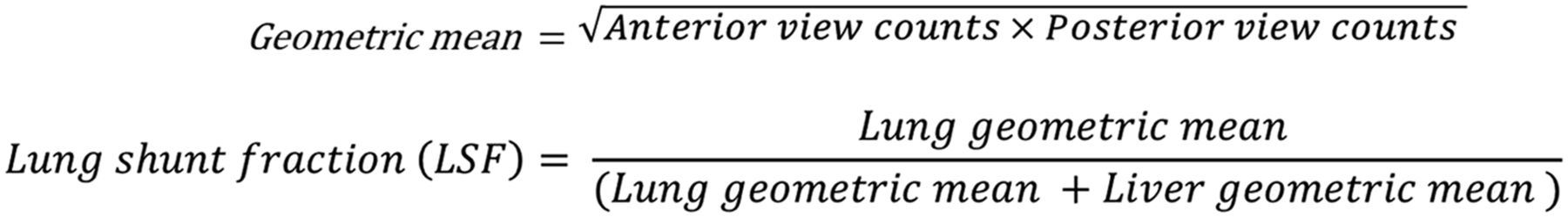
Radiation pneumonitis is a known complication of 90Y radioembolization, and the risk of this complication is related to radiation delivered unintentionally to pulmonary tissue via hepatopulmonary shunting (3). 99mTc-MAA imaging is therefore critical for 90Y patient selection, a requirement for shunt reduction intervention and radioembolization dosing. After 99mTc-MAA injection, anterior and posterior planar imaging is performed. The geometric mean is calculated as the square root of the product of counts for regions of interest (ROIs) from anterior and posterior planar images (Fig. 1). Many vendors’ software generates a single geometric mean image, which is a composite of the anterior and (flipped) posterior images, and from this the geometric mean counts for each ROI are obtained. The lung shunt fraction (LSF) is calculated as the counts from the lung ROI divided by the total counts for the lung and liver ROI (from the geometric mean image) (Fig. 1). For radioembolization therapy, an LSF of more than 20% or an LSF that results in an estimated lung radiation exposure of more than 30 Gy (based on the planned 90Y dose) is considered a contraindication (4) or requires a significant dose reduction at the risk of reduced treatment efficacy.

FIGURE 1.
  • Download figure
  • Open in new tab
  • Download powerpoint
FIGURE 1.

Formulas used for calculating geometric mean (for designated ROI) and LSF.

CASE REPORT

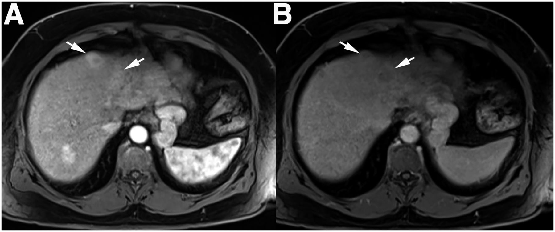
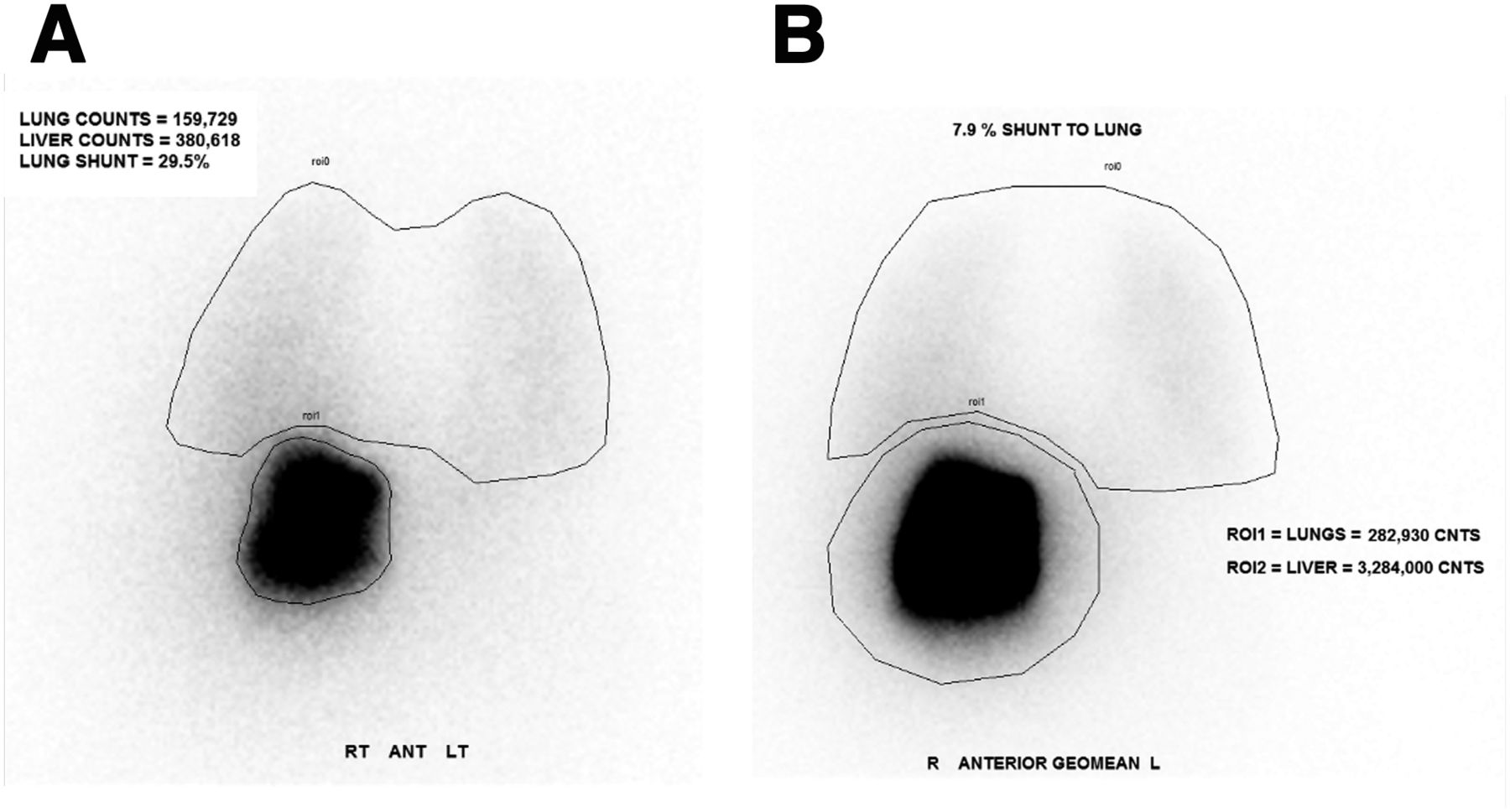
A 65-y-old man with multifocal hepatocellular carcinoma (Fig. 2) underwent evaluation for potential treatment with 90Y radioembolization. Pretreatment 99mTc-MAA imaging was performed to assess hepatopulmonary shunting. ROI determination and postprocessing were completed per standard procedures, and the LSF was initially calculated as 29.5% (Fig. 3A). The nuclear medicine physician noted a potential error in shunt fraction based on visual assessment and requested that the image be reprocessed. After reprocessing, the LSF was recalculated as 7.9% (Fig. 3B). The patient ultimately received a successful 90Y radioembolization.

FIGURE 2.
  • Download figure
  • Open in new tab
  • Download powerpoint
FIGURE 2.

Contrast-enhanced axial T1-weighted images during arterial (A) and delayed (B) phases demonstrate washout within 2 adjacent hepatocellular carcinomas (arrows) located anteriorly within segment 4A of liver.

FIGURE 3.
  • Download figure
  • Open in new tab
  • Download powerpoint
FIGURE 3.

(A) Initial 99mTc-MAA (flipped) posterior planar image with lung and liver ROIs. Incorrect LSF calculated was 29.5%. (B) 99mTc-MAA geometric mean image with lung and liver ROIs. Correct LSF calculated was 7.9%.

DISCUSSION

This case highlights a critical source of error that can occur during LSF calculation by 99mTc-MAA imaging. Initially, the nuclear medicine technologist mistakenly labeled the raw data from a posterior flipped view as the geometric mean image; thus, the ROI contained counts from only the posterior view. The initial incorrect LSF (using only the flipped posterior planar image) was 159,7289 ÷ (159,7289 + 380,618) = 0.295 (Fig. 3A). The correct LSF (based on counts from the geometric mean image) was 282,930 ÷ (282,930 + 3,284,000) = 0.079 (Fig. 3B).

The initial shunt fraction of 29.5% would be a contraindication to 90Y radioembolization, and the error thus would have mistakenly precluded the patient from therapy. Although differences in ROI size can alter count totals, the variance in this case was too large to be attributed to this factor alone because the error led to a 3.5 greater discrepancy between the calculated LSFs. The initial artifactually low liver count was especially exacerbated by the anterior position of the tumor (Fig. 4), as only the (flipped) posterior planar image was used for the initial LSF calculation. Our nuclear medicine laboratory has scanners from multiple vendors, and this error likely occurred because of confusion over how the geometric mean is displayed or calculated among different vendors.

FIGURE 4.
  • Download figure
  • Open in new tab
  • Download powerpoint
FIGURE 4.

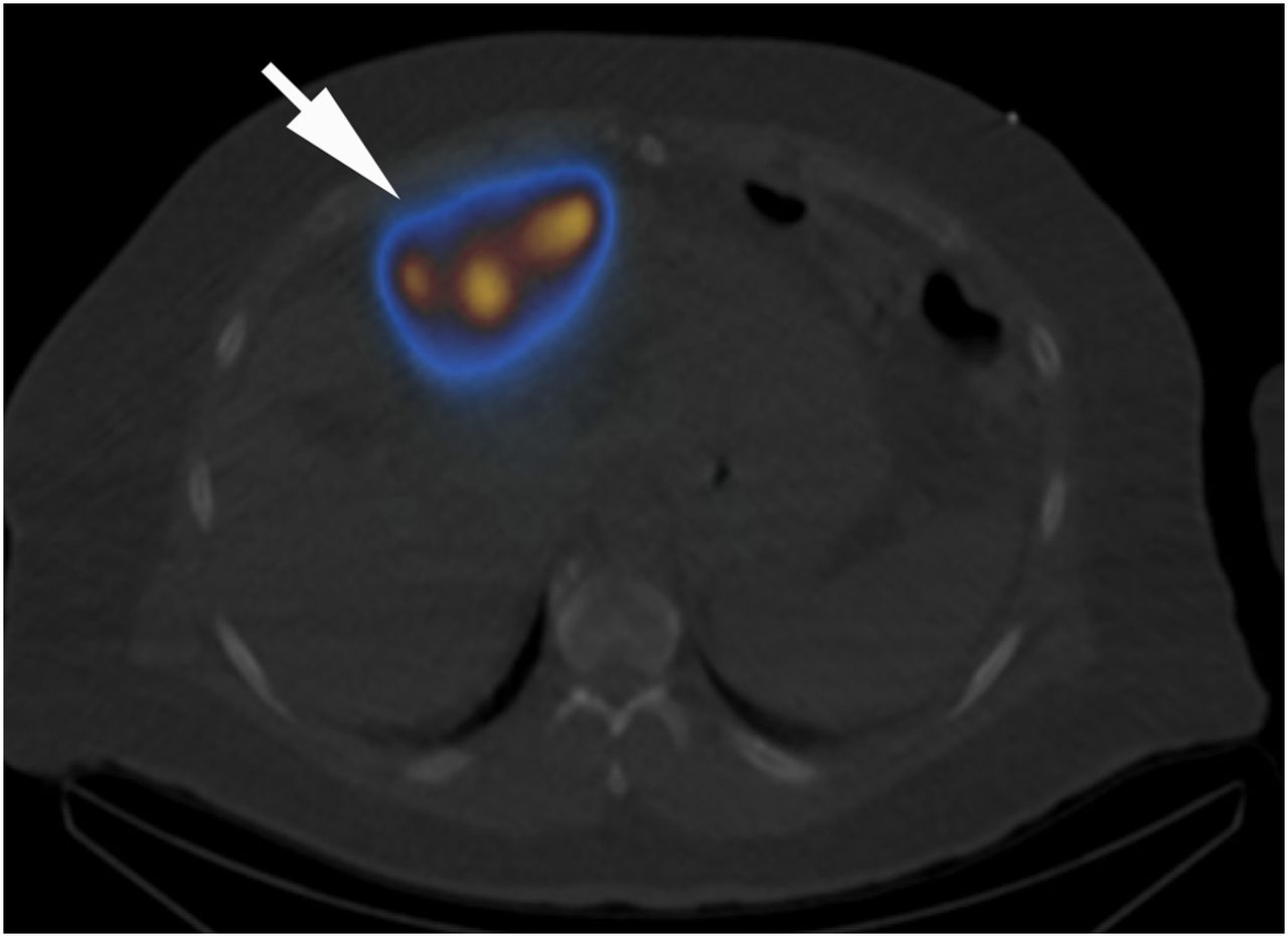
Selected axial 99mTc-MAA SPECT/CT image confirming radiotracer delivery to anteriorly positioned hepatocellular carcinomas (arrow).

To overcome this potential error, the nuclear medicine technologist may benefit from having easy access to a vendor-specific guide for the accurate calculation of LSF. Labeling the images with “geometric mean” instead of “ROI” or “counts,” as in our case (Fig. 3), may act as a reminder to use the correct data source. Further, if the nuclear medicine technologist (or nuclear medicine physician) needs to actively window the raw data to discriminate the lung ROI (or liver ROI) from the background, then the LSF should be low. Nuclear medicine technologists and nuclear medicine physicians may also use the provided LSF visual reference (Fig. 5) as a guide when visually assessing the adequacy of the LSF from the raw data images.

FIGURE 5.
  • Download figure
  • Open in new tab
  • Download powerpoint
FIGURE 5.

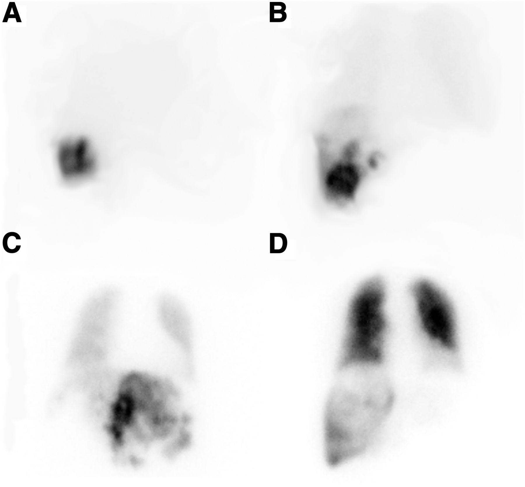
Lung shunt fraction visual reference for LSFs of 3% (A), 15% (B), 37% (C), and 79% (D).

CONCLUSION

99mTc-MAA imaging and LSF calculations are important steps in pretreatment assessment for patients before 90Y radioembolization for hepatocellular carcinoma. Confirming that the visual assessment and calculated LSF are concordant ensures that the LSF has been calculated appropriately from the geometric mean image during processing.

DISCLOSURE

No potential conflict of interest relevant to this article was reported.

Footnotes

  • Published online Dec. 6, 2019.

REFERENCES

  1. 1.↵
    1. Saini A,
    2. Wallace A,
    3. Alzubaidi S,
    4. et al
    . History and evolution of yttrium-90 radioembolization for hepatocellular carcinoma. J Clin Med. 2019;8:55.
    OpenUrl
  2. 2.↵
    1. Leung WT,
    2. Lau WY,
    3. Ho SK,
    4. et al
    . Measuring lung shunting in hepatocellular carcinoma with intrahepatic-arterial technetium-99m macroaggregated albumin. J Nucl Med. 1994;35:70–73.
    OpenUrlAbstract/FREE Full Text
  3. 3.↵
    1. Leung TW,
    2. Lau WY,
    3. Ho SK,
    4. et al
    . Radiation pneumonitis after selective internal radiation treatment with intraarterial 90yttrium-microspheres for inoperable hepatic tumors. Int J Radiat Oncol Biol Phys. 1995;33:919–924.
    OpenUrlCrossRefPubMed
  4. 4.↵
    1. Coldwell D,
    2. Sangro B,
    3. Wasan H,
    4. Salem R,
    5. Kennedy A
    . General selection criteria of patients for radioembolization of liver tumors: an international working group report. Am J Clin Oncol. 2011;34:337–341.
    OpenUrlCrossRefPubMed
  • Received for publication July 29, 2019.
  • Accepted for publication October 7, 2019.
PreviousNext
Back to top

In this issue

Journal of Nuclear Medicine Technology: 48 (2)
Journal of Nuclear Medicine Technology
Vol. 48, Issue 2
June 1, 2020
  • Table of Contents
  • About the Cover
  • Index by author
Print
Download PDF
Article Alerts
Sign In to Email Alerts with your Email Address
Email Article

Thank you for your interest in spreading the word on Journal of Nuclear Medicine Technology.

NOTE: We only request your email address so that the person you are recommending the page to knows that you wanted them to see it, and that it is not junk mail. We do not capture any email address.

Enter multiple addresses on separate lines or separate them with commas.
Miscalculated Lung Shunt Fraction for Planning of Hepatic Radioembolization
(Your Name) has sent you a message from Journal of Nuclear Medicine Technology
(Your Name) thought you would like to see the Journal of Nuclear Medicine Technology web site.
Citation Tools
Miscalculated Lung Shunt Fraction for Planning of Hepatic Radioembolization
Justin S. Caskey, Matthew D. Kay, Natalie A. McMillan, Phillip H. Kuo, Gregory J. Woodhead
Journal of Nuclear Medicine Technology Jun 2020, 48 (2) 184-186; DOI: 10.2967/jnmt.119.234385

Citation Manager Formats

  • BibTeX
  • Bookends
  • EasyBib
  • EndNote (tagged)
  • EndNote 8 (xml)
  • Medlars
  • Mendeley
  • Papers
  • RefWorks Tagged
  • Ref Manager
  • RIS
  • Zotero
Share
Miscalculated Lung Shunt Fraction for Planning of Hepatic Radioembolization
Justin S. Caskey, Matthew D. Kay, Natalie A. McMillan, Phillip H. Kuo, Gregory J. Woodhead
Journal of Nuclear Medicine Technology Jun 2020, 48 (2) 184-186; DOI: 10.2967/jnmt.119.234385
Twitter logo Facebook logo LinkedIn logo Mendeley logo
  • Tweet Widget
  • Facebook Like
  • Google Plus One
Bookmark this article

Jump to section

  • Article
    • Abstract
    • CASE REPORT
    • DISCUSSION
    • CONCLUSION
    • DISCLOSURE
    • Footnotes
    • REFERENCES
  • Figures & Data
  • Info & Metrics
  • PDF

Related Articles

  • No related articles found.
  • PubMed
  • Google Scholar

Cited By...

  • PET/CT and SPECT/CT imaging of 90Y hepatic radioembolization at therapeutic and diagnostic activity levels: anthropomorphic phantom study
  • Google Scholar

More in this TOC Section

  • High-Sensitivity Troponin Elevation in a Young Woman with Typical Chest Pain: The Heart of the Matter
  • Pulmonary Adenocarcinoma Revealed by Parathyroid Scintigraphy: An Incidental Case to Remember
  • Prominent Right Ventricular Tracer Uptake: A Harbinger of Multivessel Coronary Artery Disease
Show more Teaching Case Studies

Similar Articles

Keywords

  • 90Y radioembolization
  • lung shunt fraction
  • 99mTc-labeled macroaggregated albumin
  • hepatocellular carcinoma
SNMMI

© 2025 SNMMI

Powered by HighWire