


Article Figures & Data
Figures
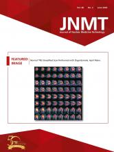
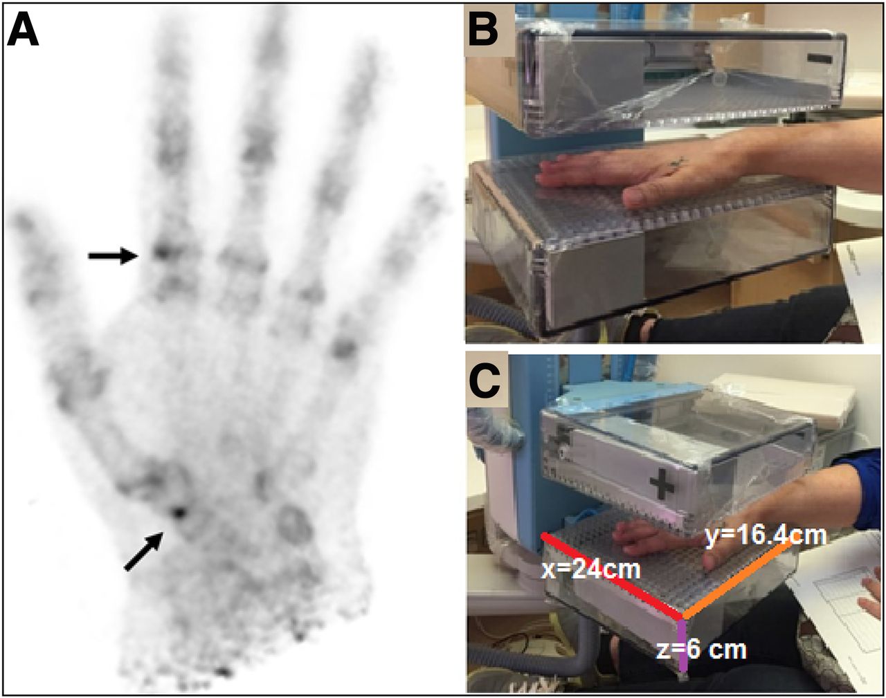
- FIGURE 1.
(A) Maximum-intensity projections of hand showing focal increased tracer activity at first carpometacarpal, trapezium-scaphoid, and second metacarpophalangeal joints (arrows). (B and C) Photographs of GE Healthcare Naviscan PEM scanner.

Tables
Parameter Characteristic Acquisition Views Anterior and posterior images of hands and feet Field of view (x–y plane) 24 × 16.8 cm (maximum) Field of view (z direction) Patient-dependent (maximum up to 19 cm) Compression force 67 newton for breast view, patient-dependent for hand and foot view Scan duration Variable (typically 10 min) Reconstruction Coincidence timing window 6 ns Energy window 350–750 keV Acceptance angle 25 crystals; angle varies with paddle separation Algorithm Iterative 3-dimensional maximum-likelihood expectation maximization Number of iterations 5 Corrections Detector normalization and geometric efficiency; no corrections for randoms, dead-time, attenuation, scatter, or intrascan decay Reconstruction time Depends on number of counts (typically <15 min) Images Image matrix 136 × 200 Pixel size 1.2 × 1.2 mm Resolution 2.4 mm in full width at half maximum Number of slices 12 Slice thickness 1/12 detector separation Units μCi/cm3 or PEM uptake value


Jump to section
Related Articles
Cited By...
- No citing articles found.