
Main menu

  • Home
  • Content
    • Current
      • JNMT Supplement
    • Ahead of print
    • Past Issues
    • Continuing Education
    • JNMT Podcast
    • SNMMI Annual Meeting Abstracts
  • Subscriptions
    • Subscribers
    • Rates
    • Journal Claims
    • Institutional and Non-member
  • Authors
    • Submit to JNMT
    • Information for Authors
    • Assignment of Copyright
    • AQARA Requirements
  • Info
    • Reviewers
    • Permissions
    • Advertisers
    • Corporate & Special Sales
  • About
    • About Us
    • Editorial Board
    • Contact Information
  • More
    • Alerts
    • Feedback
    • Help
    • SNMMI Journals
  • SNMMI
    • JNMT
    • JNM
    • SNMMI Journals
    • SNMMI

User menu

  • Subscribe
  • My alerts
  • Log in
  • My Cart

Search

  • Advanced search
Journal of Nuclear Medicine Technology
  • SNMMI
    • JNMT
    • JNM
    • SNMMI Journals
    • SNMMI
  • Subscribe
  • My alerts
  • Log in
  • My Cart
Journal of Nuclear Medicine Technology

Advanced Search

  • Home
  • Content
    • Current
    • Ahead of print
    • Past Issues
    • Continuing Education
    • JNMT Podcast
    • SNMMI Annual Meeting Abstracts
  • Subscriptions
    • Subscribers
    • Rates
    • Journal Claims
    • Institutional and Non-member
  • Authors
    • Submit to JNMT
    • Information for Authors
    • Assignment of Copyright
    • AQARA Requirements
  • Info
    • Reviewers
    • Permissions
    • Advertisers
    • Corporate & Special Sales
  • About
    • About Us
    • Editorial Board
    • Contact Information
  • More
    • Alerts
    • Feedback
    • Help
    • SNMMI Journals
  • Watch or Listen to JNMT Podcast
  • Visit SNMMI on Facebook
  • Join SNMMI on LinkedIn
  • Follow SNMMI on Twitter
  • Subscribe to JNMT RSS feeds
Research ArticleImaging

Automated Quantitative Analysis of American College of Radiology PET Phantom Images

Frank P. DiFilippo, Meghal Patel and Sagar Patel
Journal of Nuclear Medicine Technology September 2019, 47 (3) 249-254; DOI: https://doi.org/10.2967/jnmt.118.221317
Frank P. DiFilippo
Department of Nuclear Medicine, Cleveland Clinic, Cleveland, Ohio
  • Find this author on Google Scholar
  • Find this author on PubMed
  • Search for this author on this site
Meghal Patel
Department of Nuclear Medicine, Cleveland Clinic, Cleveland, Ohio
  • Find this author on Google Scholar
  • Find this author on PubMed
  • Search for this author on this site
Sagar Patel
Department of Nuclear Medicine, Cleveland Clinic, Cleveland, Ohio
  • Find this author on Google Scholar
  • Find this author on PubMed
  • Search for this author on this site
  • Article
  • Figures & Data
  • Info & Metrics
  • PDF
Loading

Article Figures & Data

Figures

  • Tables
  • FIGURE 1.
    • Download figure
    • Open in new tab
    • Download powerpoint
    FIGURE 1.

    Example of standard quantitative measurements specified for ACR PET phantom evaluation. Transaxial slice of 10-mm thickness is generated that includes hot and cold vials. Circular ROIs of 25-mm diameter are drawn, centered over each vial. Circular ROI (diameter between 60 and 70 mm) is drawn over background region. Some pass/fail criteria are based on SUV measurements of these regions, whereas other pass/fail criteria depend on qualitative assessment of hot vial visibility, cold rod visibility, and background uniformity. SUVbw = SUV normalized to body weight. Color version of this figure is available as supplemental file at http://tech.snmjournals.org.
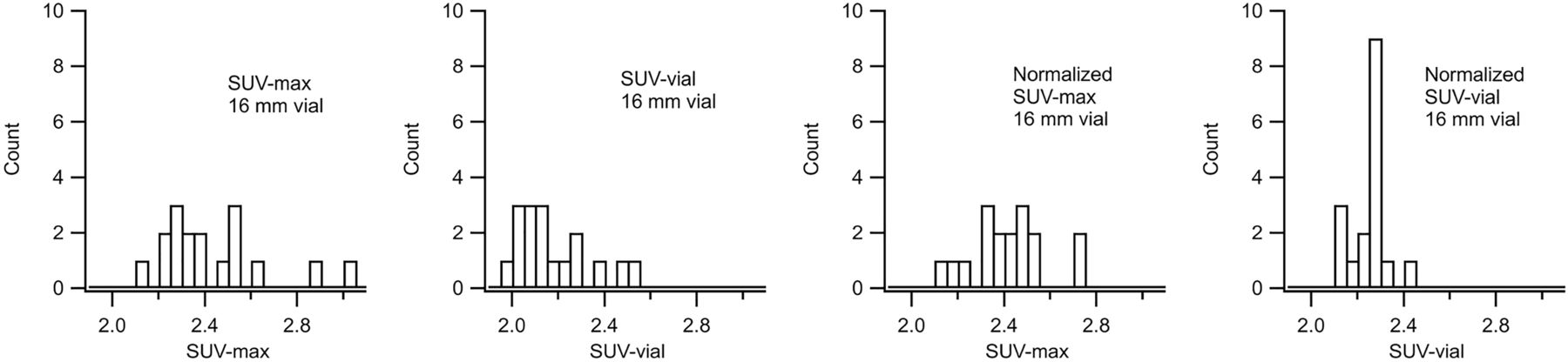
  • FIGURE 2.
    • Download figure
    • Open in new tab
    • Download powerpoint
    FIGURE 2.

    Histograms of SUV and normalized SUV measurements of 16-mm hot vial for 17 phantom studies. From left to right: SUVmax using ACR ROI, SUVvial using cylindric VOI, normalized SUVmax using ACR ROI, and normalized SUVvial using cylindric VOI. Most consistent results are obtained with normalized SUVvial measurements.
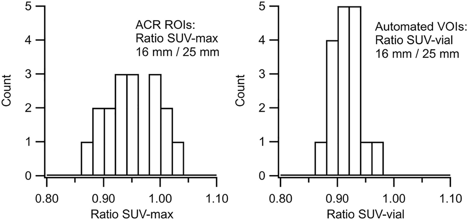
  • FIGURE 3.
    • Download figure
    • Open in new tab
    • Download powerpoint
    FIGURE 3.

    Histograms of SUVmax ratio (left) and SUVvial ratio (right) for 16- to 25-mm hot vials. SUVvial ratios were more consistent and, unlike SUVmax ratios, did not have outliers greater than 1.0.
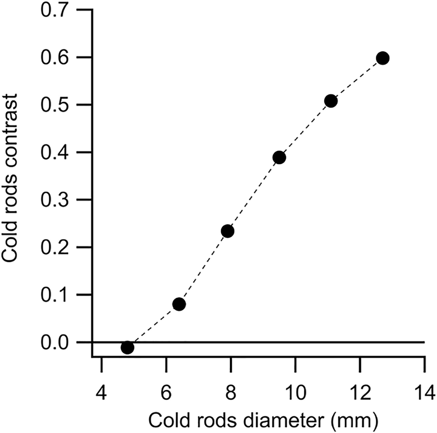
  • FIGURE 4.
    • Download figure
    • Open in new tab
    • Download powerpoint
    FIGURE 4.

    Cold rod contrast versus rod diameter (mean values for 17 phantom studies analyzed). SD for each point is approximately 0.01; error bars are smaller than size of markers.

Tables

  • Figures
    • View popup
    TABLE 1

    Hot Vial SUVmax and SUVvial Measured With and Without Normalization Using ACR ROIs and Cylindric VOIs

    Hot vial diameter (mm)SUVmaxSUVvialSUVmax, NormalizedSUVvial, Normalized
    252.56 ± 6.7%2.37 ± 6.2%2.54 ± 3.6%2.46 ± 3.1%
    162.43 ± 9.7%2.16 ± 7.2%2.41 ± 7.0%2.24 ± 3.8%
    122.02 ± 6.0%1.82 ± 5.6%2.01 ± 5.2%1.89 ± 2.4%
    81.35 ± 9.4%1.29 ± 8.3%1.34 ± 6.3%1.34 ± 3.7%
    • SUVmax using 25-mm circular ROI in 10-mm slice. SUVvial using cylindric VOI. Normalized SUVs account for actual syringe activities and dilution vessel used during phantom preparation.

    • View popup
    TABLE 2

    Hot Vial SUV Ratios Measured Using ACR ROIs and Cylindric VOIs

    Hot vial ratioSUVmax ratioSUVvial ratio
    16 mm/25 mm0.95 ± 5.0%0.91 ± 2.7%
    12 mm/25 mm0.79 ± 6.7%0.77 ± 3.3%
    8 mm/25 mm0.53 ± 5.2%0.54 ± 3.7%
    • SUVmax using 25-mm circular ROI in 10-mm slice. SUVvial using cylindric VOI.

    • View popup
    TABLE 3

    Cold Rod Contrast for 6 Sectors of ACR PET Phantom

    ParameterRod diameter (mm)
    12.711.19.57.96.44.8
    Mean contrast0.5980.5080.3890.2340.080−0.011
    SD0.0110.0070.0080.0120.0050.007
    • Data are for 17 studies.

PreviousNext
Back to top

In this issue

Journal of Nuclear Medicine Technology: 47 (3)
Journal of Nuclear Medicine Technology
Vol. 47, Issue 3
September 1, 2019
  • Table of Contents
  • About the Cover
  • Index by author
Print
Download PDF
Article Alerts
Sign In to Email Alerts with your Email Address
Email Article

Thank you for your interest in spreading the word on Journal of Nuclear Medicine Technology.

NOTE: We only request your email address so that the person you are recommending the page to knows that you wanted them to see it, and that it is not junk mail. We do not capture any email address.

Enter multiple addresses on separate lines or separate them with commas.
Automated Quantitative Analysis of American College of Radiology PET Phantom Images
(Your Name) has sent you a message from Journal of Nuclear Medicine Technology
(Your Name) thought you would like to see the Journal of Nuclear Medicine Technology web site.
Citation Tools
Automated Quantitative Analysis of American College of Radiology PET Phantom Images
Frank P. DiFilippo, Meghal Patel, Sagar Patel
Journal of Nuclear Medicine Technology Sep 2019, 47 (3) 249-254; DOI: 10.2967/jnmt.118.221317

Citation Manager Formats

  • BibTeX
  • Bookends
  • EasyBib
  • EndNote (tagged)
  • EndNote 8 (xml)
  • Medlars
  • Mendeley
  • Papers
  • RefWorks Tagged
  • Ref Manager
  • RIS
  • Zotero
Share
Automated Quantitative Analysis of American College of Radiology PET Phantom Images
Frank P. DiFilippo, Meghal Patel, Sagar Patel
Journal of Nuclear Medicine Technology Sep 2019, 47 (3) 249-254; DOI: 10.2967/jnmt.118.221317
Twitter logo Facebook logo LinkedIn logo Mendeley logo
  • Tweet Widget
  • Facebook Like
  • Google Plus One
Bookmark this article

Jump to section

  • Article
    • Abstract
    • MATERIALS AND METHODS
    • RESULTS
    • DISCUSSION
    • CONCLUSION
    • DISCLOSURE
    • Footnotes
    • REFERENCES
  • Figures & Data
  • Info & Metrics
  • PDF

Related Articles

  • No related articles found.
  • PubMed
  • Google Scholar

Cited By...

  • No citing articles found.
  • Google Scholar

More in this TOC Section

  • Early 10-Minute Postinjection [18F]F-FAPI-42 uEXPLORER Total-Body PET/CT Scanning Protocol for Staging Lung Cancer Using HYPER Iterative Reconstruction
  • Single- Versus Dual-Time-Point Imaging for Transthyretin Cardiac Amyloid Using 99mTc-Pyrophosphate
  • Does Arthrography Improve Accuracy of SPECT/CT for Diagnosis of Aseptic Loosening in Patients with Painful Knee Arthroplasty: A Systematic Review and Metaanalysis
Show more Imaging

Similar Articles

Keywords

  • PET
  • ACR phantom
  • quality assurance
  • quantitative
SNMMI

© 2025 SNMMI

Powered by HighWire