


Article Figures & Data
Figures
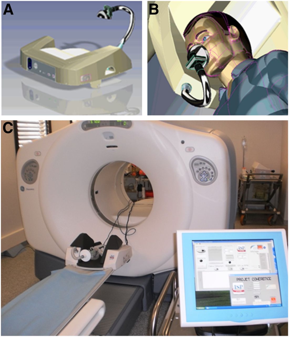
- FIGURE 1.
SGD PET/CT setup. (A) SGD device. (B) Patient positioning by computer-assisted design. (C) SGD device position on PET/CT camera with screen monitoring.
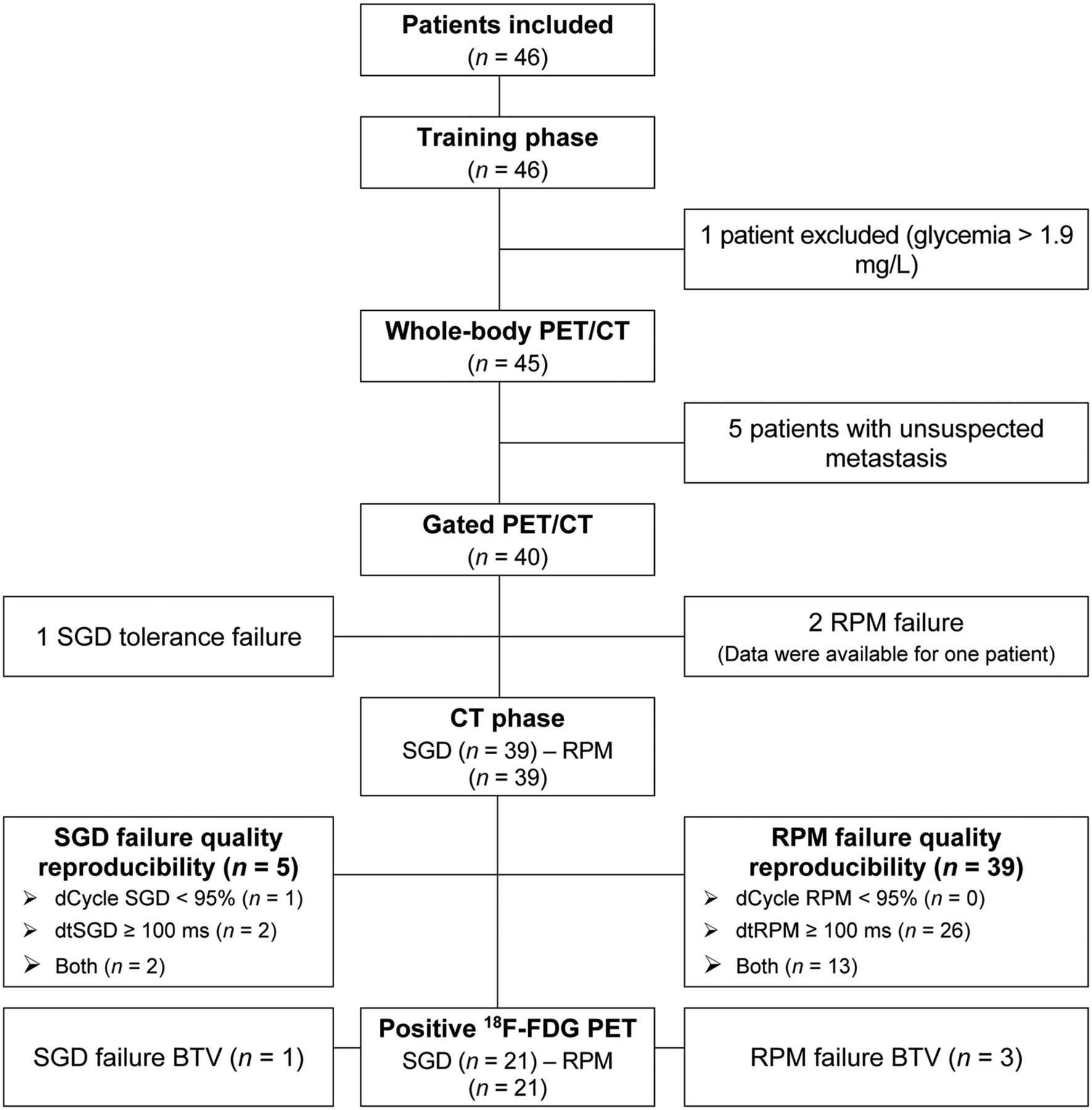
- FIGURE 2.
Patient and data flowchart. dCycle (cutoff set at 95%) and dt (cutoff set at 100 ms) assess reproducibility of time binning operated by SGD and RPM devices. Time lag represents difference between synchronization signals sent to PET and maximum inspiratory amplitude for each cycle detected by device (RPM or SGD). It is a parameter for accuracy of detection of maximal inspiration amplitude and time needed for system to send this information to PET.
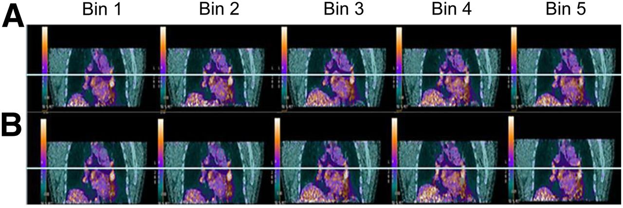
- FIGURE 3.
Simultaneous respiratory signal processing with SGD and RPM PET/CT. Patient was referred for suspected colorectal cancer recurrence with suggestive CT findings (1 left perihilar lung nodule). Shown are 2 reconstructed image data sets (SGD [A] and RPM [B] gated bins using AdvantageSim 4D software on Advantage Windows workstation). 18F-FDG PET/CT showed increased, nonspecific uptake bilaterally in (peri)hilar (L > R) and subcarinal lymph node regions on both SGD and RPM image datasets without significant differences in SUVmax, but without significant respiratory motion.
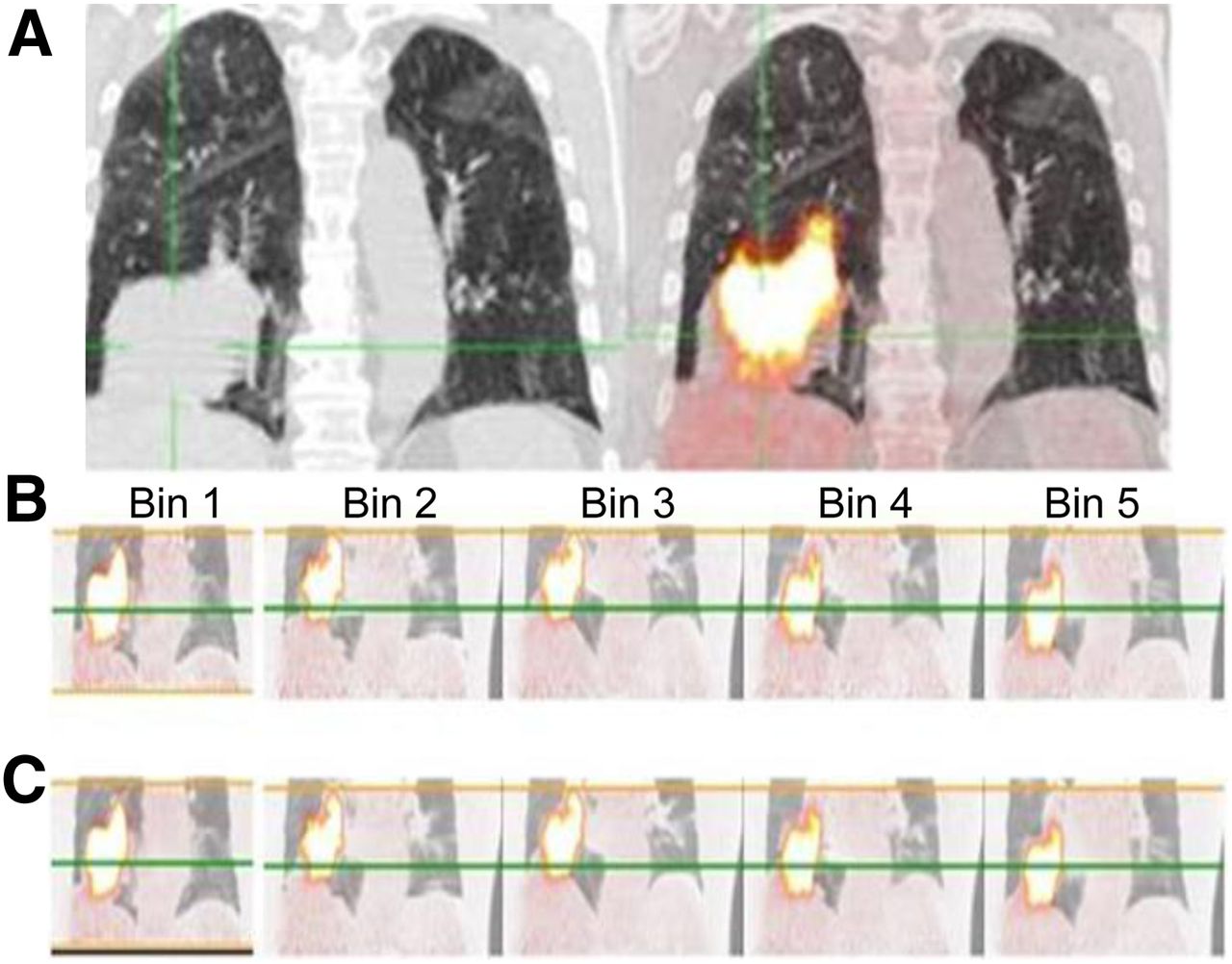
- FIGURE 4.
Simultaneous respiratory signal processing with SGD and RPM 18F-FDG PET/CT in 70-y-old man with T3N0 squamous cell carcinoma of inferior lobe of right lung who underwent imaging for staging. (A) Ungated PET/CT image (right) showing substantial misregistration due to respiratory artifacts and high tumoral uptake (body-weight–normalized SUVmax of lesion is 14.6). (B and C) SGD and RPM-gated PET/CT images, respectively, with body-weight–normalized SUVmax of 19.4 for SGD and 18.3 for RPM. Green line is reference level highlighting respiratory motion between bins. Supplemental Video 1 shows the displacement of the tumor associated with the patient’s breathing (top scanner, PET in the middle, and fused image at the bottom) on transverse views passing through the lung (left), in profile (at center), and coronal (right).


Tables
Criterion Description Inclusion Indication for 18F-FDG PET/CT Characterization of solitary pulmonary nodule Staging of non–small cell lung carcinoma Definition of biologic target volume for radiotherapy Patients able to maintain supine position for 60 min Age ≥ 18 y WHO status ≤ 1 Well-informed written consent Exclusion Lung tumor histology with classically low 18F-FDG avidity (bronchial carcinoid; lepidic and mucinous adenocarcinoma) Infectious or any other active severe bronchopneumopathy, respiratory pain, or distress; alteration in vital parameters; pneumothorax, peripheral lung biopsy or puncture, or hemoptysis within previous month Poorly controlled diabetes mellitus, pregnancy, or breast feeding WHO = World Health Organization.
Characteristic Data Age at inclusion (y) 63.5 (33.0–84.0) Sex Male 31 (67.4%) Female 15 (32.6%) WHO status 0 40 (87.0%) 1 6 (13.0%) Body mass index 25.8 (15.8–43.8) Tobacco abuse Active 26 (57.8%) Pack-years 46.5 (16.0–80.0) Significant cardiac history No 20 (43.5%) Yes 26 (56.5%) Significant lung history No 35 (76.1%) Yes 11 (23.9%) Qualitative data are expressed as numbers followed by percentages in parentheses; continuous data are expressed as median followed by range in parentheses.
WHO = World Health Organization.
Indication Data Evaluation of solitary lung nodule (n) 43 (93.5%) Nodule location Left superior lobe 9 (20.9%) Right superior lobe 17 (39.5%) Right inferior lobe 7 (16.3%) Left inferior lobe 6 (14.0%) Mid lobe 4 (9.3%) Staging of lung cancer (n) 3 (6.5%) CT largest lesion diameter (mm) Median 12.5 Range 5.0–90.0 Performance parameter SGD (n = 39) RPM (n = 39) P Respiratory gating accuracy (dCycle) Inhalation peak detection (%) 100.0 (86.3–102.1) 92.0 (26.7–101.0) <0.0001 Time lag (ms) 25.0 (5.7–146.4) 285.4 (137.7–595.5) <0.0001 Time lag shift (classes) (dtSGD) <0.0001 <100 ms 35 (89.7%) 0 (0.0%) ≥100 ms 4 (10.3%) 39 (100.0%) Deviation of end-expiration baseline 8.6 mL (−2.5–53.3) 18.6 mm (−17.9–342.2) 0.0018 Gated BTV Gated BTV availability 0.1250 No 1 (2.6%) 5 (12.8%) Yes 38 (97.4%) 34 (87.2%) BTV (mL) 7.2 (0.2–188.0) 5.4 (0.8–167.0) 0.1767 Gated SUVmax (g/mL) 2.8 (1.0–43.1) 2.0 (0.9–19.6) 0.1966 Qualitative data are expressed as numbers followed by percentages in parentheses; continuous data are expressed as median followed by range in parentheses.
SUVmax (g/mL) Ungated (n = 9) RPM gated (n = 8) SGD (n = 9) Median 1.9 2.0 2.6 Range 1.0–44.4 0.9–18.3 0.9–43.1 SUVs of 9 18F-FDG–positive lesions in lower part of lung (1 patient is missing because of RPM failure to provide BTV).


Jump to section
Related Articles
Cited By...
- No citing articles found.