
Main menu

  • Home
  • Content
    • Current
      • JNMT Supplement
    • Ahead of print
    • Past Issues
    • Continuing Education
    • JNMT Podcast
    • SNMMI Annual Meeting Abstracts
  • Subscriptions
    • Subscribers
    • Rates
    • Journal Claims
    • Institutional and Non-member
  • Authors
    • Submit to JNMT
    • Information for Authors
    • Assignment of Copyright
    • AQARA Requirements
  • Info
    • Reviewers
    • Permissions
    • Advertisers
    • Corporate & Special Sales
  • About
    • About Us
    • Editorial Board
    • Contact Information
  • More
    • Alerts
    • Feedback
    • Help
    • SNMMI Journals
  • SNMMI
    • JNMT
    • JNM
    • SNMMI Journals
    • SNMMI

User menu

  • Subscribe
  • My alerts
  • Log in
  • My Cart

Search

  • Advanced search
Journal of Nuclear Medicine Technology
  • SNMMI
    • JNMT
    • JNM
    • SNMMI Journals
    • SNMMI
  • Subscribe
  • My alerts
  • Log in
  • My Cart
Journal of Nuclear Medicine Technology

Advanced Search

  • Home
  • Content
    • Current
    • Ahead of print
    • Past Issues
    • Continuing Education
    • JNMT Podcast
    • SNMMI Annual Meeting Abstracts
  • Subscriptions
    • Subscribers
    • Rates
    • Journal Claims
    • Institutional and Non-member
  • Authors
    • Submit to JNMT
    • Information for Authors
    • Assignment of Copyright
    • AQARA Requirements
  • Info
    • Reviewers
    • Permissions
    • Advertisers
    • Corporate & Special Sales
  • About
    • About Us
    • Editorial Board
    • Contact Information
  • More
    • Alerts
    • Feedback
    • Help
    • SNMMI Journals
  • Watch or Listen to JNMT Podcast
  • Visit SNMMI on Facebook
  • Join SNMMI on LinkedIn
  • Follow SNMMI on Twitter
  • Subscribe to JNMT RSS feeds
Research ArticleImaging

Improving the Spatial Alignment in PET/CT Using Amplitude-Based Respiration-Gated PET and Patient-Specific Breathing–Instructed CT

Charlotte S. van der Vos, Antoi P.W. Meeuwis, Willem Grootjans, Lioe-Fee de Geus-Oei and Eric P. Visser
Journal of Nuclear Medicine Technology June 2019, 47 (2) 154-159; DOI: https://doi.org/10.2967/jnmt.118.215970
Charlotte S. van der Vos
1Department of Radiology and Nuclear Medicine, Radboud University Medical Center, Nijmegen, The Netherlands
2University of Twente, Enschede, The Netherlands; and
  • Find this author on Google Scholar
  • Find this author on PubMed
  • Search for this author on this site
Antoi P.W. Meeuwis
1Department of Radiology and Nuclear Medicine, Radboud University Medical Center, Nijmegen, The Netherlands
  • Find this author on Google Scholar
  • Find this author on PubMed
  • Search for this author on this site
Willem Grootjans
3Department of Radiology, Leiden University Medical Center, Leiden, The Netherlands
  • Find this author on Google Scholar
  • Find this author on PubMed
  • Search for this author on this site
Lioe-Fee de Geus-Oei
2University of Twente, Enschede, The Netherlands; and
3Department of Radiology, Leiden University Medical Center, Leiden, The Netherlands
  • Find this author on Google Scholar
  • Find this author on PubMed
  • Search for this author on this site
Eric P. Visser
1Department of Radiology and Nuclear Medicine, Radboud University Medical Center, Nijmegen, The Netherlands
  • Find this author on Google Scholar
  • Find this author on PubMed
  • Search for this author on this site
  • Article
  • Figures & Data
  • Info & Metrics
  • PDF
Loading

Article Figures & Data

Figures

  • Tables
  • FIGURE 1.
    • Download figure
    • Open in new tab
    • Download powerpoint
    FIGURE 1.

    Example of respiratory signal before and during CT acquisition. Patients were instructed to hold their breath between amplitude limits of optimal PET gate (between horizontal lines). To determine whether patient complied with instructions, amplitude range of respiratory signal before start of CT scan (mean amplitude range over several respiratory cycles) and during breathing instructions was determined. Ratio between those two was used to determine whether patients were able to hold their breath.
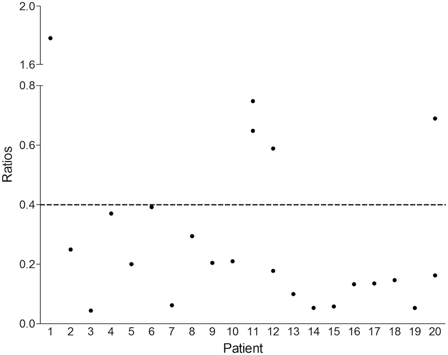
  • FIGURE 2.
    • Download figure
    • Open in new tab
    • Download powerpoint
    FIGURE 2.

    Ratios between average amplitude range of respiratory signal during breathing instructions and before CT acquisition, for each patient. Patients 1, 11, 12, and 20 could not comply with breathing instructions and showed higher ratio than other patients. When amplitude during CT acquisition of only lungs (not including upper abdomen region) was considered, ratios of patients 12 and 20 improved; therefore, they could be included in data analysis. Patients 1 and 11 showed only slight or no improvement and were excluded from data analysis.
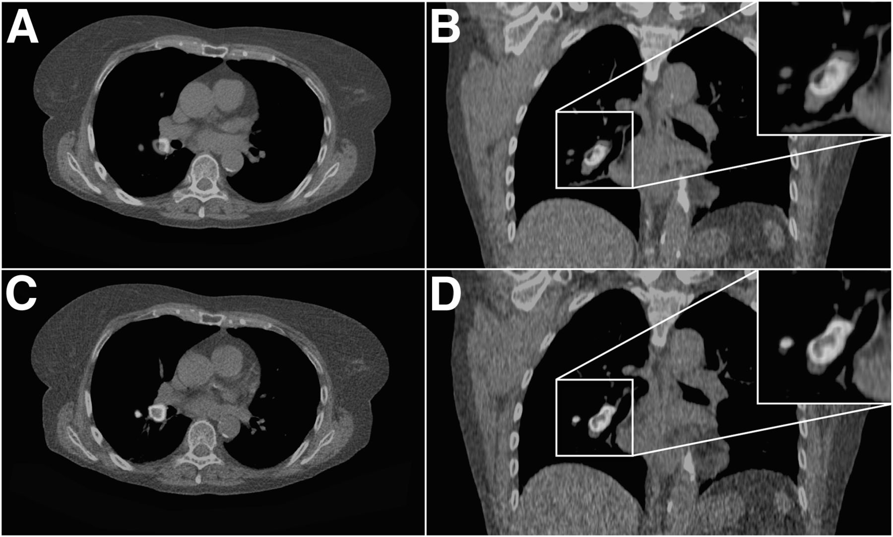
  • FIGURE 3.
    • Download figure
    • Open in new tab
    • Download powerpoint
    FIGURE 3.

    Patient with small cell lung cancer. (A and B) Transaxial (A) and coronal (B) planes of standard CT fused with respiration-gated PET image showed mismatch. (C and D) Same transaxial (C) and coronal (D) plane of CT with breathing instructions and corresponding gated PET images showed improved match. (A color version of this figure is available as a supplemental file at http://tech.snmjournals.org.)
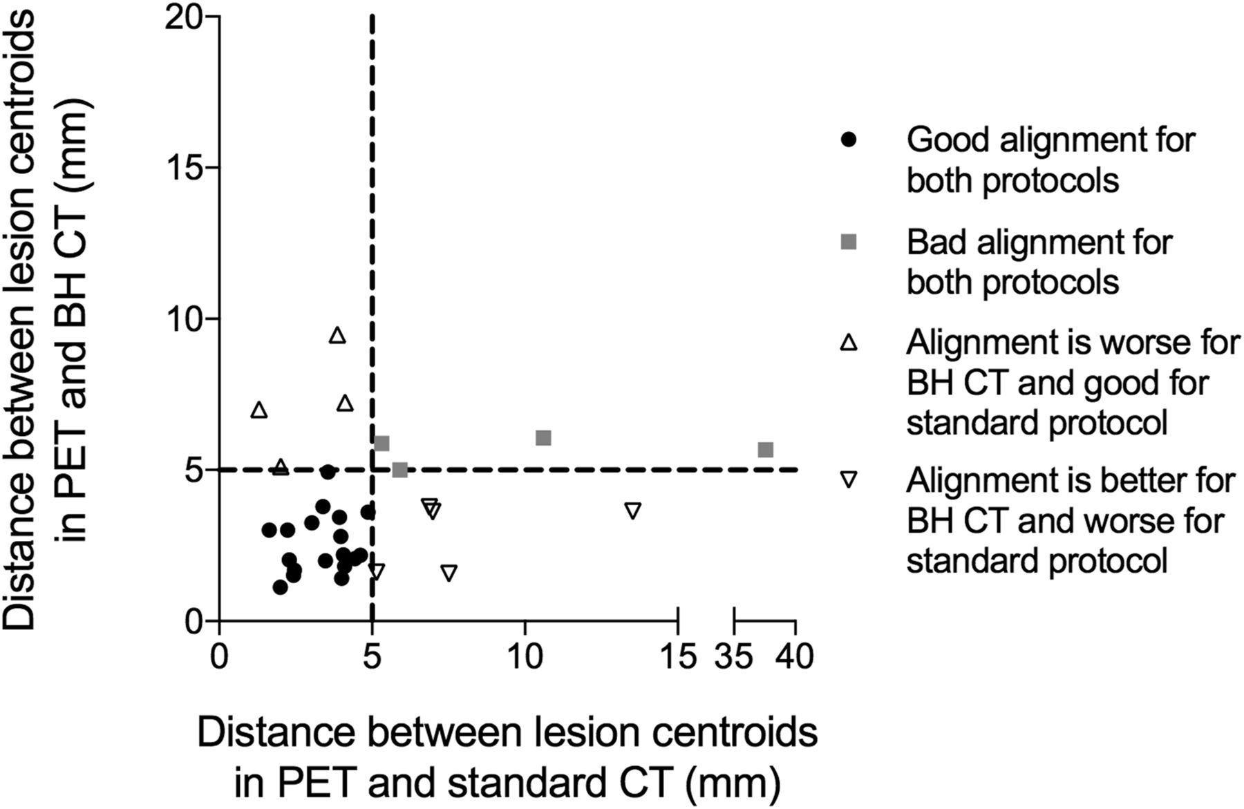
  • FIGURE 4.
    • Download figure
    • Open in new tab
    • Download powerpoint
    FIGURE 4.

    Scatterplot showing results of distance between centroids for lesions between PET and breath-hold (BH) CT and between PET and standard CT.

Tables

  • Figures
    • View popup
    TABLE 1

    Patient Characteristics

    CharacteristicData
    Sex (n)
     Male12
     Female8
    Mean age (y)64.2 (SD, 9.2)
    Mean weight (kg)76.3 (SD, 18.1)
    Mean administered activity (MBq)210 (SD, 105)
    Diagnosis (n)
     Primary lung cancer10
     Metastasis6
     Other and unconfirmed4
    Location of lesion (n)
     Upper lobes16
     Middle and lower lobes9
     Lung hilum6
    • View popup
    TABLE 2

    Results of Analyses of Spatial Alignment for Both Patient Groups

    ParameterStandard CT and PETBreath-hold CT and PETP
    Mismatch of lung–liver boundary (mm)5.6 ± 7.31.7 ± 2.10.049
    Average distance between lesion centroids (mm)5.5 ± 6.53.6 ± 2.00.040
    Jaccard similarity coefficient0.32 ± 0.160.36 ± 0.160.176
    SUVmax (g/cm3)10.3 ± 6.410.6 ± 6.60.104
    SUVmean (g/cm3)6.1 ± 3.86.3 ± 3.90.044
    Metabolic tumor volume6.73 ± 15.66.69 ± 15.70.930
    TLG54.50 ± 143.454.55 ± 141.90.018
    • Data are mean and SD.

PreviousNext
Back to top

In this issue

Journal of Nuclear Medicine Technology: 47 (2)
Journal of Nuclear Medicine Technology
Vol. 47, Issue 2
June 1, 2019
  • Table of Contents
  • About the Cover
  • Index by author
Print
Download PDF
Article Alerts
Sign In to Email Alerts with your Email Address
Email Article

Thank you for your interest in spreading the word on Journal of Nuclear Medicine Technology.

NOTE: We only request your email address so that the person you are recommending the page to knows that you wanted them to see it, and that it is not junk mail. We do not capture any email address.

Enter multiple addresses on separate lines or separate them with commas.
Improving the Spatial Alignment in PET/CT Using Amplitude-Based Respiration-Gated PET and Patient-Specific Breathing–Instructed CT
(Your Name) has sent you a message from Journal of Nuclear Medicine Technology
(Your Name) thought you would like to see the Journal of Nuclear Medicine Technology web site.
Citation Tools
Improving the Spatial Alignment in PET/CT Using Amplitude-Based Respiration-Gated PET and Patient-Specific Breathing–Instructed CT
Charlotte S. van der Vos, Antoi P.W. Meeuwis, Willem Grootjans, Lioe-Fee de Geus-Oei, Eric P. Visser
Journal of Nuclear Medicine Technology Jun 2019, 47 (2) 154-159; DOI: 10.2967/jnmt.118.215970

Citation Manager Formats

  • BibTeX
  • Bookends
  • EasyBib
  • EndNote (tagged)
  • EndNote 8 (xml)
  • Medlars
  • Mendeley
  • Papers
  • RefWorks Tagged
  • Ref Manager
  • RIS
  • Zotero
Share
Improving the Spatial Alignment in PET/CT Using Amplitude-Based Respiration-Gated PET and Patient-Specific Breathing–Instructed CT
Charlotte S. van der Vos, Antoi P.W. Meeuwis, Willem Grootjans, Lioe-Fee de Geus-Oei, Eric P. Visser
Journal of Nuclear Medicine Technology Jun 2019, 47 (2) 154-159; DOI: 10.2967/jnmt.118.215970
Twitter logo Facebook logo LinkedIn logo Mendeley logo
  • Tweet Widget
  • Facebook Like
  • Google Plus One
Bookmark this article

Jump to section

  • Article
    • Abstract
    • MATERIALS AND METHODS
    • RESULTS
    • DISCUSSION
    • CONCLUSION
    • DISCLOSURE
    • Footnotes
    • REFERENCES
  • Figures & Data
  • Info & Metrics
  • PDF

Related Articles

  • No related articles found.
  • PubMed
  • Google Scholar

Cited By...

  • No citing articles found.
  • Google Scholar

More in this TOC Section

  • Early 10-Minute Postinjection [18F]F-FAPI-42 uEXPLORER Total-Body PET/CT Scanning Protocol for Staging Lung Cancer Using HYPER Iterative Reconstruction
  • Single- Versus Dual-Time-Point Imaging for Transthyretin Cardiac Amyloid Using 99mTc-Pyrophosphate
  • Does Arthrography Improve Accuracy of SPECT/CT for Diagnosis of Aseptic Loosening in Patients with Painful Knee Arthroplasty: A Systematic Review and Metaanalysis
Show more Imaging

Similar Articles

Keywords

  • amplitude-based optimal respiratory gating
  • lung tumors
  • image quantification in PET
  • spatial alignment
  • breath-hold CT
SNMMI

© 2025 SNMMI

Powered by HighWire