


Article Figures & Data
Figures
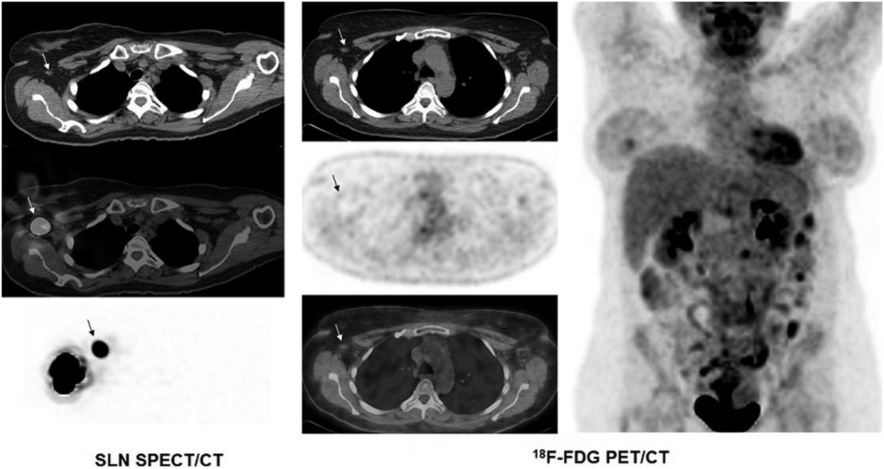
- FIGURE 1.
A 50-y-old woman with bilateral grade 2 invasive lobular carcinoma (left breast, pT3 N3a Mx; right breast, pT1c N3a Mx). (Left) SLN scintigraphic study from right breast with selected transaxial CT, SPECT/CT, and maximum-intensity-projection images demonstrates SLN in right axilla (arrows). (Middle) Selected 18F-FDG transaxial PET, CT, and PET/CT images of bilateral axillae demonstrate mild focal uptake in SLN in right axilla (SUVmax, 1.4) (arrows) and multiple mildly hypermetabolic left axillary lymph nodes (SUVmax, 2.9). Histopathologic analysis demonstrated metastasis in right SLN and bilaterally in multiple axillary lymph nodes. (Right) 18F-FDG PET whole-body maximum-intensity-projection image demonstrates bilateral diffuse uptake in breasts, focal mild tumoral uptake in left breast (SUVmax, 2.3), and bilateral mildly hypermetabolic axillary lymph nodes, more prominent on left side. Abnormal hypermetabolic activity in right lobe of thyroid is suspicious for malignancy and large myomatous uterus is also seen.
- FIGURE 2.
A 53-y-old woman with grade 1 invasive ductal carcinoma of right breast (pT1c N0(sln) Mx). (Left) SLN scintigraphic study from right breast with selected SPECT/CT and maximum-intensity-projection images demonstrates SLN in right axilla (arrows). (Middle) Selected 18F-FDG transaxial PET, CT, and PET/CT images demonstrate only faint uptake in SLN in right axilla (SUVmax, 0.7) (arrows). Mild but slightly more prominent 18F-FDG uptake is seen in contralateral left axillary lymph nodes. Histopathologic analysis demonstrated no evidence of metastasis in right SLN. (Right) 18F-FDG PET whole-body maximum-intensity-projection image demonstrates bilateral diffuse uptake in breasts and focal tumoral uptake in right breast (SUVmax, 2.1).
- FIGURE 3.
ROC curve for 18F-FDG PET/CT in detecting metastasis in SLN.


Tables
- TABLE 1
Histologic Subtype, Tumor Grade, Pathologic Stage, 18F-FDG Uptake in SLN, and Pathology Result
Age (y) Subtype Grade Stage SLN SUVmax SLN metastasis 53 IDC 2 pT1c N1a Mx 1.2 Positive 35 IDC 3 pT2 N2a Mx 1.5 Positive 74 IDC 2 pT2 N1a Mx 0.8 Positive 45 IDC 2 pT2 N1a Mx 4.1 Positive 53 IDC 2 pT3 N2a Mx 2.8 Positive 67 IDC 2 pT2 N1a Mx 2 Positive 50 ILC (left)* 2 pT3 N3a Mx – – ILC (right) 2 pT1c N3a Mx 1.4 Positive 47 IDC 2 pT2 N0 Mx 1.4 Negative 62 IDC 3 pT2 N0(sln) Mx 0.9 Negative 61 IDC 2 pT2 N0(sln) Mx 0.6 Negative 70 IDC 2 pT2 N0 Mx 0.8 Negative 68 IDC 3 pT2 N0 Mx 5.9 Negative 49 IDC 2 pT1a N0(sln) Mx 1.8 Negative 78 IDC 1 pT2p N0(sln) Mx 0.8 Negative 68 IDC 2 pT2 N0(sln) Mx 0.9 Negative 53 IDC 1 pT1c N0(sln) Mx 0.7 Negative 76 DCIS NA Bilateral 0.9 Negative 70 ILC and IDC 3 and 2 pmT2 N0(sln) Mx 0.9 Negative 81 ILC 2 pT1c N0 Mx 0.8 Negative 65 IMC 3 pT1c N0(sln) 1.8 Negative ↵* SLN study not done for left breast.
IDC = invasive ductal carcinoma; DCIS = ductal carcinoma in situ; ILC = invasive lobular carcinoma; NA = not applicable; IMC = invasive mammary carcinoma.


