


Article Figures & Data
Figures
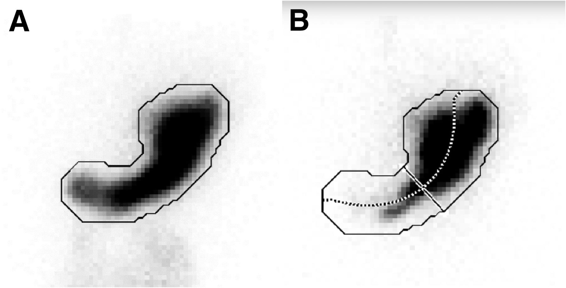
- FIGURE 1.
Anatomic division of stomach into proximal and distal halves by semiautomated software. (A) Demonstration of how software automatically contours outer border for total-stomach ROI (solid line) from summed set of all static anterior images. (B) Application of total-stomach ROI to each static image. In this case, image is anterior image acquired immediately after meal ingestion. By finding midpoint between opposing points of total-stomach ROI, longitudinal axis through stomach is generated (dotted line). Line perpendicular to longitudinal axis is then defined at point dividing longitudinal axis into equal halves to separate upper and lower segments of stomach.
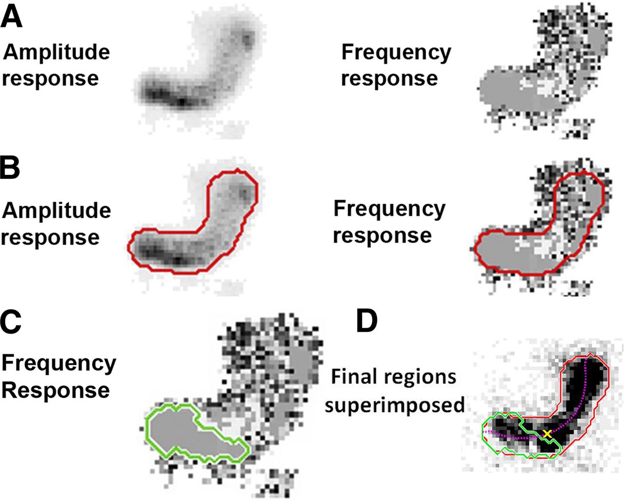
- FIGURE 2.
Fourier frequency and amplitude analysis used to segment antrum (distal stomach) from proximal stomach based on antral contractions. (A) Fast Fourier transformation of all pixels in DACS image set: amplitude and frequency response. (B) Red border on amplitude response indicating threshold of high-amplitude pixels. Same border is applied to frequency response. (C) Region of dominant frequency. Green border indicates contiguous region of pixels that have same frequency (gray color). (D) Starting location of antral contractions. Antrum-defined ROI (green border) using region of dominant frequency from C and red border from B is applied to one image of gastric emptying study. Purple dotted line is longitudinal axis as shown in Figure 1B. Yellow X is intersection of longitudinal axis, and convex boundary around green border is starting location of antral contractions.
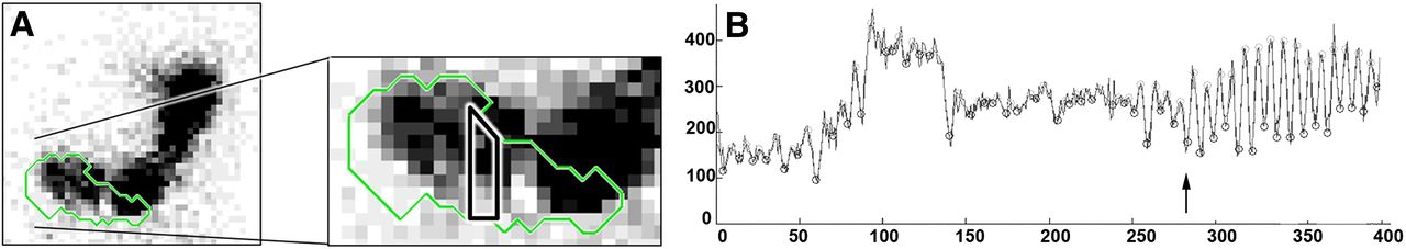
- FIGURE 3.
Time to onset of antral contractions. (A) Vertical rectangular ROI is placed over midportion of gastric antrum from one frame of serial anterior dynamic images used to generate time–activity curves shown in B. (B) y-axis is counts generated in antral ROI shown in A over time. x-axis is frame number, with 400 frames at 3 s per image. Time to onset of regular antral contractions is seen at image 278 or at 13.9 min (arrow) after immediately-after-meal static images.
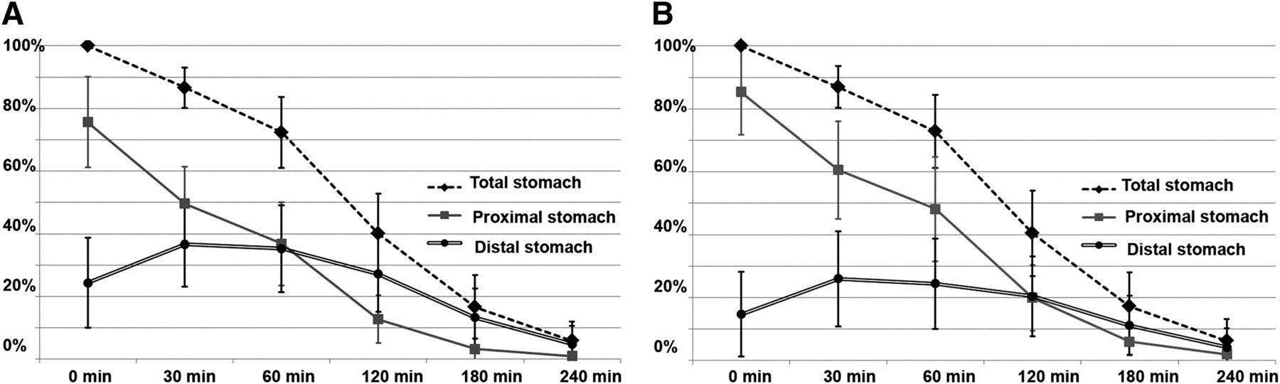
- FIGURE 4.
(A) Total and regional gastric retention over time using anatomic division of stomach into halves. (B) Total and regional gastric retention over time using Fourier frequency and amplitude to separate antrum from remaining proximal stomach.
Tables
Parameter 0 min 30 min 60 min 120 min 180 min 240 min IMD: gastric division into proximal and distal halves 0.76 ± 0.14 0.50 ± 0.12 0.37 ± 0.13 0.13 ± 0.08 0.03 ± 0.04 0.01 ± 0.01 IMD: gastric division into proximal and distal segments with Fourier-defined antral region 0.85 ± 0.14 0.61 ± 0.16 0.48 ± 0.17 0.20 ± 0.10 0.06 ± 0.04 0.02 ± 0.02 Total gastric retention (%) 100.0 ± 0.0 86.2 ± 6.9 72.1 ± 11.5 39.8 ± 12.8 16.4 ± 9.9 5.8 ± 6.0 Antral frequency (cycles/min) 3.09 ± 0.31 3.30 ± 0.73 3.15 ± 0.28 2.96 ± 0.35 NA NA Antral ejection fraction (%) 26.28 ± 13.74 29.43 ± 13.42 27.96 ± 15.31 26.95 ± 9.38 NA NA Contraction speed (mm/s) 2.91 ± 1.89 3.04 ± 1.65 3.54 ± 0.90 2.67 ± 1.37 NA NA IMD = ratio of count in proximal stomach to total gastric count immediately after meal ingestion; NA = not available because of low count caused by majority of meal’s having left stomach.
Data are mean ± SD.


Jump to section
Related Articles
Cited By...
- Distinct subgroups in gastroparesis defined by simultaneous body surface gastric mapping and gastric emptying breath testing
- Fourier Phase Analysis of Dynamic Antral Contraction Scintigraphy: New Software, Reference Values, and Comparisons to Conventional Gastric Emptying
- Abnormal gastrointestinal motility is a major factor in explaining symptoms and a potential therapeutic target in patients with disorders of gut-brain interaction
- Fourier Phase Analysis of Dynamic Antral Contraction Scintigraphy: New Software, Reference Values, and Comparisons to Conventional Gastric Emptying
- Actionable biomarkers: the key to resolving disorders of gastrointestinal function
- Gastroparesis: a turning point in understanding and treatment
- Readers Help to Shape JNMT Content
- Using Intragastric Meal Distribution and Antral Contractility for Enhanced Gastric Emptying Analysis