


Article Figures & Data
Figures
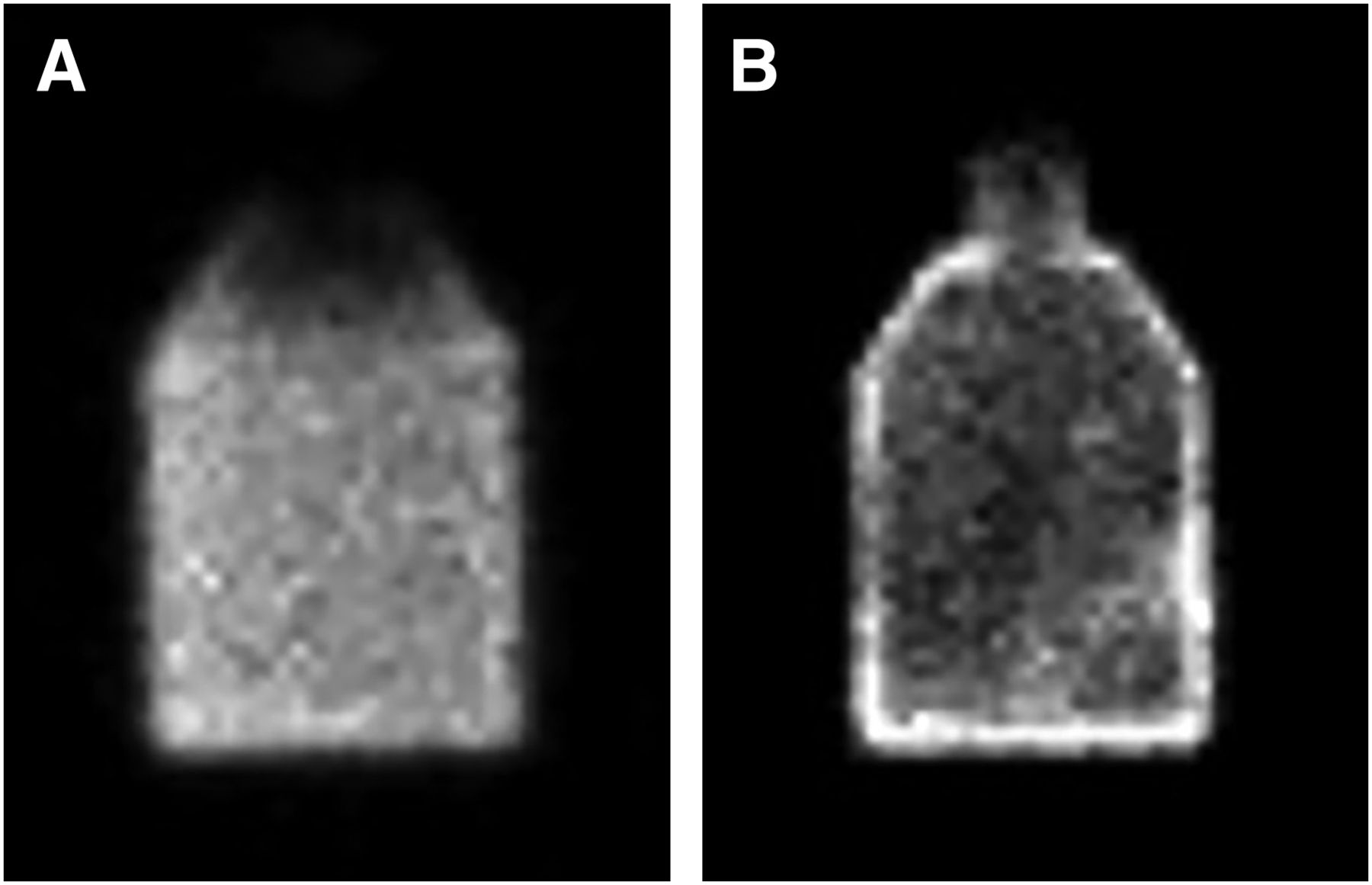
- FIGURE 1.
Examples of 99mTc-pertechnetate and 99mTc-sestamibi in plastic flask. (A) 99mTc-pertechnetate mixture is homogeneous within flask. (B) Distribution of 99mTc-sestamibi mixture is nonuniform, and there are increased counts along surface of flask.
- FIGURE 2.
Proper positioning for uniformity flood acquisition using refillable 99mTc source. Source is positioned between upper and lower detectors of dual-head cadmium-zinc-telluride system. Gantry is angled to ensure that any air bubbles in phantom are outside field of view.
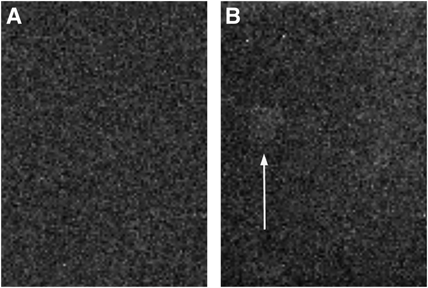
- FIGURE 3.
Potential of new 57Co sheet sources to create artifacts. (A) Uniformity flood acquisition obtained using 57Co sheet source and 57Co correction map. (B) Uniformity flood acquisition obtained using 99mTc flood source and 57Co correction map immediately after acquisition of image in A. Artifact (arrow) can be seen over one module in middle of detector.
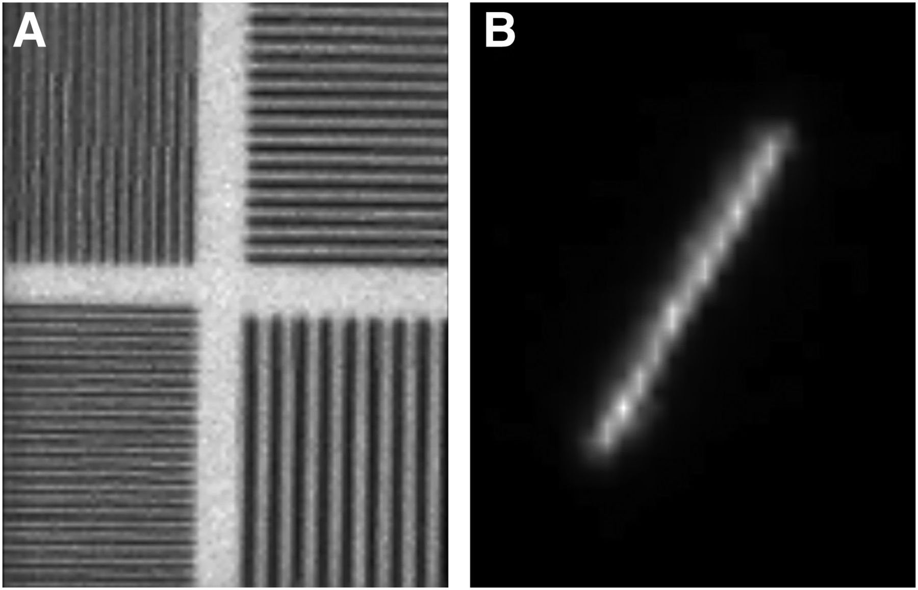
- FIGURE 4.
Examples of aliasing artifacts on pixelated imaging system. (A) Aliasing artifact from imaging bar phantom. (B) Zoomed image of artifact from line source for evaluating spatial resolution.
- FIGURE 5.
Recommended setup for measurement of system sensitivity. (A) For single-head system, flask is filled with just enough water to cover surface area of flask when lying on its side. (B) For dual-head system, flask is filled completely with water to minimize differences between detector geometry. Both images can be acquired simultaneously.
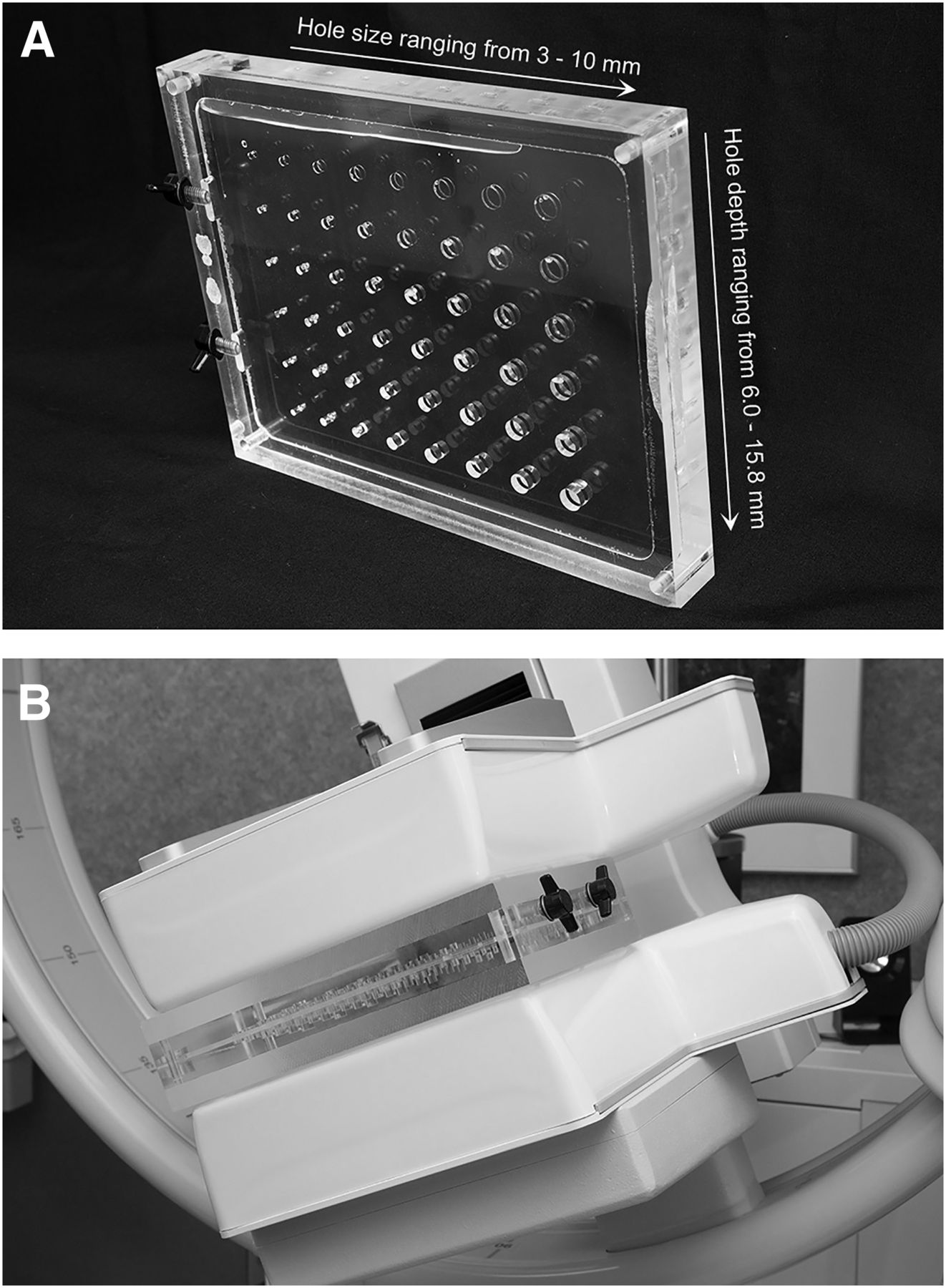
- FIGURE 6.
Contrast-detail phantom. (A) Phantom contains 3-cm-thick central section with multiple hole sizes and depths. (B) Display of image setup using two 1.5-cm-thick acrylic plates on either side of contrast-detail phantom. Camera is angled to move air bubbles out of field of view.
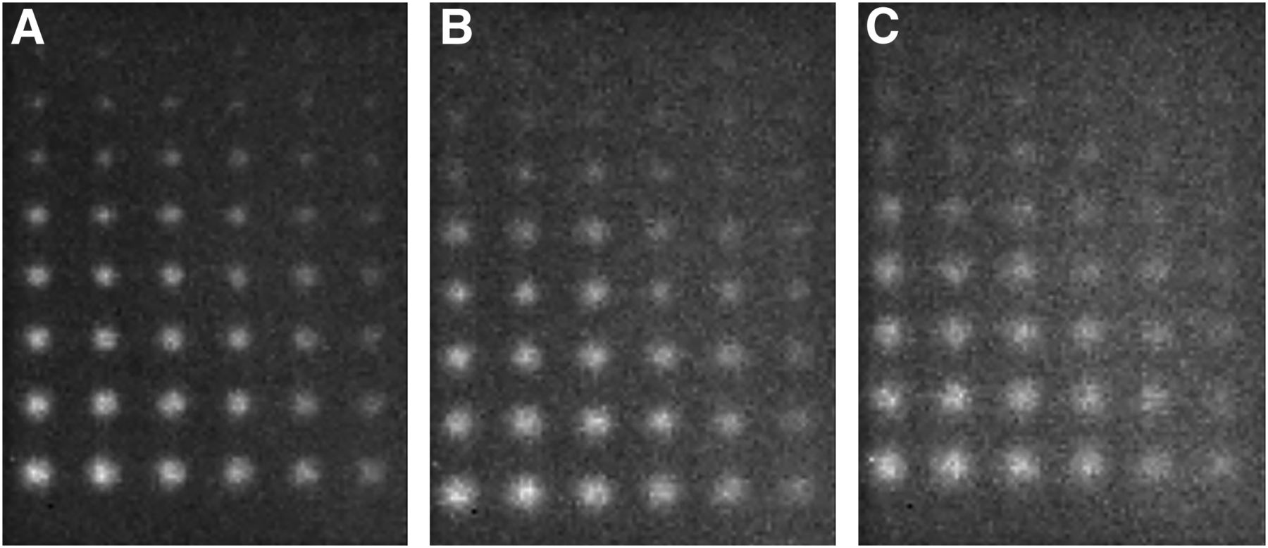
- FIGURE 7.
Sample contrast-detail phantom images. These images were acquired at distances of 1.5 cm (A), 3.0 cm (B), and 4.5 cm (C) from collimator face.


Tables
Test Equipment Frequency Acquisition details Passing criterion Uniformity 57Co sheet source or fillable phantom Daily 7.5 million counts ≤5% integral uniformity Spatial resolution 4-quadrant bar phantom Semiannually 7.5 million counts; phantom angled across field of view Meets manufacturer’s specifications Sensitivity Flask Annually 120-s images ≤10% difference between 2 detectors Energy resolution Point source Annually 2-keV energy windows; 1-min images Full width at half maximum ≤ 10% Lesion contrast Contrast-detail phantom Quarterly 1 million counts; images at 3 depths CNR > 3; count number of visible lesions at each depth * All tests should be performed at acceptance testing and after major service work.
CNR = contrast-to-noise ratio.
- TABLE 2
Suggested Number of Lesions Visualized in Contrast-Detail Phantom for Satisfactory, Marginal, and Fail Criteria
Distance Satisfactory Marginal Fail 1.5 cm 42 40 <40 3.0 cm 37 35 <35 4.5 cm 32 30 <30


