
Main menu

  • Home
  • Content
    • Current
      • JNMT Supplement
    • Ahead of print
    • Past Issues
    • Continuing Education
    • JNMT Podcast
    • SNMMI Annual Meeting Abstracts
  • Subscriptions
    • Subscribers
    • Rates
    • Journal Claims
    • Institutional and Non-member
  • Authors
    • Submit to JNMT
    • Information for Authors
    • Assignment of Copyright
    • AQARA Requirements
  • Info
    • Reviewers
    • Permissions
    • Advertisers
    • Corporate & Special Sales
  • About
    • About Us
    • Editorial Board
    • Contact Information
  • More
    • Alerts
    • Feedback
    • Help
    • SNMMI Journals
  • SNMMI
    • JNMT
    • JNM
    • SNMMI Journals
    • SNMMI

User menu

  • Subscribe
  • My alerts
  • Log in
  • Log out
  • My Cart

Search

  • Advanced search
Journal of Nuclear Medicine Technology
  • SNMMI
    • JNMT
    • JNM
    • SNMMI Journals
    • SNMMI
  • Subscribe
  • My alerts
  • Log in
  • Log out
  • My Cart
Journal of Nuclear Medicine Technology

Advanced Search

  • Home
  • Content
    • Current
    • Ahead of print
    • Past Issues
    • Continuing Education
    • JNMT Podcast
    • SNMMI Annual Meeting Abstracts
  • Subscriptions
    • Subscribers
    • Rates
    • Journal Claims
    • Institutional and Non-member
  • Authors
    • Submit to JNMT
    • Information for Authors
    • Assignment of Copyright
    • AQARA Requirements
  • Info
    • Reviewers
    • Permissions
    • Advertisers
    • Corporate & Special Sales
  • About
    • About Us
    • Editorial Board
    • Contact Information
  • More
    • Alerts
    • Feedback
    • Help
    • SNMMI Journals
  • Watch or Listen to JNMT Podcast
  • Visit SNMMI on Facebook
  • Join SNMMI on LinkedIn
  • Follow SNMMI on Twitter
  • Subscribe to JNMT RSS feeds
Review ArticleTeaching Case Studies

18F-FDG PET/CT for the Evaluation of Primary Eosinophilic Granuloma of the Hypothalamus

Anna Eshghi, Robert Klein, Naghmehossadat Eshghi and Phillip H. Kuo
Journal of Nuclear Medicine Technology September 2018, 46 (3) 290-291; DOI: https://doi.org/10.2967/jnmt.118.209957
Anna Eshghi
1Department of Molecular and Cellular Biology, University of Arizona, Tucson, Arizona
  • Find this author on Google Scholar
  • Find this author on PubMed
  • Search for this author on this site
Robert Klein
2Department of Pathology, Banner University Medical Center, Tucson, Arizona
  • Find this author on Google Scholar
  • Find this author on PubMed
  • Search for this author on this site
Naghmehossadat Eshghi
3Department of Medical Imaging, University of Arizona, Tucson, Arizona; and
  • Find this author on Google Scholar
  • Find this author on PubMed
  • Search for this author on this site
Phillip H. Kuo
3Department of Medical Imaging, University of Arizona, Tucson, Arizona; and
  • Find this author on Google Scholar
  • Find this author on PubMed
  • Search for this author on this site
  • Article
  • Figures & Data
  • Info & Metrics
  • PDF
Loading

Abstract

A 21-y-old man who presented with polyuria and polydipsia was discovered to have diabetes insipidus due to eosinophilic granuloma of the hypothalamus. 18F-FDG PET/CT, which was performed as a metastatic work-up, revealed an intensely 18F-FDG–avid hypothalamic mass and no other sites of disease.

  • molecular imaging
  • oncology
  • 18F-FDG PET/CT
  • eosinophilic granuloma
  • Langerhans cell histiocytosis

Eosinophilic granuloma, a form of Langerhans cell histiocytosis, is very rare in the central nervous system. Langerhans cell histiocytosis can occur as a unicentric tumor or as multicentric aggressive disease. Therefore, Langerhans cell histiocytosis can present with various clinical manifestations, depending on the organ systems involved (1).

CASE REPORT

A 21-y-old man was referred to endocrinology for evaluation of diabetes insipidus with polyuria and polydipsia and associated hypernatremia. After initiation of desmopressin therapy and correction of his sodium level, T1-weighted brain MRI was performed with intravenous contrast material. The unenhanced sequence showed a hypothalamic mass with a signal intensity similar to that of gray matter (Fig. 1A), and the contrast-enhanced sequence showed strong enhancement of a 2-cm hypothalamic mass with smooth borders (Fig. 1B). Unenhanced CT was also performed, revealing that the mass was isodense to gray matter and had no calcification or hemorrhage (Fig. 1C). Given its pattern of enhancement and borders, a low-grade glioma or ganglioglioma was the primary consideration for the differential diagnosis (2).

FIGURE 1.
  • Download figure
  • Open in new tab
  • Download powerpoint
FIGURE 1.

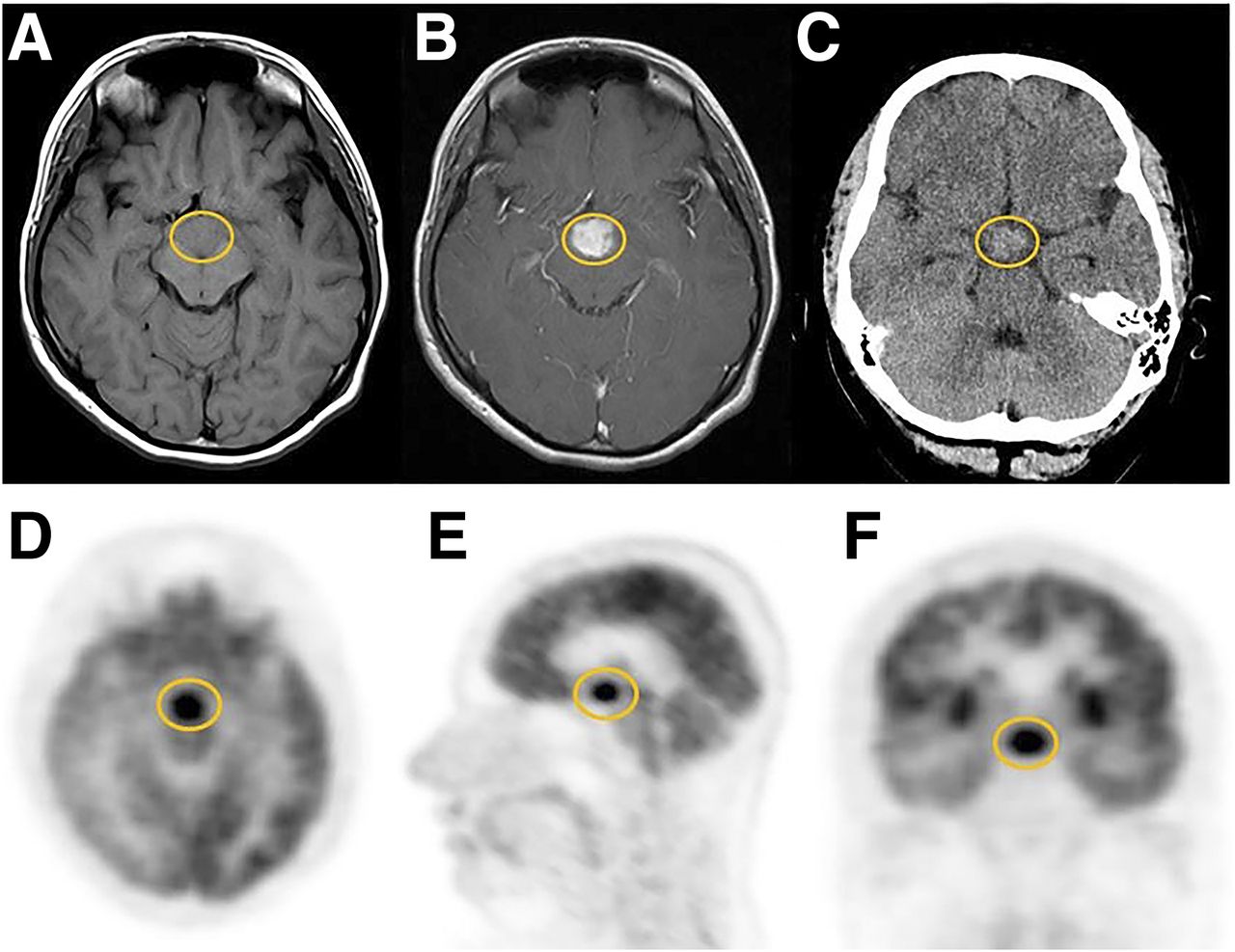
(A–C) Transaxial T1-weighted unenhanced MR (A), T1-weighted gadolinium-enhanced MR (B), and unenhanced CT (C) images show hypothalamic lesion (encircled in yellow) to be strongly enhanced and isodense to gray matter. (D–F) Axial (D), sagittal (E), and coronal (F) 18F-FDG PET images reveal intense uptake in hypothalamic mass.

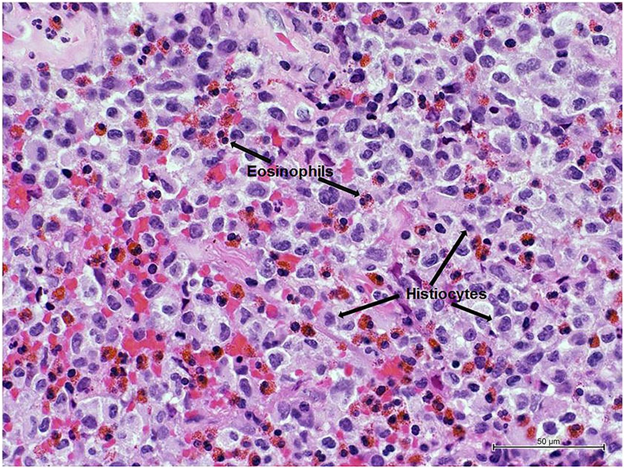
Because of the potentially devastating complications of surgery in this region, other treatment options such as radiation therapy and chemotherapy were discussed. A biopsy of the mass revealed numerous eosinophils with histiocytes, which were confirmed to be Langerhans-type cells (Fig. 2). Furthermore, immunohistochemical staining showed that the tumor was CD1a-positive, S100 protein–positive, CD117-negative, and CD34-negative. Overall, the pathologic findings were consistent with eosinophilic granuloma/histiocytosis. To evaluate for possible multicentric disease, 18F-FDG PET/CT was performed and showed a unicentric, intensely 18F-FDG–avid mass of the hypothalamus (Figs. 1D–1F), with no other 18F-FDG–avid lesions (Fig. 3).

FIGURE 2.
  • Download figure
  • Open in new tab
  • Download powerpoint
FIGURE 2.

Histopathologic photomicrograph (hematoxylin and eosin, ×400) shows numerous eosinophils and histiocytes (arrows).

FIGURE 3.
  • Download figure
  • Open in new tab
  • Download powerpoint
FIGURE 3.

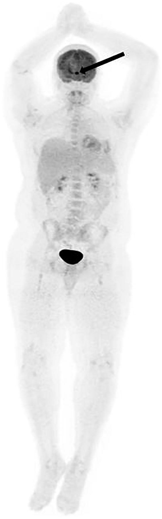
18F-FDG PET anterior whole-body maximum-intensity projection shows intensely 18F-FDG–avid hypothalamic lesion (arrow) and no other sites of disease.

DISCUSSION

The biopsy of the hypothalamic mass in this symptomatic patient revealed eosinophilic granuloma. This disease has a wide spectrum of clinical manifestations that can make diagnosis and therapeutic response assessment challenging. 18F-FDG PET/CT has been shown to identify lesions in addition to those found on plain radiography, CT, and MRI (3,4). Furthermore, the utility of 18F-FDG PET/CT for monitoring response to therapy in a small number of patients has been reported (5). After multidisciplinary discussion by the tumor board, the patient was treated with radiation therapy.

CONCLUSION

Eosinophilic granuloma can present as unicentric or multicentric disease. In this very rare case of hypothalamic eosinophilic granuloma, 18F-FDG PET/CT was particularly critical given the high-risk location of the tumor, which made detection of distant metastases—requiring systemic rather than local therapy—even more crucial.

DISCLOSURE

No potential conflict of interest relevant to this article was reported.

Acknowledgments

We thank Carol Stuehm for her assistance in preparing the figures.

Footnotes

  • Published online May 3, 2018.

REFERENCES

  1. 1.↵
    1. Girschikofsky M,
    2. Arico M,
    3. Castillo D,
    4. et al
    . Management of adult patients with Langerhans cell histiocytosis: recommendations from an expert panel on behalf of Euro-Histio-Net. Orphanet J Rare Dis. 2013;8:72.
    OpenUrlCrossRefPubMed
  2. 2.↵
    1. Saleem SN,
    2. Ahmed-Hashem S,
    3. Lee DH
    . Lesions of the hypothalamus: MR imaging diagnostic features. Radiographics. 2007;27:1087–1108.
    OpenUrlCrossRefPubMed
  3. 3.↵
    1. Phillips M,
    2. Allen C,
    3. Gerson P,
    4. et al
    . Comparison of FDG-PET scans to conventional radiography and bone scans in management of Langerhans cell histiocytosis. Pediatr Blood Cancer. 2009;52:97–101.
    OpenUrlCrossRefPubMed
  4. 4.↵
    1. Albano D,
    2. Bosio G,
    3. Giubbini R,
    4. et al
    . Role of 18F-FDG PET/CT in patients affected by Langerhans cell histiocytosis. Jpn J Radiol. 2017;35:574–583.
    OpenUrl
  5. 5.↵
    1. Lee HJ,
    2. Ahn BC,
    3. Lee SW,
    4. et al
    . The usefulness of F-18 fluorodeoxyglucose positron emission tomography/computed tomography in patients with Langerhans cell histiocytosis. Ann Nucl Med. 2012;26:730–737.
    OpenUrlCrossRefPubMed
  • Received for publication February 15, 2018.
  • Accepted for publication March 29, 2018.
PreviousNext
Back to top

In this issue

Journal of Nuclear Medicine Technology: 46 (3)
Journal of Nuclear Medicine Technology
Vol. 46, Issue 3
September 1, 2018
  • Table of Contents
  • About the Cover
  • Index by author
Print
Download PDF
Article Alerts
Sign In to Email Alerts with your Email Address
Email Article

Thank you for your interest in spreading the word on Journal of Nuclear Medicine Technology.

NOTE: We only request your email address so that the person you are recommending the page to knows that you wanted them to see it, and that it is not junk mail. We do not capture any email address.

Enter multiple addresses on separate lines or separate them with commas.
18F-FDG PET/CT for the Evaluation of Primary Eosinophilic Granuloma of the Hypothalamus
(Your Name) has sent you a message from Journal of Nuclear Medicine Technology
(Your Name) thought you would like to see the Journal of Nuclear Medicine Technology web site.
Citation Tools
18F-FDG PET/CT for the Evaluation of Primary Eosinophilic Granuloma of the Hypothalamus
Anna Eshghi, Robert Klein, Naghmehossadat Eshghi, Phillip H. Kuo
Journal of Nuclear Medicine Technology Sep 2018, 46 (3) 290-291; DOI: 10.2967/jnmt.118.209957

Citation Manager Formats

  • BibTeX
  • Bookends
  • EasyBib
  • EndNote (tagged)
  • EndNote 8 (xml)
  • Medlars
  • Mendeley
  • Papers
  • RefWorks Tagged
  • Ref Manager
  • RIS
  • Zotero
Share
18F-FDG PET/CT for the Evaluation of Primary Eosinophilic Granuloma of the Hypothalamus
Anna Eshghi, Robert Klein, Naghmehossadat Eshghi, Phillip H. Kuo
Journal of Nuclear Medicine Technology Sep 2018, 46 (3) 290-291; DOI: 10.2967/jnmt.118.209957
Twitter logo Facebook logo LinkedIn logo Mendeley logo
  • Tweet Widget
  • Facebook Like
  • Google Plus One
Bookmark this article

Jump to section

  • Article
    • Abstract
    • CASE REPORT
    • DISCUSSION
    • CONCLUSION
    • DISCLOSURE
    • Acknowledgments
    • Footnotes
    • REFERENCES
  • Figures & Data
  • Info & Metrics
  • PDF

Related Articles

  • No related articles found.
  • PubMed
  • Google Scholar

Cited By...

  • No citing articles found.
  • Google Scholar

More in this TOC Section

  • High-Sensitivity Troponin Elevation in a Young Woman with Typical Chest Pain: The Heart of the Matter
  • Pulmonary Adenocarcinoma Revealed by Parathyroid Scintigraphy: An Incidental Case to Remember
  • Prominent Right Ventricular Tracer Uptake: A Harbinger of Multivessel Coronary Artery Disease
Show more Teaching Case Studies

Similar Articles

Keywords

  • molecular imaging
  • oncology
  • 18F-FDG PET/CT
  • Eosinophilic Granuloma
  • Langerhans cell histiocytosis
SNMMI

© 2025 SNMMI

Powered by HighWire