  • Main menu
  • User menu
  • Search
  • English ▼
    • English
    • Afrikaans
    • Albanian
    • Amharic
    • Arabic
    • Armenian
    • Azerbaijani
    • Basque
    • Belarusian
    • Bengali
    • Bosnian
    • Bulgarian
    • Catalan
    • Cebuano
    • Chichewa
    • Chinese (Simplified)
    • Chinese (Traditional)
    • Corsican
    • Croatian
    • Czech
    • Danish
    • Dutch
    • Esperanto
    • Estonian
    • Filipino
    • Finnish
    • French
    • Frisian
    • Galician
    • Georgian
    • German
    • Greek
    • Gujarati
    • Haitian Creole
    • Hausa
    • Hawaiian
    • Hebrew
    • Hindi
    • Hmong
    • Hungarian
    • Icelandic
    • Igbo
    • Indonesian
    • Irish
    • Italian
    • Japanese
    • Javanese
    • Kannada
    • Kazakh
    • Khmer
    • Korean
    • Kurdish (Kurmanji)
    • Kyrgyz
    • Lao
    • Latin
    • Latvian
    • Lithuanian
    • Luxembourgish
    • Macedonian
    • Malagasy
    • Malay
    • Malayalam
    • Maltese
    • Maori
    • Marathi
    • Mongolian
    • Myanmar (Burmese)
    • Nepali
    • Norwegian
    • Pashto
    • Persian
    • Polish
    • Portuguese
    • Punjabi
    • Romanian
    • Russian
    • Samoan
    • Scottish Gaelic
    • Serbian
    • Sesotho
    • Shona
    • Sindhi
    • Sinhala
    • Slovak
    • Slovenian
    • Somali
    • Spanish
    • Sudanese
    • Swahili
    • Swedish
    • Tajik
    • Tamil
    • Telugu
    • Thai
    • Turkish
    • Ukrainian
    • Urdu
    • Uzbek
    • Vietnamese
    • Welsh
    • Xhosa
    • Yiddish
    • Yoruba
    • Zulu

Main menu

  • Home
  • Content
    • Current
      • JNMT Supplement
    • Ahead of print
    • Past Issues
    • Continuing Education
    • JNMT Podcast
    • SNMMI Annual Meeting Abstracts
  • Subscriptions
    • Subscribers
    • Rates
    • Journal Claims
    • Institutional and Non-member
  • Authors
    • Submit to JNMT
    • Information for Authors
    • Assignment of Copyright
    • AQARA Requirements
  • Info
    • Reviewers
    • Permissions
    • Advertisers
    • Corporate & Special Sales
  • About
    • About Us
    • Editorial Board
    • Contact Information
  • More
    • Alerts
    • Feedback
    • Help
    • SNMMI Journals
  • SNMMI
    • JNMT
    • JNM
    • SNMMI Journals
    • SNMMI

User menu

  • Subscribe
  • My alerts
  • Log in
  • My Cart

Search

  • Advanced search
Journal of Nuclear Medicine Technology
  • SNMMI
    • JNMT
    • JNM
    • SNMMI Journals
    • SNMMI
  • Subscribe
  • My alerts
  • Log in
  • My Cart
Journal of Nuclear Medicine Technology

Advanced Search

English ▼
  • English
  • Afrikaans
  • Albanian
  • Amharic
  • Arabic
  • Armenian
  • Azerbaijani
  • Basque
  • Belarusian
  • Bengali
  • Bosnian
  • Bulgarian
  • Catalan
  • Cebuano
  • Chichewa
  • Chinese (Simplified)
  • Chinese (Traditional)
  • Corsican
  • Croatian
  • Czech
  • Danish
  • Dutch
  • Esperanto
  • Estonian
  • Filipino
  • Finnish
  • French
  • Frisian
  • Galician
  • Georgian
  • German
  • Greek
  • Gujarati
  • Haitian Creole
  • Hausa
  • Hawaiian
  • Hebrew
  • Hindi
  • Hmong
  • Hungarian
  • Icelandic
  • Igbo
  • Indonesian
  • Irish
  • Italian
  • Japanese
  • Javanese
  • Kannada
  • Kazakh
  • Khmer
  • Korean
  • Kurdish (Kurmanji)
  • Kyrgyz
  • Lao
  • Latin
  • Latvian
  • Lithuanian
  • Luxembourgish
  • Macedonian
  • Malagasy
  • Malay
  • Malayalam
  • Maltese
  • Maori
  • Marathi
  • Mongolian
  • Myanmar (Burmese)
  • Nepali
  • Norwegian
  • Pashto
  • Persian
  • Polish
  • Portuguese
  • Punjabi
  • Romanian
  • Russian
  • Samoan
  • Scottish Gaelic
  • Serbian
  • Sesotho
  • Shona
  • Sindhi
  • Sinhala
  • Slovak
  • Slovenian
  • Somali
  • Spanish
  • Sudanese
  • Swahili
  • Swedish
  • Tajik
  • Tamil
  • Telugu
  • Thai
  • Turkish
  • Ukrainian
  • Urdu
  • Uzbek
  • Vietnamese
  • Welsh
  • Xhosa
  • Yiddish
  • Yoruba
  • Zulu
  • Home
  • Content
    • Current
    • Ahead of print
    • Past Issues
    • Continuing Education
    • JNMT Podcast
    • SNMMI Annual Meeting Abstracts
  • Subscriptions
    • Subscribers
    • Rates
    • Journal Claims
    • Institutional and Non-member
  • Authors
    • Submit to JNMT
    • Information for Authors
    • Assignment of Copyright
    • AQARA Requirements
  • Info
    • Reviewers
    • Permissions
    • Advertisers
    • Corporate & Special Sales
  • About
    • About Us
    • Editorial Board
    • Contact Information
  • More
    • Alerts
    • Feedback
    • Help
    • SNMMI Journals
  • Watch or Listen to JNMT Podcast
  • Visit SNMMI on Facebook
  • Join SNMMI on LinkedIn
  • Follow SNMMI on Twitter
  • Subscribe to JNMT RSS feeds
Research ArticleTeaching Case Studies

Recognizing Pinhole Parallax Error for Accurate Localization of Thyroid and Parathyroid Disease

Ismet Sarikaya, Zaina Al-Banna and Ali Baqer
Journal of Nuclear Medicine Technology September 2018, 46 (3) 288-289; DOI: https://doi.org/10.2967/jnmt.118.207928
Ismet Sarikaya
1Department of Nuclear Medicine, Kuwait University Faculty of Medicine, Safat, Kuwait; and
  • Find this author on Google Scholar
  • Find this author on PubMed
  • Search for this author on this site
Zaina Al-Banna
2Department of Nuclear Medicine, Mubarak Al-Kabeer Hospital, Jabriya, Kuwait
  • Find this author on Google Scholar
  • Find this author on PubMed
  • Search for this author on this site
Ali Baqer
2Department of Nuclear Medicine, Mubarak Al-Kabeer Hospital, Jabriya, Kuwait
  • Find this author on Google Scholar
  • Find this author on PubMed
  • Search for this author on this site
  • Article
  • Figures & Data
  • Info & Metrics
  • PDF
Loading

Abstract

Parallax error is a pitfall of pinhole scintigraphy that causes mislocalization of the findings. It is important to notice this error on pinhole thyroid and parathyroid scintigraphy and obtain additional images with a parallel-hole collimator to accurately determine the upper and lower margins of large goiters and nodules and the location of ectopic thyroid tissue and parathyroid adenomas. In this teaching case report, we revisit pinhole parallax error, present pinhole and SPECT/CT images of a patient with a large, hyperactive thyroid nodule, and review the literature and potential solutions to this important problem.

  • parallax error
  • pinhole collimator
  • thyroid scintigraphy
  • parathyroid scintigraphy
  • SPECT/CT

Use of a pinhole collimator is highly recommended for thyroid scintigraphy because it provides the only method of magnification, which is associated with better intrinsic resolution, contrast, and sensitivity than are obtainable with a parallel-hole collimator (1,2). However, pinhole collimators can cause geometric distortion of objects with depth (3–5). The upper and lower margins of a large goiter or nodule, and the location of ectopic thyroid tissue and parathyroid adenomas, can be incorrect on routine pinhole thyroid and parathyroid scintigraphy. Pinhole parallax error was previously reported in a case of substernal goiter and another of parathyroid adenoma (3,4). In this teaching case report, we present a case of pinhole parallax error in a patient with a large hyperactive thyroid nodule, and we briefly review the literature and discuss the potential solutions to this important problem.

CASE REPORT

A 24-y-old man with subclinical hyperthyroidism was referred to the Nuclear Medicine Department for a scintigraphic thyroid uptake study. Twenty minutes after intravenous administration of 185 MBq (5 mCi) of 99mTc-pertechnetate, routine pinhole images of the thyroid gland were obtained in right anterior and left anterior oblique views using an anterior marker. The duration of the pinhole acquisition was 150 kilo counts for each view, and the matrix size was 256 × 256. The distance from the pinhole collimator to the patient was adjusted so that the gland occupied two thirds of the field of view. Thyroid scintigraphy was performed using a SoloMobile γ-camera (DDD-Diagnostic A/S) equipped with a pinhole collimator (3-mm insert). An additional SPECT/CT image of the neck and upper mediastinum was also obtained using a GE Healthcare Discovery 670 16-slice SPECT/CT camera. First, low-dose CT was performed for attenuation correction and anatomic localization. Afterward, SPECT images were obtained at 25 s per view and 60 views. Iterative reconstruction was used for image processing. The SPECT, CT, and SPECT/CT images were reviewed in the transaxial, sagittal, and coronal planes, and a maximum-intensity projection was reviewed as well.

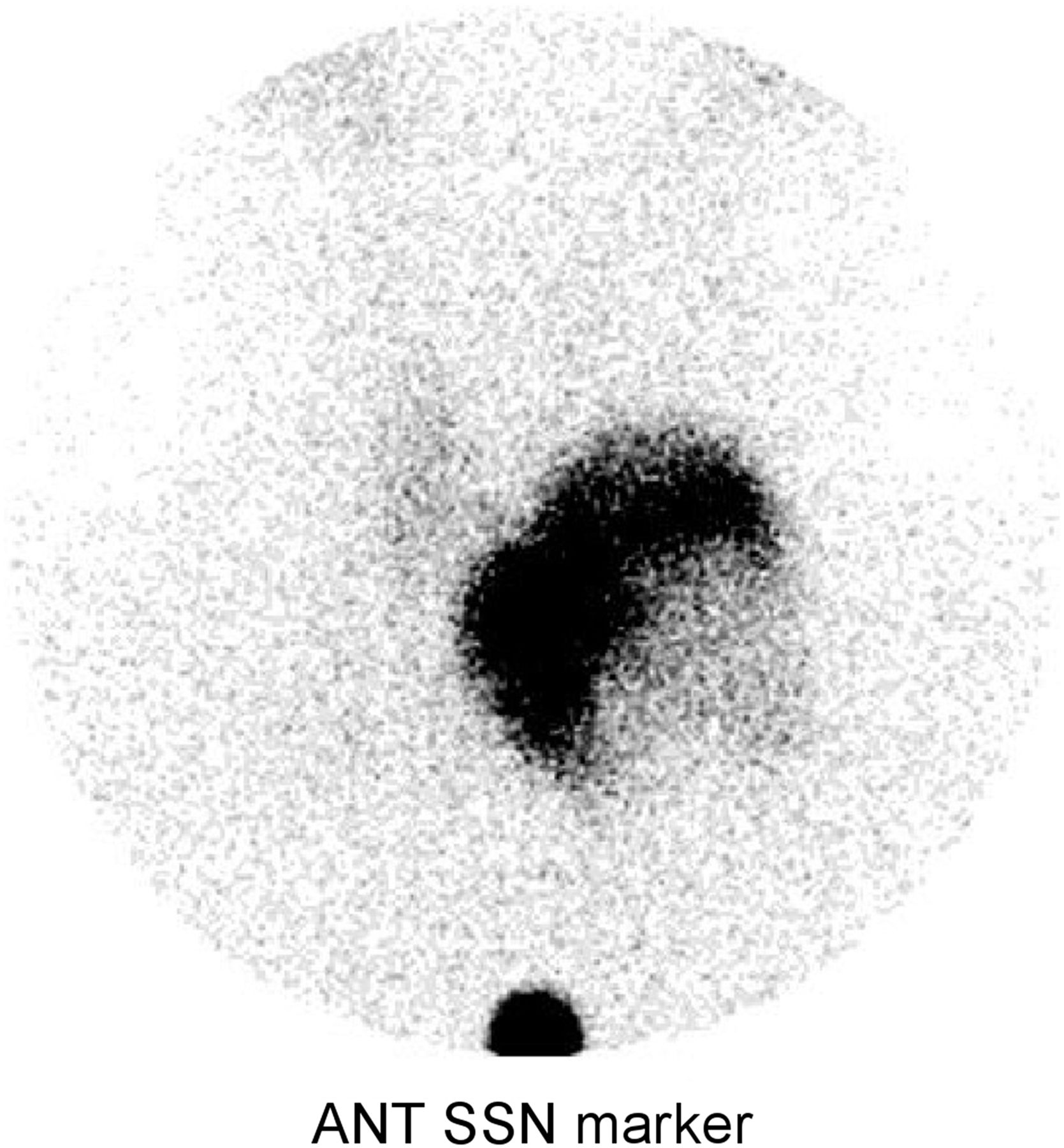
The pinhole images showed a large, heterogeneous, hyperactive nodule in the left lobe and suppression of uptake in the right lobe (Fig. 1). The lower margin of the nodule was far above the suprasternal notch. However, the SPECT/CT images showed that the lower portion of the nodule was at the level of the suprasternal notch (Fig. 2).

FIGURE 1.
  • Download figure
  • Open in new tab
  • Download powerpoint
FIGURE 1.

99mTc-pertechnetate anterior (ANT) pinhole image of thyroid gland with marker at suprasternal notch (SSN) reveals large, hyperactive nodule in left lobe. Nodule shows cold areas laterally, likely because of degeneration or necrosis. Remainder of thyroid gland is suppressed, with only mild activity seen in right lobe. Lower margin of this nodule is far above suprasternal notch.

FIGURE 2.
  • Download figure
  • Open in new tab
  • Download powerpoint
FIGURE 2.

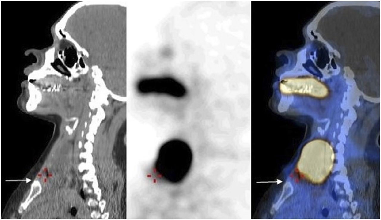
Selected 99mTc-pertechnetate sagittal CT, SPECT, and SPECT/CT images of thyroid gland show that lower margin of left thyroid nodule is at level of suprasternal notch. Arrow indicates upper margin of sternum.

DISCUSSION

The parallax error is caused by the combination of off-center positioning of the pinhole in relation to the object and the inherent minification effect of the pinhole collimator with increasing distance (3). McKitrick et al. reported the parallax error in a patient with substernal goiter who was imaged using a pinhole collimator positioned at the usual thyroid bed between the thyroid cartilage and the suprasternal notch (3). On the resulting image, the substernal portion of the thyroid appeared as if it were within the normal thyroid bed. An additional image obtained with a rectilinear scanner showed that the lower margin of the goiter was substernal. To demonstrate this parallax phenomenon, McKitrick et al. also performed a study using a thyroid phantom. Serial images were obtained at increasing depths of the phantom, which was intentionally placed off center from the pinhole. As the pinhole-to-phantom distance was increased, the thyroid image was “pulled up” to the normal location.

In another report, parathyroid adenoma was wrongly localized by pinhole 99mTc-sestamibi scintigraphy to the anterior mediastinum near a lower pole of the thyroid gland (4). After a failed surgical attempt to resect the adenoma, the patient was reimaged with a parallel-hole collimator and SPECT, which showed that the adenoma was near the heart, anterior to the carina.

In our patient, pinhole imaging erroneously showed the lower margin of the thyroid nodule to be far above the suprasternal notch. SPECT/CT helped to correctly locate the lower margin of the nodule.

This error can be avoided by centering the pinhole over the large goiter or nodule (3). In addition, using a parallel-hole collimator, a planar, SPECT, or SPECT/CT image of the thyroid and chest region (with markers at the thyroid cartilage and suprasternal notch) should also be obtained to better identify the upper and lower margins of the large goiter or nodule, determine whether it extends to the mediastinum, and locate ectopic thyroid tissue and parathyroid adenomas.

CONCLUSION

Pinhole scintigraphy is still commonly used to image the thyroid gland and is recommended for imaging parathyroid diseases. Hence, recognition of parallax error on pinhole thyroid and parathyroid scintigraphy is important, and if present, additional images should obtained with a parallel-hole collimator to accurately determine the upper and lower margins of a large goiter or nodule and the location of ectopic thyroid tissue and parathyroid adenomas.

DISCLOSURE

No potential conflict of interest relevant to this article was reported.

Footnotes

  • Published online May 3, 2018.

REFERENCES

  1. 1.↵
    1. Ghanem MA,
    2. Elgazzar AH,
    3. Elsaid MM,
    4. Shehab F
    . Comparison of pinhole and high-resolution parallel-hole imaging for nodular thyroid disease. Clin Nucl Med. 2011;36:770–771.
    OpenUrlPubMedGoogle Scholar
  2. 2.↵
    1. Keyes JW Jr.,
    2. Thrall JH,
    3. Carey JE
    . Technical considerations in vivo thyroid studies. Semin Nucl Med. 1978;8:43–57.
    OpenUrlPubMedGoogle Scholar
  3. 3.↵
    1. McKitrick WL,
    2. Park HM,
    3. Kosegi JE
    . Parallax error in pinhole thyroid scintigraphy: a critical consideration in the evaluation of substernal goiters. J Nucl Med. 1985;26:418–420.
    OpenUrlAbstract/FREE Full TextGoogle Scholar
  4. 4.↵
    1. Mavai R,
    2. Caride VJ
    . Pin-hole collimator, parallax, and the localization of mediastinal parathyroid adenoma. Clin Nucl Med. 1996;21:527–529.
    OpenUrlPubMedGoogle Scholar
  5. 5.↵
    1. Delbeke D,
    2. Coleman RE,
    3. Guiberteau MJ,
    4. et al
    .; Society of Nuclear Medicine (SNM). Procedure guideline for SPECT/CT imaging 1.0. J Nucl Med. 2006;47:1227–1234.
    OpenUrlFREE Full TextGoogle Scholar
  • Received for publication January 8, 2018.
  • Accepted for publication March 5, 2018.
PreviousNext
Back to top

In this issue

Journal of Nuclear Medicine Technology: 46 (3)
Journal of Nuclear Medicine Technology
Vol. 46, Issue 3
September 1, 2018
  • Table of Contents
  • About the Cover
  • Index by author
Print
Download PDF
Article Alerts
Email Article
Citation Tools
Share
Recognizing Pinhole Parallax Error for Accurate Localization of Thyroid and Parathyroid Disease
Ismet Sarikaya, Zaina Al-Banna, Ali Baqer
Journal of Nuclear Medicine Technology Sep 2018, 46 (3) 288-289; DOI: 10.2967/jnmt.118.207928
Twitter logo Facebook logo LinkedIn logo Mendeley logo
  • Tweet Widget
Bookmark this article

Jump to section

  • Article
    • Abstract
    • CASE REPORT
    • DISCUSSION
    • CONCLUSION
    • DISCLOSURE
    • Footnotes
    • REFERENCES
  • Figures & Data
  • Info & Metrics
  • PDF

Related Articles

  • No related articles found.
  • PubMed
  • Google Scholar

Cited By...

  • No citing articles found.
  • Google Scholar

More in this TOC Section

  • High-Sensitivity Troponin Elevation in a Young Woman with Typical Chest Pain: The Heart of Matter
  • Prominent Right Ventricular Tracer Uptake: A Harbinger of Multivessel Coronary Artery Disease
  • SPECT/CT for Discrimination Between Active and Inactive Os Trigonum in Posterior Ankle Syndrome
Show more Teaching Case Studies

Similar Articles

Keywords

  • Parallax error
  • Pinhole collimator
  • thyroid scintigraphy
  • parathyroid scintigraphy
  • SPECT/CT
SNMMI

© 2025 SNMMI

Powered by HighWire
Alerts for this Article
Sign In to Email Alerts with your Email Address
Email this Article

Thank you for your interest in spreading the word on Journal of Nuclear Medicine Technology.

NOTE: We only request your email address so that the person you are recommending the page to knows that you wanted them to see it, and that it is not junk mail. We do not capture any email address.

Enter multiple addresses on separate lines or separate them with commas.
Recognizing Pinhole Parallax Error for Accurate Localization of Thyroid and Parathyroid Disease
(Your Name) has sent you a message from Journal of Nuclear Medicine Technology
(Your Name) thought you would like to see the Journal of Nuclear Medicine Technology web site.
Citation Tools
Recognizing Pinhole Parallax Error for Accurate Localization of Thyroid and Parathyroid Disease
Ismet Sarikaya, Zaina Al-Banna, Ali Baqer
Journal of Nuclear Medicine Technology Sep 2018, 46 (3) 288-289; DOI: 10.2967/jnmt.118.207928

Citation Manager Formats

  • BibTeX
  • Bookends
  • EasyBib
  • EndNote (tagged)
  • EndNote 8 (xml)
  • Medlars
  • Mendeley
  • Papers
  • RefWorks Tagged
  • Ref Manager
  • RIS
  • Zotero

We use cookies on this site to enhance your user experience

By clicking any link on this page you are giving your consent for us to set cookies.