


Article Figures & Data
Figures
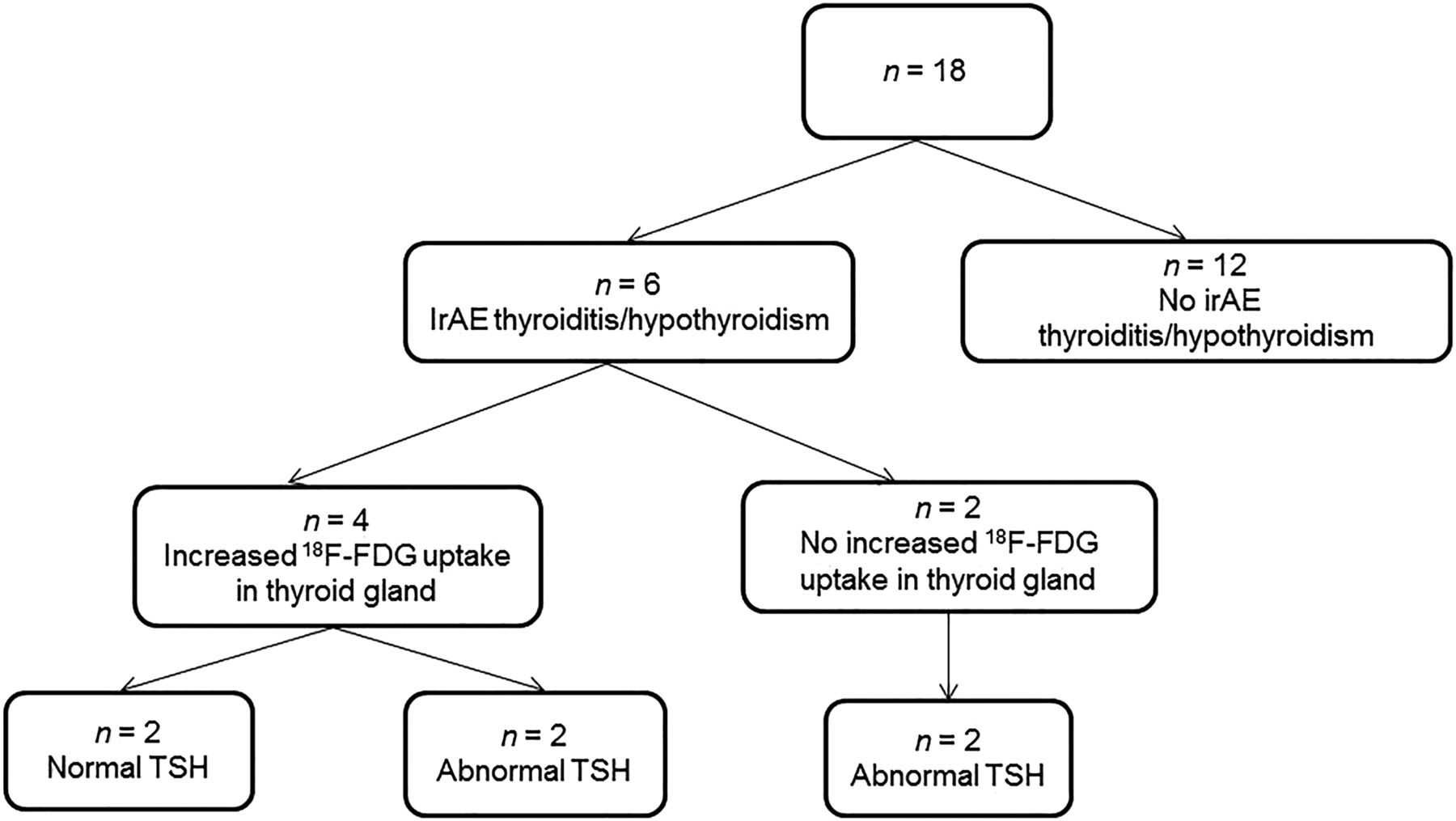
- FIGURE 1.
Flow chart showing distribution of patients by development of thyroiditis, 18F-FDG uptake in thyroid gland, and value of TSH at time of PET/CT.
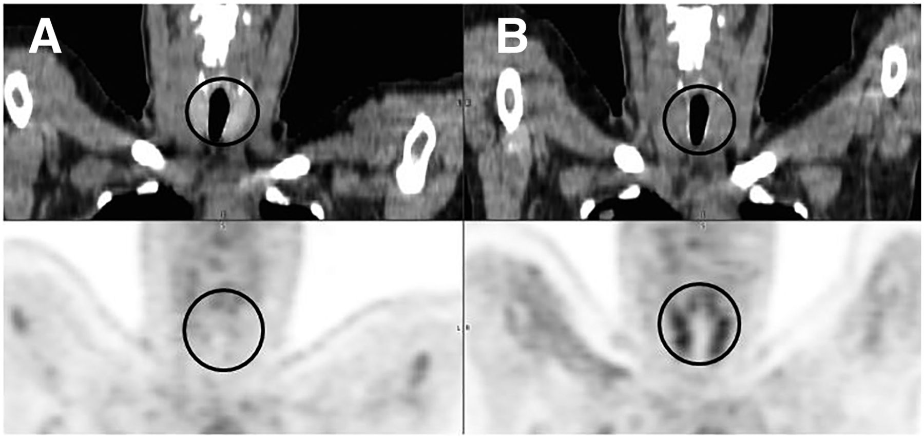
- FIGURE 2.
Patient with increased 18F-FDG uptake in thyroid gland during nivolumab therapy. (A) Coronal CT (top) and 18F-FDG PET (left) images before therapy show normal uptake in thyroid gland, with SUVmax of 1.7. (B) Coronal CT (top) and 18F-FDG PET (bottom) images during therapy show increased thyroid uptake, with SUVmax of 4.3. Region of thyroid gland is encircled.
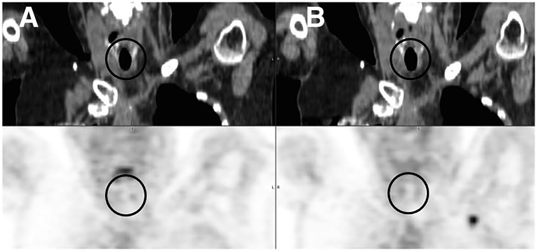
- FIGURE 3.
Patient with normal 18F-FDG uptake in thyroid gland during nivolumab therapy. (A) Coronal CT (top) and 18F-FDG PET (left) images before therapy show normal uptake in thyroid gland, with SUVmax of 2.2. (B) Coronal CT (top) and 18F-FDG PET (bottom) images during therapy show stable, normal thyroid uptake, with SUVmax of 1.8. Region of thyroid gland is encircled.

Tables
Characteristic Total (n = 18) Thyroid irAE (n = 6) No thyroid irAE (n = 12) Age (y) Mean 69 65 72 Range 31–86 31–79 51–86 Sex (n) Female 11 (61%) 5 (83%) 6 (50%) Male 7 (39%) 1 (17%) 6 (50%) Therapy cycles (n) Mean 8.6 10.6 7.6 Range 3–20 6–20 3–15 Before therapy During therapy Difference Patient no. SUVmean SUVmax TLG SUVmean SUVmax TLG SUVmean SUVmax TLG 1 1.5 1.5 0.7 3.3 4.1 3.5 1.8 2.6 2.8 2 1.7 2.1 1.5 3.7 4.5 2.9 2 2.4 1.4 3 1.3 1.7 0.9 3.5 4.3 2.3 2.2 2.6 1.4 4 0.9 1.1 0.7 1.9 2.4 1.6 1 1.3 0.9 5 2.4 2.8 1.9 1.6 1.8 1.6 −0.8 −1 −0.3 6 2 2.2 0.2 1.6 1.8 0.3 −0.4 −0.4 0.1 - TABLE 3
Comparison of Thyroid 18F-FDG Uptake During Therapy Between Groups That Did and Did Not Develop Thyroiditis
Parameter Thyroid irAE (n = 6) No thyroid irAE (n = 12) Difference SUVmean 2.41 (1.04) 1.64 (0.44) 0.77 (P = 0.04) SUVmax 2.96 (1.28) 2.00 (0.5) 0.96 (P = 0.04) TLG 1.96 (1.05) 1.00 (0.47) 0.96 (P = 0.02) Data are mean followed by SD in parentheses.


Jump to section
Related Articles
Cited By...
- No citing articles found.