


Article Figures & Data
Figures
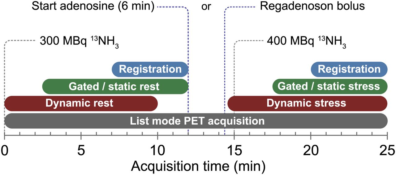
- FIGURE 1.
Components of rest and stress acquisitions (adenosine or regadenoson).
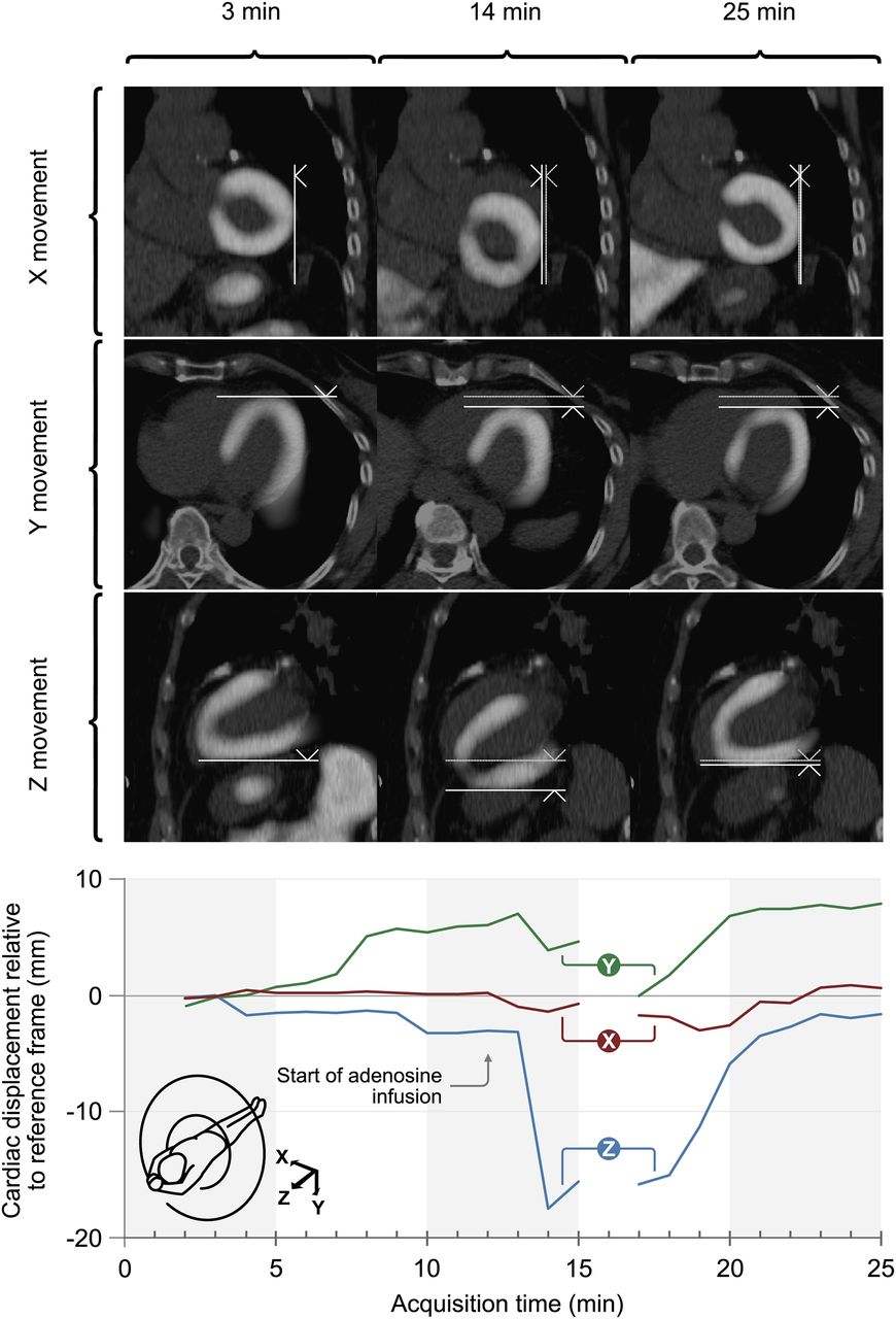
- FIGURE 2.
(Top) Example of cardiac displacement during adenosine stress. Displacement is depicted in coronal, transverse, and sagittal planes in x, y, and z directions, respectively. Images represent data obtained at 3 min after scan initiation (frame 3; reference frame), at 14 min (2 min after initiation of adenosine), and at 25 min (last frame of stress acquisition). Bolder vertical and horizontal lines in each tile of 14- and 25-min series represent displacement relative to initial position of heart (fainter lines). (Bottom) Displacement in x, y, and z directions in this patient during entire scan, relative to reference frame (at 3 min after initiation of scan).
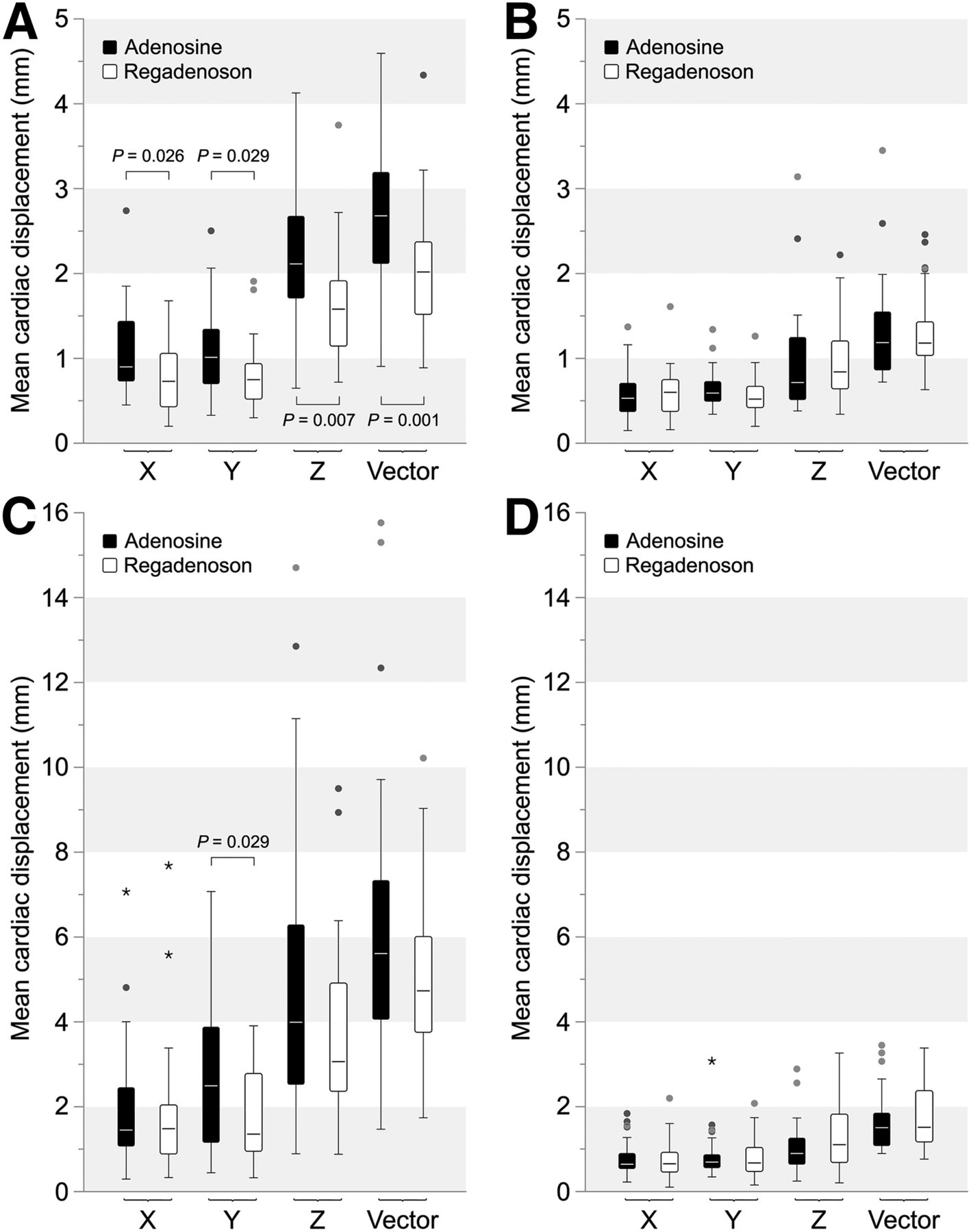
- FIGURE 3.
Median with interquartile ranges of mean cardiac displacement in x, y, and z directions and total displacement vector length for both adenosine and regadenoson. (A) Displacement during stress relative to previous available frame. (B) Displacement at rest. (C) Displacement relative to reference frame (frame 3) during stress. (D) Displacement relative to reference frame at rest. Statistical analysis was performed using Mann–Whitney tests.
- FIGURE 4.
Cardiac displacement during stress and rest acquisitions in x, y, and z directions and total displacement vector length (dotted line). Data represent mean of all patients relative to reference frame (frame 3) (A and B) or relative to previous frame (C and D). (A and C) Displacement during adenosine stress. (B and D) Displacement during regadenoson stress. Gaps in plots are result of exclusion of frames because of high blood-pool activity after injection of 13NH3.


Tables
- TABLE 1
Baseline Characteristics, Known Risk Factors, Stress Test Parameters, and Clinical PET/CT Diagnosis
Parameter Adenosine Regadenoson P Baseline characteristics Sex 0.901 Male 15 (50%) 15 (48%) Female 15 (50%) 16 (52%) Age (y) 68 ± 10 67 ± 9 0.608† Body mass index 28.1 ± 4.8 27.1 ± 4.9 0.116 Duke Clinical Score (%)* 62 ± 33 55 ± 32 0.243 Risk factors Diabetes mellitus 0 (0.0%) 6 (19.4%) 0.012 Family history of coronary artery disease 7 (23.3%) 8 (25.8%) 0.824 Hypertension 18 (60.0%) 14 (45.2%) 0.250 Smoking 4 (13.3%) 6 (19.4%) 0.529 Previous myocardial infarction 13 (43.3%) 5 (16.1%) 0.021 Previous percutaneous coronary intervention 12 (40.0%) 6 (19.4%) 0.080 Previous coronary artery bypass grafting 2 (6.7%) 2 (6.5%) 0.973 Stress test parameters Maximum heart rate during stress (bpm) 91 ± 18 94 ± 18 0.704 Percentage heart rate of maximum 59.1 ± 9.9 61.1 ± 12.0 0.367 Systole at peak stress (mm Hg) 125.1 ± 15.8 136.1 ± 14.8 0.602 Diastole at peak stress (mm Hg) 67.5 ± 10.5 71.0 ± 9.5 0.611 PET/CT results No ischemia or infarction 17 (56.7%) 22 (70.9%) Ischemia 10 (33.3%) 3 (9.7%) 0.141‡ Infarction 3 (10.0%) 6 (19.4%) Motion artifacts, static stress 14 (46.7%) 9 (30.0%) 0.192 ↵* In both groups, Duke Clinical Score was missing in 2 patients.
↵† Independent-samples t tests.
↵‡ χ2 tests.
Qualitative data are expressed as numbers followed by percentages in parentheses; continuous data are expressed as mean ± SD. Statistical analyses were done using Mann–Whitney tests by default, or as indicated.
- TABLE 2
Maximal Displacement in 3 Axes During Stress Acquisitions* Using Previous Frame or Frame 3 as Reference
Relative to previous frame Relative to frame 3 Direction Adenosine Regadenoson P Adenosine Regadenoson P Negative x −2.9 ± 1.8 −2.3 ± 1.5 0.083 −2.5 ± 1.9 −2.9 ± 2.3 0.435 Negative y −3.1 ± 1.8 −2.3 ± 1.5 0.024 −4.8 ± 3.3 −3.1 ± 2.0 0.012 Negative z −6.4 ± 3.7 −4.9 ± 2.2 0.123 −9.9 ± 5.3 −7.1 ± 3.6 0.048 Positive x 2.7 ± 1.4 1.8 ± 1.0 0.007 2.2 ± 2.6 0.7 ± 1.5 0.063 Positive y 2.6 ± 1.4 2.2 ± 1.7 0.082 0.2 ± 2.3 0.7 ± 1.3 0.030 Positive z 4.9 ± 2.5 3.7 ± 1.7 0.034 0.4 ± 3.2 0.6 ± 2.3 0.229 ↵* Frames 13–25 for adenosine and frames 15–25 for regadenoson.
Data are mean millimeters ± SD. Analysis for displacement relative to previous frame was done using Mann–Whitney tests. Analysis for displacement relative to frame 3 was done using independent-samples t tests.
- TABLE 3
Maximal Displacement as Vector Length During Rest and Stress Acquisitions Using Previous Frame or Frame 3 as Reference
Relative to previous frame Relative to frame 3 Vector length during… Adenosine Regadenoson P Adenosine Regadenoson P Rest 3.8 ± 1.9 3.8 ± 1.5 0.593 4.1 ± 1.7 4.4 ± 2.6 0.971 Stress 8.1 ± 3.7 6.1 ± 2.3 0.022 11.6 ± 5.2 8.6 ± 3.0 0.014 Data are mean millimeters ± SD. Analysis was done using Mann–Whitney tests.
- TABLE 4
Total Number of Frames of All Patients with Minor, Medium, and Large Displacement During Stress Acquisition Using Previous Frame or Frame 3 as Reference
Relative to previous frame Relative to frame 3 Parameter Adenosine Regadenoson P Adenosine Regadenoson P Frames with <5 mm stress 1,025 (94.9%) 1,088 (97.5%) 861 (79.7%) 991 (88.8%) Frames with 5–10 mm stress 51 (4.7%) 27 (2.4%) 0.005 174 (16.1%) 112 (10.0%) <0.001 Frames with >10 mm stress 4 (0.4%) 1 (0.1%) 45 (4.2%) 13 (1.2%) Minor displacement (<5 mm) 10 (33.3%) 12 (38.7%) 4 (13.3%) 7 (22.6%) Medium displacement (5–10 mm) 16 (53.3%) 18 (58.1%) 0.352 11 (36.7%) 20 (64.5%) 0.007 Large displacement (>10 mm) 4 (13.3%) 1 (3.2%) 15 (50.0%) 4 (12.9%) Data are n (of patients) and in percentage. Analysis was done using χ2 tests.
Symptoms during stress acquisitions Adenosine* Regadenoson P None 6 (19.4%) 9 (15.3%) 0.357 Typical chest pain 10 (33.3%) 8 (25.8%) 0.409 Respiratory 16 (53.3%) 11 (35.5%) 0.095 Gastrointestinal 5 (16.7%) 7 (22.6%) 0.653 Vasodilation 12 (40.0%) 14 (45.2%) 0.859 Other 3 (10.0%) 3 (9.7%) 0.895 General degree of discomfort† 2.9 ± 1.1 2.6 ± 1.1 0.428 ↵* Two surveys were missing from adenosine group.
↵† General degree of discomfort was expressed as a number from 1 (no discomfort at all) to 5 (very inconvenient). One patient in each group gave a deviant answer, and both of these patients were excluded from the analysis.
Data are n and percentage, or mean ± SD. Analysis was done using χ2 tests to identify differences between stress and rest acquisitions.


Jump to section
Related Articles
Cited By...
- No citing articles found.