
Main menu

  • Home
  • Content
    • Current
      • JNMT Supplement
    • Ahead of print
    • Past Issues
    • Continuing Education
    • JNMT Podcast
    • SNMMI Annual Meeting Abstracts
  • Subscriptions
    • Subscribers
    • Rates
    • Journal Claims
    • Institutional and Non-member
  • Authors
    • Submit to JNMT
    • Information for Authors
    • Assignment of Copyright
    • AQARA Requirements
  • Info
    • Reviewers
    • Permissions
    • Advertisers
    • Corporate & Special Sales
  • About
    • About Us
    • Editorial Board
    • Contact Information
  • More
    • Alerts
    • Feedback
    • Help
    • SNMMI Journals
  • SNMMI
    • JNMT
    • JNM
    • SNMMI Journals
    • SNMMI

User menu

  • Subscribe
  • My alerts
  • Log in
  • My Cart

Search

  • Advanced search
Journal of Nuclear Medicine Technology
  • SNMMI
    • JNMT
    • JNM
    • SNMMI Journals
    • SNMMI
  • Subscribe
  • My alerts
  • Log in
  • My Cart
Journal of Nuclear Medicine Technology

Advanced Search

  • Home
  • Content
    • Current
    • Ahead of print
    • Past Issues
    • Continuing Education
    • JNMT Podcast
    • SNMMI Annual Meeting Abstracts
  • Subscriptions
    • Subscribers
    • Rates
    • Journal Claims
    • Institutional and Non-member
  • Authors
    • Submit to JNMT
    • Information for Authors
    • Assignment of Copyright
    • AQARA Requirements
  • Info
    • Reviewers
    • Permissions
    • Advertisers
    • Corporate & Special Sales
  • About
    • About Us
    • Editorial Board
    • Contact Information
  • More
    • Alerts
    • Feedback
    • Help
    • SNMMI Journals
  • Watch or Listen to JNMT Podcast
  • Visit SNMMI on Facebook
  • Join SNMMI on LinkedIn
  • Follow SNMMI on Twitter
  • Subscribe to JNMT RSS feeds
Review ArticleTeaching Case Studies

Utility of 111In-Cl3 Scintigraphy for Differentiating Between Bone Marrow Reconversion and Bone Metastasis with Esophageal Adenocarcinoma

Shoji Okuda, Hironori Nishibori and Hiroaki Hoshi
Journal of Nuclear Medicine Technology March 2018, 46 (1) 63-64; DOI: https://doi.org/10.2967/jnmt.117.197913
Shoji Okuda
1Department of Radiology, Kizawa Memorial Hospital, Gifu, Japan; and
  • Find this author on Google Scholar
  • Find this author on PubMed
  • Search for this author on this site
Hironori Nishibori
2Department of Radiology, Kizawa Memorial Hospital, Gifu, Japan
  • Find this author on Google Scholar
  • Find this author on PubMed
  • Search for this author on this site
Hiroaki Hoshi
2Department of Radiology, Kizawa Memorial Hospital, Gifu, Japan
  • Find this author on Google Scholar
  • Find this author on PubMed
  • Search for this author on this site
  • Article
  • Figures & Data
  • Info & Metrics
  • PDF
Loading

Abstract

Indium chloride (111In-Cl3) scintigraphy has been used to evaluate various hematologic diseases for many years. However, there have been few reports on patients with bone marrow reconversion showing high uptake in 111In-Cl3 scintigraphy. Here, we report the case of a 68-y-old man with esophageal cancer who underwent 18F-FDG PET/CT for staging of the disease. In the first lumbar vertebral body, 18F-FDG PET/CT demonstrated high uptake that made it difficult to distinguish bone metastasis from bone marrow reconversion. 111In-Cl3 scintigraphy demonstrated specific findings with high uptake in the lesion, indicating bone marrow hyperplasia or reconversion.

  • 111In-Cl3 scintigraphy
  • bone marrow reconversion
  • chemical shift study

Indium chloride (111In-Cl3) scintigraphy is a useful modality for bone marrow imaging and has been used for evaluating various hematologic diseases for many years. However, there are few reports in which high uptake helped differentiate bone marrow reconversion from bone metastasis.

This report highlights the utility of 111In-Cl3 scintigraphy for differentiating bone marrow reconversion from bone metastasis.

CASE REPORT

A 68-y-old man with esophageal cancer presented to our hospital for further evaluation and surgical resection. Findings on physical examination were normal. Laboratory examination showed anemia (hemoglobin, 9.7 mg/μL) and a high level of blood glucose but was negative for tumor markers.

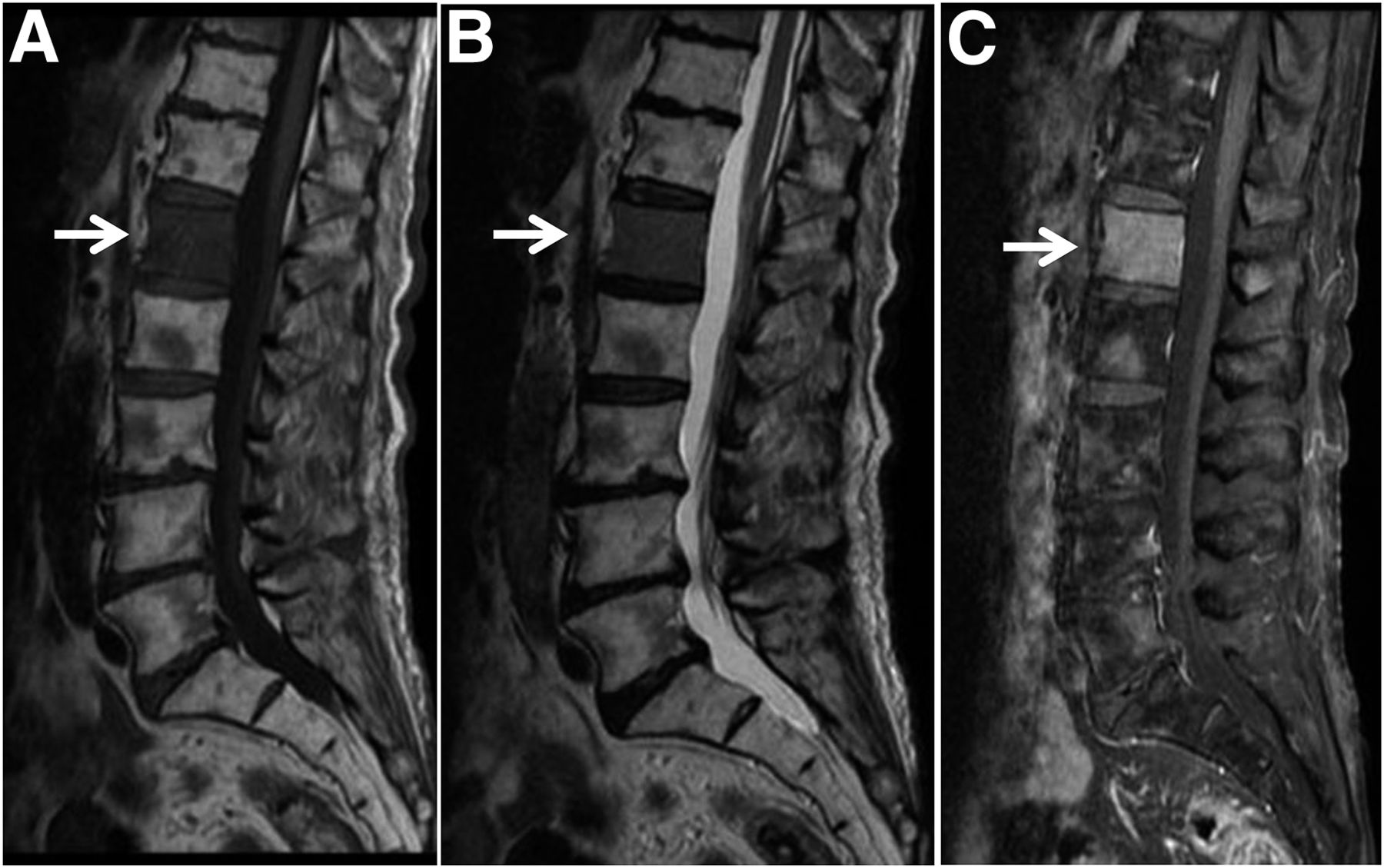
18F-FDG PET/CT showed relatively high uptake (SUVmax, 3.33 for the early phase and 4.46 for the delayed phase) in the first lumbar vertebral body, in addition to slightly high uptake in the primary lesion (chest to lower esophagus). CT showed a slightly sclerotic lesion in the first lumbar vertebral body (Fig. 1). On both T1- and T2-weighted imaging, MRI demonstrated decreased signal intensity, which showed enhancement with gadolinium. MRI also showed several patchy signal changes in the thoracic vertebral bodies (Fig. 2).

FIGURE 1.
  • Download figure
  • Open in new tab
  • Download powerpoint
FIGURE 1.

18F-FDG PET/CT showing relatively high uptake in L1 vertebral body: anterior maximum-intensity projection (A), fused axial delayed-phase images of primary lesion (B) and L1 vertebral body (C), and sagittal CT image of slightly sclerotic lesion in L1 vertebral body (D).

FIGURE 2.
  • Download figure
  • Open in new tab
  • Download powerpoint
FIGURE 2.

L1 vertebral body (arrows) showing decreased signal intensity on both T1-weighted (A) and T2-weighted (B) MRI and enhancement with gadolinium (C).

It was difficult to distinguish bone metastasis from bone marrow reconversion; therefore, an MRI chemical shift study and bone marrow scintigraphy (48 h after intravenous injection of 74 MBq of 111In-Cl3) were performed. We used chemical shift imaging to assess for fatty infiltrates, which would suggest bone marrow reconversion. However, no significant decrease in signal was seen on out-of-phase images (Fig. 3). 111In-Cl3 SPECT/CT demonstrated high uptake in the first lumbar vertebral body (Fig. 4).

FIGURE 3.
  • Download figure
  • Open in new tab
  • Download powerpoint
FIGURE 3.

In-phase (A) and out-of-phase (B) MR images, with the latter showing no significant decrease in signal in L1 vertebral body (arrows).

FIGURE 4.
  • Download figure
  • Open in new tab
  • Download powerpoint
FIGURE 4.

111In-Cl3 SPECT/CT showing high uptake (arrowheads) in L1 vertebral body and lower uptake in the primary tumor: scintigraphy (A), fused axial image (B), and fused sagittal image (C). Arrows indicate lower uptake in thoracic vertebral bodies as well as bone marrow reconversion.

The high uptake of 111In-Cl3 was suggestive of bone marrow hyperplasia and not bone marrow metastasis. The patient underwent surgical resection of esophageal cancer, and the first lumbar vertebral lesion showed no changes on a follow-up MRI scan that was acquired 1 y later.

DISCUSSION

In patients with cancer, it is occasionally difficult to differentiate bone marrow reconversion from bone metastasis. There are reports in which localized bone marrow reconversion sometimes mimics malignant tumors, such as bone metastasis in both MRI and 18F-FDG PET/CT (1,2). Several reports have recommended an MRI chemical shift study (3,4). However, no significant findings were seen in the present case. Diffusion-weighted MRI is a potential imaging modality to differentiate malignant from benign lesions. However, studies on this topic have been controversial, and findings should be interpreted in line with routine marrow sequences (3).

After 111In-Cl3 is injected, it binds to transferrin in the same manner as ions and is distributed throughout the bone marrow system. The biologic behaviors of indium and iron are similar, and in many previous reports, bone marrow 111In-Cl3 uptake has been thought to reflect the distribution of erythropoietic marrow. 111In-Cl3 has been clinically used for bone marrow studies. Approximately 30% of the administered tracer is found in the bone marrow, 20% in the liver, 7% in the kidneys, and 1% in the spleen (5).

111In is a cyclotron-produced isotope with a half-life of 2.8 d emitting γ-rays with energies of 171 keV (89%) and 245 keV (94%). It decays by electron capture to stable 111Cd (5). In comparison with the 99mTc-labeled colloids, the specificity to bone marrow accumulation is thought to be higher, but radiation exposure is also higher because of relatively long half-life.

111In-Cl3 may accumulate in tumors (6). However, to our knowledge, there has been no report concerning bone metastasis in a patient with esophageal adenocarcinoma taking up 111In-Cl3.

CONCLUSION

111In-Cl3 scintigraphy in the present case demonstrated high uptake in the lesion and was effective for differentiating between bone marrow reconversion and bone metastasis from esophageal adenocarcinoma.

DISCLOSURE

No potential conflict of interest relevant to this article was reported.

Footnotes

  • Published online Nov. 10, 2017.

REFERENCES

  1. 1.↵
    1. Bordalo-Rodrigues M,
    2. Galant C,
    3. Lonneux M,
    4. Clause D,
    5. Vande Berg BC
    . Focal nodular hyperplasia of the hematopoietic marrow simulating vertebral metastasis on FDG positron emission tomography. AJR. 2003;180:669–671.
    OpenUrlCrossRefPubMed
  2. 2.↵
    1. Ulaner G,
    2. Hwang S,
    3. Lefkowitz RA,
    4. Landa J,
    5. Panicek DM
    . Musculoskeletal tumors and tumor-like conditions: common and avoidable pitfalls at imaging in patients with known or suspected cancer. Int Orthop. 2013;37:871–876.
    OpenUrl
  3. 3.↵
    1. Nouh MR,
    2. Eid AF
    . Magnetic resonance imaging of the spinal marrow: basic understanding of the normal marrow pattern and its variant. World J Radiol. 2015;7:448–458.
    OpenUrl
  4. 4.↵
    1. Zajick DC,
    2. Morrison WB,
    3. Schweitzer ME,
    4. Parellada JA,
    5. Carrino JA
    . Benign and malignant processes: normal values and differentiation with chemical shift MR imaging in vertebral marrow. Radiology. 2005;237:590–596.
    OpenUrlCrossRefPubMed
  5. 5.↵
    1. Agool A,
    2. Glaudemans AW,
    3. Boersma HH,
    4. Dierckx RA,
    5. Vellenga E,
    6. Slart RH
    . Radionuclide imaging of bone marrow disorders. Eur J Nucl Med Mol Imaging. 2011;38:166–178.
    OpenUrlPubMed
  6. 6.↵
    1. Kakite S,
    2. Tanabe Y,
    3. Kinoshita F,
    4. Harada H,
    5. Ogawa T
    . Clinical usefulness of In-111 chloride and Tc-99m Sn colloid scintigraphy in the diagnosis of intrathoracic extramedullary hematopoiesis. Ann Nucl Med. 2005;19:317–320.
    OpenUrlPubMed
  • Received for publication July 26, 2017.
  • Accepted for publication October 12, 2017.
PreviousNext
Back to top

In this issue

Journal of Nuclear Medicine Technology: 46 (1)
Journal of Nuclear Medicine Technology
Vol. 46, Issue 1
March 1, 2018
  • Table of Contents
  • About the Cover
  • Index by author
Print
Download PDF
Article Alerts
Sign In to Email Alerts with your Email Address
Email Article

Thank you for your interest in spreading the word on Journal of Nuclear Medicine Technology.

NOTE: We only request your email address so that the person you are recommending the page to knows that you wanted them to see it, and that it is not junk mail. We do not capture any email address.

Enter multiple addresses on separate lines or separate them with commas.
Utility of 111In-Cl3 Scintigraphy for Differentiating Between Bone Marrow Reconversion and Bone Metastasis with Esophageal Adenocarcinoma
(Your Name) has sent you a message from Journal of Nuclear Medicine Technology
(Your Name) thought you would like to see the Journal of Nuclear Medicine Technology web site.
Citation Tools
Utility of 111In-Cl3 Scintigraphy for Differentiating Between Bone Marrow Reconversion and Bone Metastasis with Esophageal Adenocarcinoma
Shoji Okuda, Hironori Nishibori, Hiroaki Hoshi
Journal of Nuclear Medicine Technology Mar 2018, 46 (1) 63-64; DOI: 10.2967/jnmt.117.197913

Citation Manager Formats

  • BibTeX
  • Bookends
  • EasyBib
  • EndNote (tagged)
  • EndNote 8 (xml)
  • Medlars
  • Mendeley
  • Papers
  • RefWorks Tagged
  • Ref Manager
  • RIS
  • Zotero
Share
Utility of 111In-Cl3 Scintigraphy for Differentiating Between Bone Marrow Reconversion and Bone Metastasis with Esophageal Adenocarcinoma
Shoji Okuda, Hironori Nishibori, Hiroaki Hoshi
Journal of Nuclear Medicine Technology Mar 2018, 46 (1) 63-64; DOI: 10.2967/jnmt.117.197913
Twitter logo Facebook logo LinkedIn logo Mendeley logo
  • Tweet Widget
  • Facebook Like
  • Google Plus One
Bookmark this article

Jump to section

  • Article
    • Abstract
    • CASE REPORT
    • DISCUSSION
    • CONCLUSION
    • DISCLOSURE
    • Footnotes
    • REFERENCES
  • Figures & Data
  • Info & Metrics
  • PDF

Related Articles

  • No related articles found.
  • PubMed
  • Google Scholar

Cited By...

  • No citing articles found.
  • Google Scholar

More in this TOC Section

  • High-Sensitivity Troponin Elevation in a Young Woman with Typical Chest Pain: The Heart of the Matter
  • Pulmonary Adenocarcinoma Revealed by Parathyroid Scintigraphy: An Incidental Case to Remember
  • Prominent Right Ventricular Tracer Uptake: A Harbinger of Multivessel Coronary Artery Disease
Show more Teaching Case Studies

Similar Articles

Keywords

  • 111In-Cl3 scintigraphy
  • bone marrow reconversion
  • chemical shift study
SNMMI

© 2025 SNMMI

Powered by HighWire