
Main menu

  • Home
  • Content
    • Current
      • JNMT Supplement
    • Ahead of print
    • Past Issues
    • Continuing Education
    • JNMT Podcast
    • SNMMI Annual Meeting Abstracts
  • Subscriptions
    • Subscribers
    • Rates
    • Journal Claims
    • Institutional and Non-member
  • Authors
    • Submit to JNMT
    • Information for Authors
    • Assignment of Copyright
    • AQARA Requirements
  • Info
    • Reviewers
    • Permissions
    • Advertisers
    • Corporate & Special Sales
  • About
    • About Us
    • Editorial Board
    • Contact Information
  • More
    • Alerts
    • Feedback
    • Help
    • SNMMI Journals
  • SNMMI
    • JNMT
    • JNM
    • SNMMI Journals
    • SNMMI

User menu

  • Subscribe
  • My alerts
  • Log in
  • My Cart

Search

  • Advanced search
Journal of Nuclear Medicine Technology
  • SNMMI
    • JNMT
    • JNM
    • SNMMI Journals
    • SNMMI
  • Subscribe
  • My alerts
  • Log in
  • My Cart
Journal of Nuclear Medicine Technology

Advanced Search

  • Home
  • Content
    • Current
    • Ahead of print
    • Past Issues
    • Continuing Education
    • JNMT Podcast
    • SNMMI Annual Meeting Abstracts
  • Subscriptions
    • Subscribers
    • Rates
    • Journal Claims
    • Institutional and Non-member
  • Authors
    • Submit to JNMT
    • Information for Authors
    • Assignment of Copyright
    • AQARA Requirements
  • Info
    • Reviewers
    • Permissions
    • Advertisers
    • Corporate & Special Sales
  • About
    • About Us
    • Editorial Board
    • Contact Information
  • More
    • Alerts
    • Feedback
    • Help
    • SNMMI Journals
  • Watch or Listen to JNMT Podcast
  • Visit SNMMI on Facebook
  • Join SNMMI on LinkedIn
  • Follow SNMMI on Twitter
  • Subscribe to JNMT RSS feeds
Research ArticleImaging

Validation of Left Ventricular Ejection Fraction with the IQ•SPECT System in Small-Heart Patients

Hiroto Yoneyama, Takayuki Shibutani, Takahiro Konishi, Asuka Mizutani, Ryosuke Hashimoto, Masahisa Onoguchi, Koichi Okuda, Shinro Matsuo, Kenichi Nakajima and Seigo Kinuya
Journal of Nuclear Medicine Technology September 2017, 45 (3) 201-207; DOI: https://doi.org/10.2967/jnmt.116.188680
Hiroto Yoneyama
1Department of Radiological Technology, Kanazawa University Hospital, Kanazawa, Japan
  • Find this author on Google Scholar
  • Find this author on PubMed
  • Search for this author on this site
Takayuki Shibutani
2Department of Health Science, Graduate School of Medical Science, Kanazawa University, Kanazawa, Japan
  • Find this author on Google Scholar
  • Find this author on PubMed
  • Search for this author on this site
Takahiro Konishi
1Department of Radiological Technology, Kanazawa University Hospital, Kanazawa, Japan
  • Find this author on Google Scholar
  • Find this author on PubMed
  • Search for this author on this site
Asuka Mizutani
1Department of Radiological Technology, Kanazawa University Hospital, Kanazawa, Japan
  • Find this author on Google Scholar
  • Find this author on PubMed
  • Search for this author on this site
Ryosuke Hashimoto
2Department of Health Science, Graduate School of Medical Science, Kanazawa University, Kanazawa, Japan
  • Find this author on Google Scholar
  • Find this author on PubMed
  • Search for this author on this site
Masahisa Onoguchi
2Department of Health Science, Graduate School of Medical Science, Kanazawa University, Kanazawa, Japan
  • Find this author on Google Scholar
  • Find this author on PubMed
  • Search for this author on this site
Koichi Okuda
3Department of Physics, Kanazawa Medical University, Kanazawa, Japan; and
  • Find this author on Google Scholar
  • Find this author on PubMed
  • Search for this author on this site
Shinro Matsuo
4Department of Nuclear Medicine, Kanazawa University Hospital, Kanazawa, Japan
  • Find this author on Google Scholar
  • Find this author on PubMed
  • Search for this author on this site
Kenichi Nakajima
4Department of Nuclear Medicine, Kanazawa University Hospital, Kanazawa, Japan
  • Find this author on Google Scholar
  • Find this author on PubMed
  • Search for this author on this site
Seigo Kinuya
4Department of Nuclear Medicine, Kanazawa University Hospital, Kanazawa, Japan
  • Find this author on Google Scholar
  • Find this author on PubMed
  • Search for this author on this site
  • Article
  • Figures & Data
  • Info & Metrics
  • PDF
Loading

Article Figures & Data

Figures

  • Tables
  • FIGURE 1.
    • Download figure
    • Open in new tab
    • Download powerpoint
    FIGURE 1.

    Gated myocardial perfusion SPECT images of 67-y-old woman for whom echocardiographic measurements were 110 mL for ESV, 29 mL for EDV, and 73% for LVEF. (A) Conventional SPECT with QGS: EDV, 60 mL; ESV, 9 mL; and LVEF, 86%. (B) IQ•SPECT with QGS: EDV, 49 mL; ESV, 4 mL; and LVEF, 92%. (C) Conventional SPECT with cardioREPO: EDV, 71 mL; ESV, 12 mL; and LVEF, 84%. (D) IQ•SPECT with cardioREPO: EDV, 70 mL; ESV, 15 mL; and LVEF, 78%. HLA = horizontal long axis; SA = short axis; VLA = vertical long axis.

  • FIGURE 2.
    • Download figure
    • Open in new tab
    • Download powerpoint
    FIGURE 2.

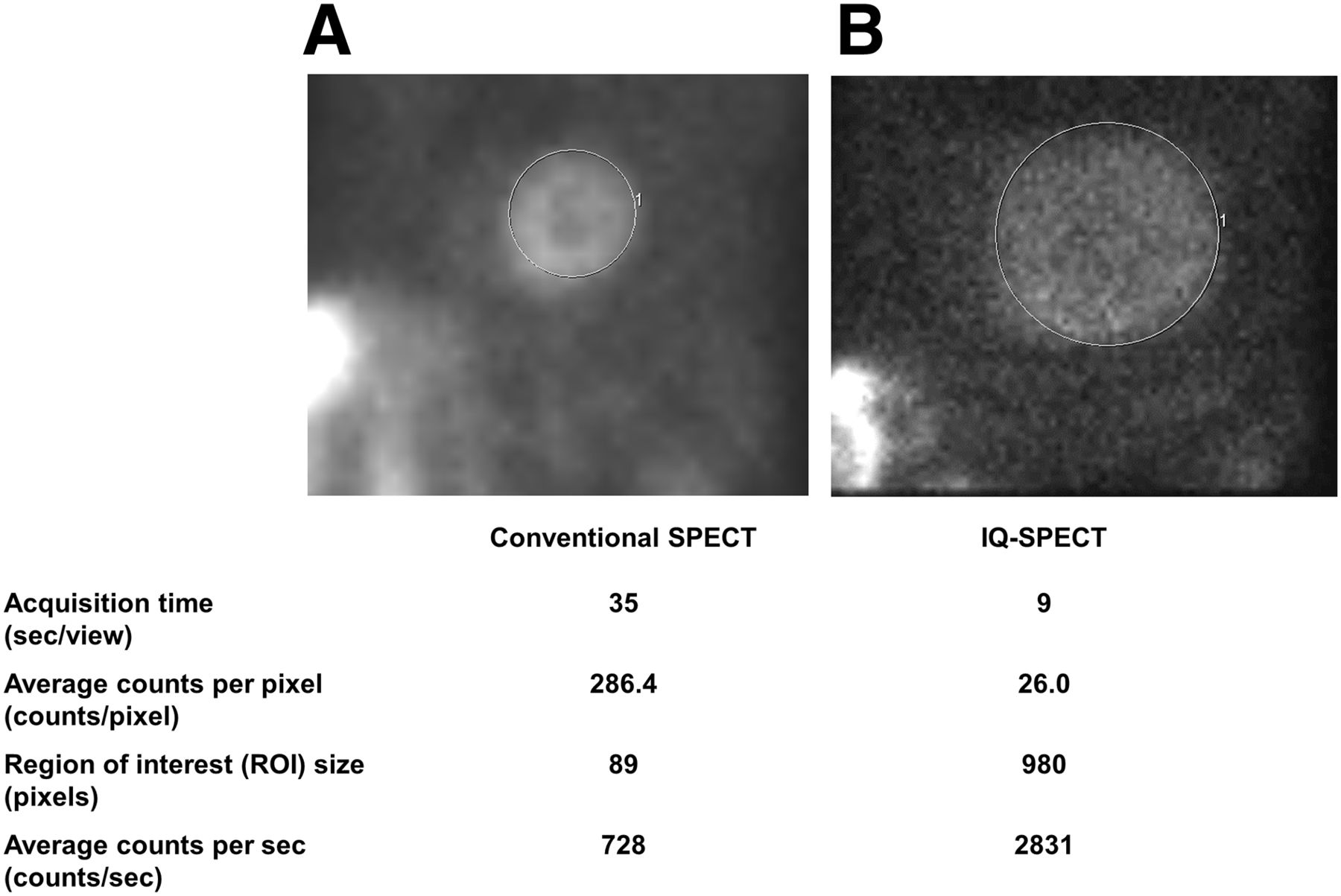
    Average myocardial counts per pixel as measured from 45° left anterior oblique projection on conventional SPECT (A) and IQ•SPECT (B).

  • FIGURE 3.
    • Download figure
    • Open in new tab
    • Download powerpoint
    FIGURE 3.

    Bland–Altman analysis of LVEF on conventional SPECT with QGS vs. IQ•SPECT with QGS (A); conventional SPECT with QGS vs. echocardiography (B); IQ•SPECT with QGS vs. echocardiography (C); conventional SPECT with cardioREPO vs. IQ•SPECT with cardioREPO (D); conventional SPECT with cardioREPO vs. echocardiography (E); and IQ•SPECT with cardioREPO vs. echocardiography (F). Gray lines are mean ± 1.96 SD. Black lines are linear regression lines.

  • FIGURE 4.
    • Download figure
    • Open in new tab
    • Download powerpoint
    FIGURE 4.

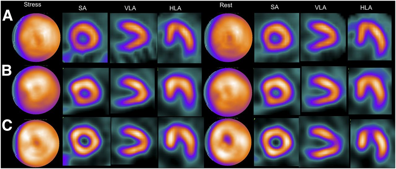
    Myocardial perfusion SPECT images showing normal findings in 85-y-old man. (A) Conventional SPECT. (B) IQ•SPECT without attenuation or scatter correction. (C) IQ•SPECT with attenuation and scatter correction. HLA = horizontal long axis; SA = short axis; VLA = vertical long axis.

  • FIGURE 5.
    • Download figure
    • Open in new tab
    • Download powerpoint
    FIGURE 5.

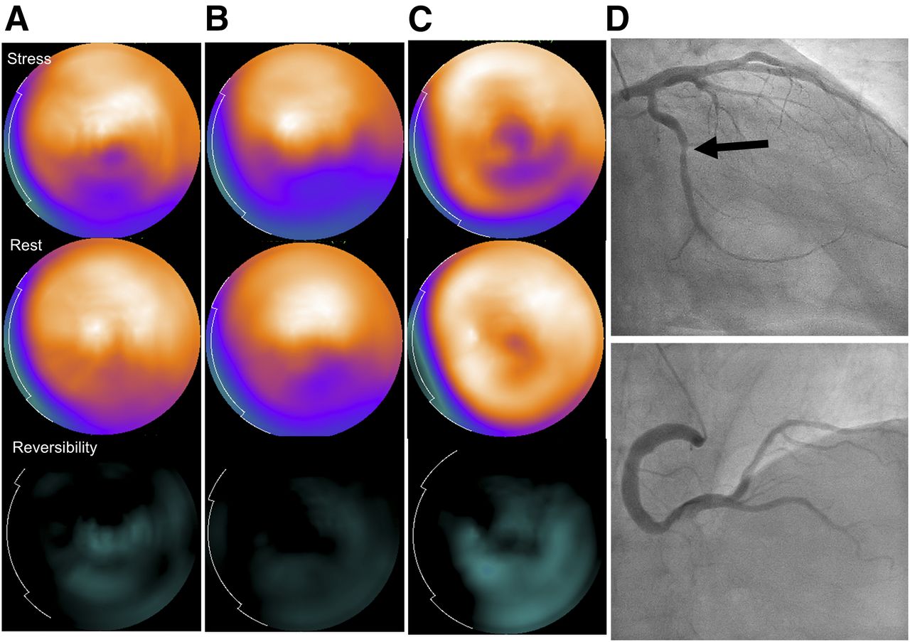
    Myocardial perfusion SPECT images showing perfusion defect in inferolateral wall of 66-y-old man. (A) Conventional SPECT. (B) IQ•SPECT without attenuation or scatter correction. (C) IQ•SPECT with attenuation and scatter correction. (D) Invasive coronary angiography showing stenosis of left circumflex coronary artery (arrow), supporting the SPECT findings.

Tables

  • Figures
    • View popup
    TABLE 1

    Patient Characteristics

    CharacteristicFinding
    Mean age ± SD (y)72.5 ± 9.4 (range, 28–88)
    Sex (n)
     Male77 (78.6%)
     Female21 (21.4%)
    Mean body mass index ± SD23.5 ± 3.1 (range, 16.4–32.8)
    Mean body height ± SD (cm)161.1 ± 9.4 (range, 134–189)
    Mean body weight ± SD (kg)61.7 ± 11.3 (range, 37–93.2)
    Tracer (n)
     Sestamibi2 (2%)
     Tetrofosmin96 (98%)
    Main risk factors (n)
     Obesity (body mass index > 25)30 (30.6%)
     Diabetes mellitus10 (10.2%)
     Abdominal aortic aneurysm37 (37.8%)
     Thoracic aortic aneurysm24 (24.5%)
     Angina pectoris10 (10.2%)
     Arteriosclerosis obliterans9 (9.2%)
     Previous coronary artery bypass surgery5 (5.1%)
     Aortic dissection5 (5.1%)
     Common iliac artery aneurysm3 (3.1%)
     Abnormal electrocardiographic pattern2 (2.0%)
     Renal artery aneurysm1 (1.0%)
     Hepatic artery aneurysm1 (1.0%)
     Aortic valve stenosis1 (1.0%)
    • View popup
    TABLE 2

    Common SPECT Acquisition and Reconstruction Parameters

    ParameterConventional SPECTIQ•SPECT
    Reconstruction algorithmFiltered backprojectionOSCGM
    CollimatorLEHRSMARTZOOM
    Energy window140 keV ± 7.5%140 keV ± 7.5%
    Number of projections60 views (30 per detector)34 views (17 per detector)
    Rotation range360°208°
    Acquisition time (min)20 (35 s per projection)5 (9 s per projection)
    Magnification×1.45×1.00
    Rotation radius (cm)24–2528
    Number of iterationsQGS: 30, QPS: 10
    Number of subsetsQGS: 1, QPS: 3
    Updates30
    Gaussian filter (mm)9.6
    Butterworth filter (cycles/cm)QGS: 0.39, QPS: 0.52
    Matrix64 × 64128 × 128
    Pixel size (mm)6.64.8
    • LEHR = low-energy high-resolution; OSCGM = ordered-subset conjugate gradient minimization.

    • View popup
    TABLE 3

    Comparison of EDV, ESV, and LVEF for Conventional SPECT, IQ•SPECT, and Echocardiography

    ParameterConventionalIQEchoConventional vs. IQConventional vs. echoIQ vs. echo
    QGS
     EDV (mL)77.0 ± 39.174.2 ± 36.8110.6 ± 38.3r = 0.98r = 0.77r = 0.77
     ESV (mL)30.0 ± 29.227.4 ± 27.340.6 ± 26.7r = 0.97r = 0.81r = 0.82
     LVEF (%)65.4 ± 13.868.4 ± 15.264.8 ± 11.6r = 0.86r = 0.63r = 0.64
    CardioREPO
     EDV (mL)87.9 ± 35.682.8 ± 34.0110.6 ± 38.3r = 0.98r = 0.77r = 0.75
     ESV (mL)28.1 ± 22.627.0 ± 22.440.6 ± 26.7r = 0.98r = 0.78r = 0.77
     LVEF (%)69.5 ± 10.669.5 ± 11.064.8 ± 11.6r = 0.80r = 0.60r = 0.65
    QGS (%)
     All LVEF65.4 ± 13.868.4 ± 15.264.8 ± 11.6P = 0.0002NSP = 0.019
     Small-heart LVEF75.0 ± 9.679.5 ± 8.370.1 ± 6.8P = 0.0005P = 0.0076P < 0.0001
    CardioREPO (%)
     All LVEF69.5 ± 10.669.5 ± 11.064.8 ± 11.6NSP < 0.0001P < 0.0001
     Small-heart LVEF72.3 ± 9.074.3 ± 8.370.1 ± 6.8NSNSP = 0.0068
    • Conventional = conventional SPECT; echo = echocardiography; IQ = IQ•SPECT; NS = not statistically significant.

    • Data are mean ± SD.

PreviousNext
Back to top

In this issue

Journal of Nuclear Medicine Technology: 45 (3)
Journal of Nuclear Medicine Technology
Vol. 45, Issue 3
September 1, 2017
  • Table of Contents
  • About the Cover
  • Index by author
Print
Download PDF
Article Alerts
Sign In to Email Alerts with your Email Address
Email Article

Thank you for your interest in spreading the word on Journal of Nuclear Medicine Technology.

NOTE: We only request your email address so that the person you are recommending the page to knows that you wanted them to see it, and that it is not junk mail. We do not capture any email address.

Enter multiple addresses on separate lines or separate them with commas.
Validation of Left Ventricular Ejection Fraction with the IQ•SPECT System in Small-Heart Patients
(Your Name) has sent you a message from Journal of Nuclear Medicine Technology
(Your Name) thought you would like to see the Journal of Nuclear Medicine Technology web site.
Citation Tools
Validation of Left Ventricular Ejection Fraction with the IQ•SPECT System in Small-Heart Patients
Hiroto Yoneyama, Takayuki Shibutani, Takahiro Konishi, Asuka Mizutani, Ryosuke Hashimoto, Masahisa Onoguchi, Koichi Okuda, Shinro Matsuo, Kenichi Nakajima, Seigo Kinuya
Journal of Nuclear Medicine Technology Sep 2017, 45 (3) 201-207; DOI: 10.2967/jnmt.116.188680

Citation Manager Formats

  • BibTeX
  • Bookends
  • EasyBib
  • EndNote (tagged)
  • EndNote 8 (xml)
  • Medlars
  • Mendeley
  • Papers
  • RefWorks Tagged
  • Ref Manager
  • RIS
  • Zotero
Share
Validation of Left Ventricular Ejection Fraction with the IQ•SPECT System in Small-Heart Patients
Hiroto Yoneyama, Takayuki Shibutani, Takahiro Konishi, Asuka Mizutani, Ryosuke Hashimoto, Masahisa Onoguchi, Koichi Okuda, Shinro Matsuo, Kenichi Nakajima, Seigo Kinuya
Journal of Nuclear Medicine Technology Sep 2017, 45 (3) 201-207; DOI: 10.2967/jnmt.116.188680
Twitter logo Facebook logo LinkedIn logo Mendeley logo
  • Tweet Widget
  • Facebook Like
  • Google Plus One
Bookmark this article

Jump to section

  • Article
    • Abstract
    • MATERIALS AND METHODS
    • RESULTS
    • DISCUSSION
    • CONCLUSION
    • DISCLOSURE
    • Acknowledgments
    • Footnotes
    • REFERENCES
  • Figures & Data
  • Info & Metrics
  • PDF

Related Articles

  • No related articles found.
  • PubMed
  • Google Scholar

Cited By...

  • No citing articles found.
  • Google Scholar

More in this TOC Section

  • Early 10-Minute Postinjection [18F]F-FAPI-42 uEXPLORER Total-Body PET/CT Scanning Protocol for Staging Lung Cancer Using HYPER Iterative Reconstruction
  • Single- Versus Dual-Time-Point Imaging for Transthyretin Cardiac Amyloid Using 99mTc-Pyrophosphate
  • Software Discrepancies in Radionuclide-Derived Left Ventricular Ejection Fraction
Show more Imaging

Similar Articles

Keywords

  • IQ-SPECT
  • echocardiographic measurement
  • cardioREPO
  • small heart
  • left ventricular ejection fraction
SNMMI

© 2025 SNMMI

Powered by HighWire