
Main menu

  • Home
  • Content
    • Current
      • JNMT Supplement
    • Ahead of print
    • Past Issues
    • Continuing Education
    • JNMT Podcast
    • SNMMI Annual Meeting Abstracts
  • Subscriptions
    • Subscribers
    • Rates
    • Journal Claims
    • Institutional and Non-member
  • Authors
    • Submit to JNMT
    • Information for Authors
    • Assignment of Copyright
    • AQARA Requirements
  • Info
    • Reviewers
    • Permissions
    • Advertisers
    • Corporate & Special Sales
  • About
    • About Us
    • Editorial Board
    • Contact Information
  • More
    • Alerts
    • Feedback
    • Help
    • SNMMI Journals
  • SNMMI
    • JNMT
    • JNM
    • SNMMI Journals
    • SNMMI

User menu

  • Subscribe
  • My alerts
  • Log in
  • My Cart

Search

  • Advanced search
Journal of Nuclear Medicine Technology
  • SNMMI
    • JNMT
    • JNM
    • SNMMI Journals
    • SNMMI
  • Subscribe
  • My alerts
  • Log in
  • My Cart
Journal of Nuclear Medicine Technology

Advanced Search

  • Home
  • Content
    • Current
    • Ahead of print
    • Past Issues
    • Continuing Education
    • JNMT Podcast
    • SNMMI Annual Meeting Abstracts
  • Subscriptions
    • Subscribers
    • Rates
    • Journal Claims
    • Institutional and Non-member
  • Authors
    • Submit to JNMT
    • Information for Authors
    • Assignment of Copyright
    • AQARA Requirements
  • Info
    • Reviewers
    • Permissions
    • Advertisers
    • Corporate & Special Sales
  • About
    • About Us
    • Editorial Board
    • Contact Information
  • More
    • Alerts
    • Feedback
    • Help
    • SNMMI Journals
  • Watch or Listen to JNMT Podcast
  • Visit SNMMI on Facebook
  • Join SNMMI on LinkedIn
  • Follow SNMMI on Twitter
  • Subscribe to JNMT RSS feeds
Research ArticleImaging

Incidence of Brain Metastases on Follow-up 18F-FDG PET/CT Scans of Non–Small Cell Lung Cancer Patients: Should We Include the Brain?

Emily S. Nia, Linda L. Garland, Naghmehossadat Eshghi, Benjamin B. Nia, Ryan J. Avery and Phillip H. Kuo
Journal of Nuclear Medicine Technology September 2017, 45 (3) 193-197; DOI: https://doi.org/10.2967/jnmt.117.194571
Emily S. Nia
1Department of Medical Imaging, University of Arizona, Tucson, Arizona
  • Find this author on Google Scholar
  • Find this author on PubMed
  • Search for this author on this site
Linda L. Garland
2Section of Hematology/Oncology, Department of Medicine, University of Arizona, Tucson, Arizona; and
  • Find this author on Google Scholar
  • Find this author on PubMed
  • Search for this author on this site
Naghmehossadat Eshghi
1Department of Medical Imaging, University of Arizona, Tucson, Arizona
  • Find this author on Google Scholar
  • Find this author on PubMed
  • Search for this author on this site
Benjamin B. Nia
3College of Medicine, University of Texas Medical Branch, Galveston, Texas
  • Find this author on Google Scholar
  • Find this author on PubMed
  • Search for this author on this site
Ryan J. Avery
1Department of Medical Imaging, University of Arizona, Tucson, Arizona
  • Find this author on Google Scholar
  • Find this author on PubMed
  • Search for this author on this site
Phillip H. Kuo
1Department of Medical Imaging, University of Arizona, Tucson, Arizona
2Section of Hematology/Oncology, Department of Medicine, University of Arizona, Tucson, Arizona; and
  • Find this author on Google Scholar
  • Find this author on PubMed
  • Search for this author on this site
  • Article
  • Figures & Data
  • Info & Metrics
  • PDF
Loading

Article Figures & Data

Figures

  • Tables
  • FIGURE 1.
    • Download figure
    • Open in new tab
    • Download powerpoint
    FIGURE 1.

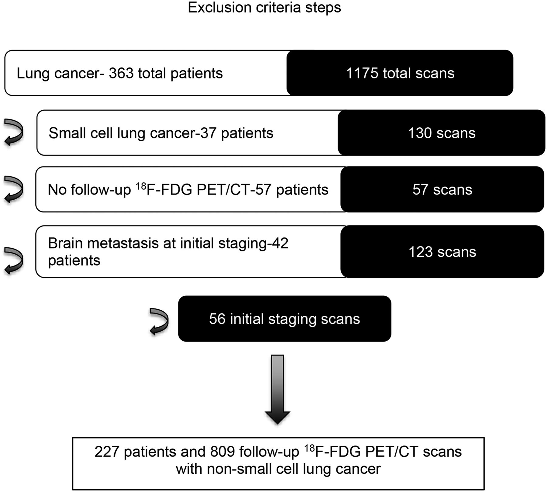
    A total of 1,175 18F-FDG PET/CT examinations in 363 patients were reviewed. Exclusion criteria included brain metastases on initial staging, histological subtype of small-cell lung cancer, and no follow up 18F-FDG PET/CT examinations. After we applied our exclusion criteria as well as eliminated all initial staging scans (only follow-up scans included), a total of 809 follow-up 18F-FDG PET/CT scans in 227 patients were included in the final analysis.

  • FIGURE 2.
    • Download figure
    • Open in new tab
    • Download powerpoint
    FIGURE 2.

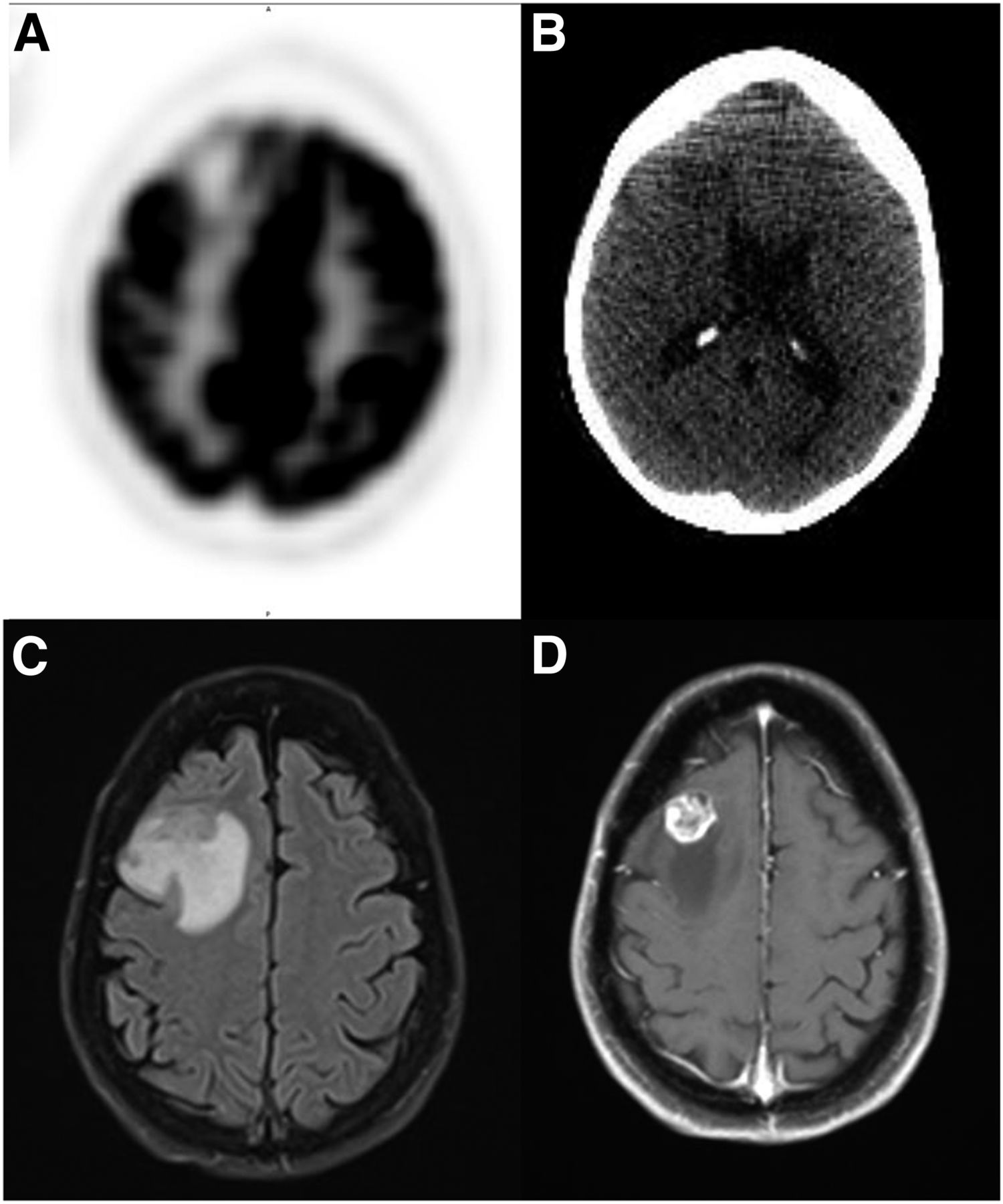
    Axial 18F-FDG PET image of brain demonstrates focal hypometabolism in the right frontal lobe (A) corresponding to vasogenic edema on low-dose CT images (B). Subsequent axial T2-FLAIR and contrast-enhanced MR images confirm metastasis with adjacent vasogenic edema (C and D).

  • FIGURE 3.
    • Download figure
    • Open in new tab
    • Download powerpoint
    FIGURE 3.

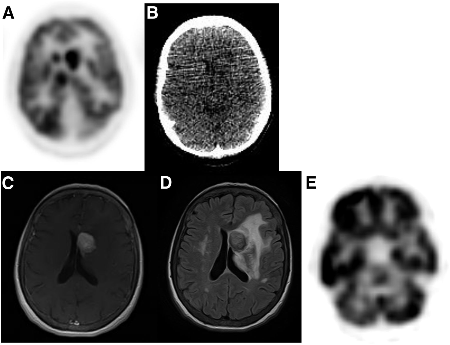
    (A and B) Axial 18F-FDG PET and low-dose CT images of brain demonstrate hypermetabolic lesion in left caudate. (C) Subsequent contrast-enhanced axial MRI of brain confirmed metastasis. (D) Axial T2 FLAIR imaging demonstrates surrounding vasogenic edema. (E) PET also shows crossed-cerebellar diaschisis with decreased activity in contralateral right cerebellum.

Tables

  • Figures
    • View popup
    TABLE 1

    Patients with Incidentally Found Brain Metastasis

    PatientAge (y)SexCancer typeTime until incidental brain metastasis (mo)No. of metastases seen on 18F-FDG PET/CTNo. of metastases seen on subsequent MRI
    Patient 160MaleAdenosquamous cell carcinoma2711
    Patient 260FemaleAdenocarcinoma1911
    Patient 367FemaleSquamous cell carcinoma53MultipleMultiple
    Patient 477FemaleAdenocarcinoma6611
    Patient 577MaleAdenocarcinoma1511
    Average6836
PreviousNext
Back to top

In this issue

Journal of Nuclear Medicine Technology: 45 (3)
Journal of Nuclear Medicine Technology
Vol. 45, Issue 3
September 1, 2017
  • Table of Contents
  • About the Cover
  • Index by author
Print
Download PDF
Article Alerts
Sign In to Email Alerts with your Email Address
Email Article

Thank you for your interest in spreading the word on Journal of Nuclear Medicine Technology.

NOTE: We only request your email address so that the person you are recommending the page to knows that you wanted them to see it, and that it is not junk mail. We do not capture any email address.

Enter multiple addresses on separate lines or separate them with commas.
Incidence of Brain Metastases on Follow-up 18F-FDG PET/CT Scans of Non–Small Cell Lung Cancer Patients: Should We Include the Brain?
(Your Name) has sent you a message from Journal of Nuclear Medicine Technology
(Your Name) thought you would like to see the Journal of Nuclear Medicine Technology web site.
Citation Tools
Incidence of Brain Metastases on Follow-up 18F-FDG PET/CT Scans of Non–Small Cell Lung Cancer Patients: Should We Include the Brain?
Emily S. Nia, Linda L. Garland, Naghmehossadat Eshghi, Benjamin B. Nia, Ryan J. Avery, Phillip H. Kuo
Journal of Nuclear Medicine Technology Sep 2017, 45 (3) 193-197; DOI: 10.2967/jnmt.117.194571

Citation Manager Formats

  • BibTeX
  • Bookends
  • EasyBib
  • EndNote (tagged)
  • EndNote 8 (xml)
  • Medlars
  • Mendeley
  • Papers
  • RefWorks Tagged
  • Ref Manager
  • RIS
  • Zotero
Share
Incidence of Brain Metastases on Follow-up 18F-FDG PET/CT Scans of Non–Small Cell Lung Cancer Patients: Should We Include the Brain?
Emily S. Nia, Linda L. Garland, Naghmehossadat Eshghi, Benjamin B. Nia, Ryan J. Avery, Phillip H. Kuo
Journal of Nuclear Medicine Technology Sep 2017, 45 (3) 193-197; DOI: 10.2967/jnmt.117.194571
Twitter logo Facebook logo LinkedIn logo Mendeley logo
  • Tweet Widget
  • Facebook Like
  • Google Plus One
Bookmark this article

Jump to section

  • Article
    • Abstract
    • MATERIALS AND METHODS
    • RESULTS
    • DISCUSSION
    • CONCLUSION
    • DISCLOSURE
    • Footnotes
    • REFERENCES
  • Figures & Data
  • Info & Metrics
  • PDF

Related Articles

  • No related articles found.
  • PubMed
  • Google Scholar

Cited By...

  • No citing articles found.
  • Google Scholar

More in this TOC Section

  • Early 10-Minute Postinjection [18F]F-FAPI-42 uEXPLORER Total-Body PET/CT Scanning Protocol for Staging Lung Cancer Using HYPER Iterative Reconstruction
  • Single- Versus Dual-Time-Point Imaging for Transthyretin Cardiac Amyloid Using 99mTc-Pyrophosphate
  • Does Arthrography Improve Accuracy of SPECT/CT for Diagnosis of Aseptic Loosening in Patients with Painful Knee Arthroplasty: A Systematic Review and Metaanalysis
Show more Imaging

Similar Articles

Keywords

  • 18F-FDG PET/CT
  • non-small cell lung cancer
  • incidental
  • brain metastasis
  • brain MRI
SNMMI

© 2025 SNMMI

Powered by HighWire