


Article Figures & Data
Figures
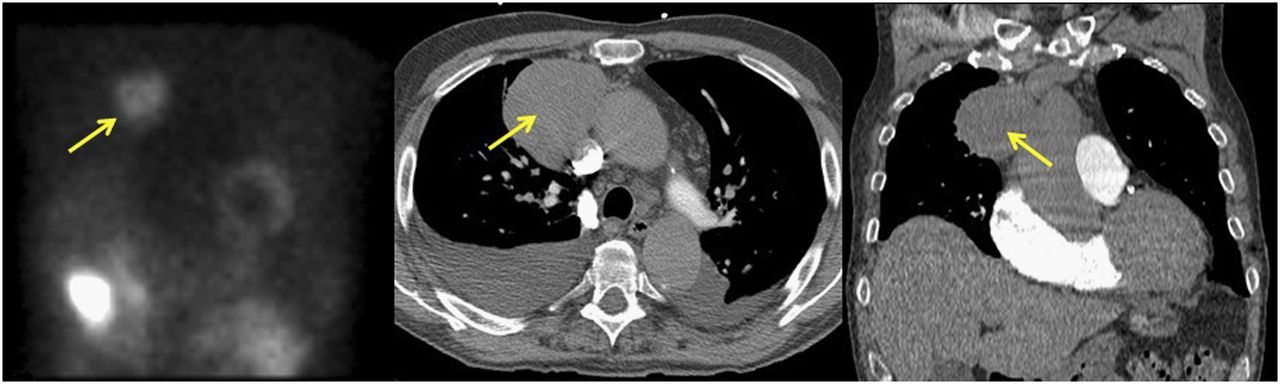
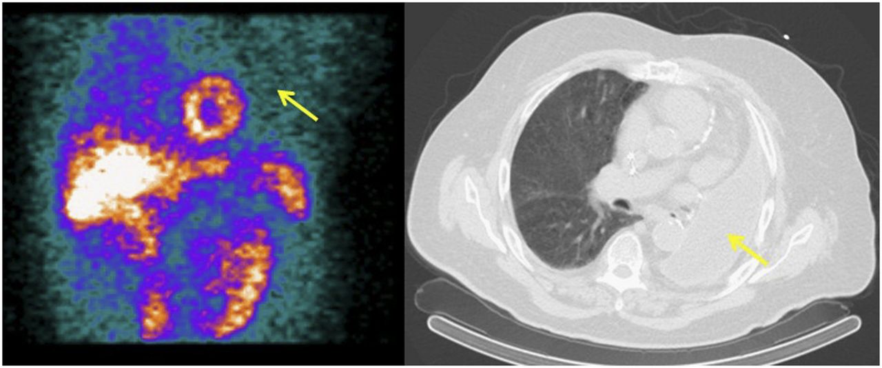
- FIGURE 1.
Patient who presented with chest pain and shortness of breath. Raw SPECT image shows small area of photopenia surrounding heart (arrows, left). CT image shows small pericardial effusion (arrows, right).
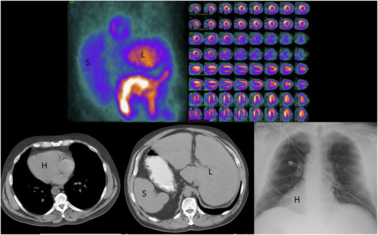
- FIGURE 2.
Patient with situs inversus who presented with chest pain. Raw SPECT image (top left) shows enlarged spleen (S) in right abdomen, and liver in left abdomen (L). Processed images (top right) show lateral wall and septum flipped from normal position as evidenced by short septum; finding is most prominent in horizontal long axis (last 2 rows of processed images). CT images (bottom left and middle) and radiograph (bottom right) show that heart (H) and major organs mirror their normal positions.
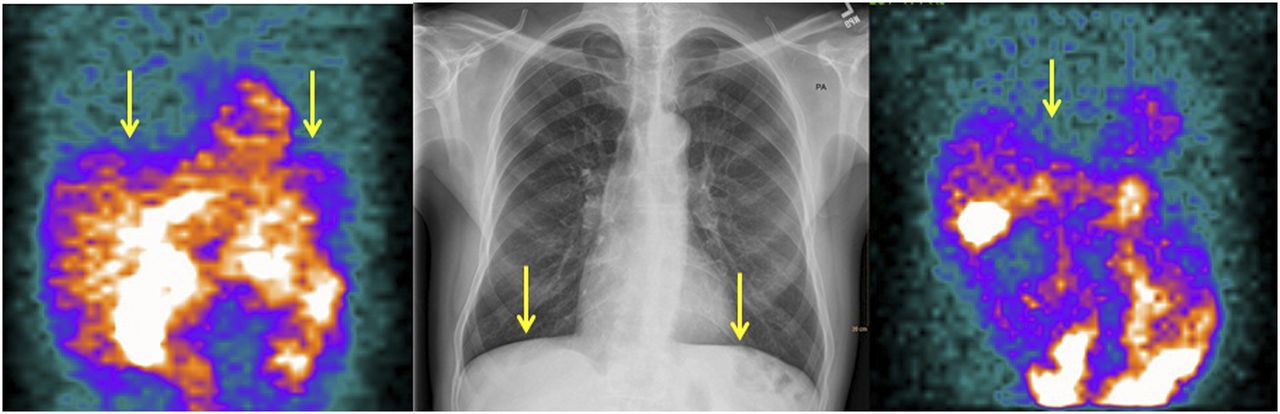
- FIGURE 3.
Patient with chronic obstructive pulmonary disease. Flattening of diaphragm is seen on raw SPECT stress (arrows, left) and rest (arrow, right) images, as well as on radiograph (arrows, center).
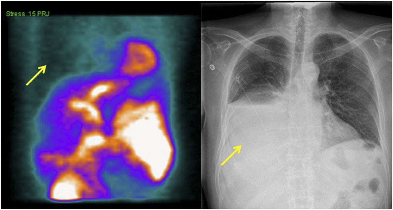
- FIGURE 4.
Patient with pleural effusion. Relative photopenia in right chest cavity on raw SPECT image (arrow, left) correlates with large pleural effusion on chest radiograph (arrow, right).
- FIGURE 5.
Lung cancer patient who was being evaluated after undergoing left pneumonectomy. Reduced counts in left chest on raw SPECT image (arrow, left) correlate with pneumonectomy changes on CT image (arrow, right).
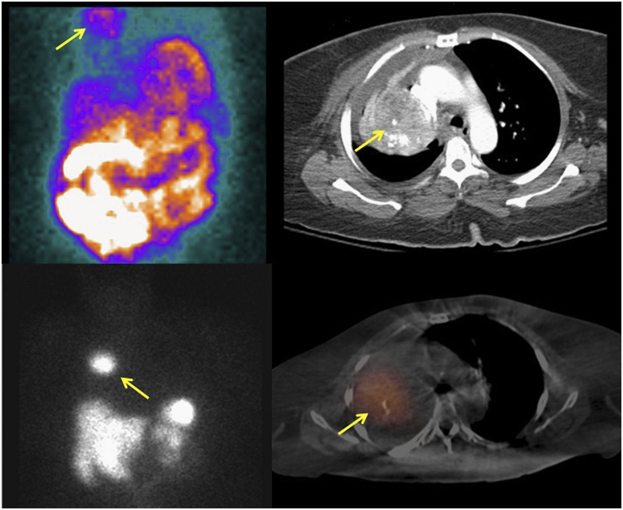
- FIGURE 6.
Patient with history of carcinoid tumor who presented with chest pain. Raw SPECT image shows mass in right upper chest (arrow, top left). Contrast-enhanced CT image shows heterogeneously enhancing mass in right lung (arrow, top right). 111In-pentetreotide SPECT image shows right-chest mass (arrow, bottom left), which fuses with right-lung mass on SPECT/CT image (arrow, bottom right).
- FIGURE 7.
Patient who presented with chest pain. Raw SPECT image shows abnormal tracer accumulation in right chest (arrow, left). CT image demonstrates, in posterior right hemidiaphragm, large hernia containing nearly all of stomach (arrow, right).
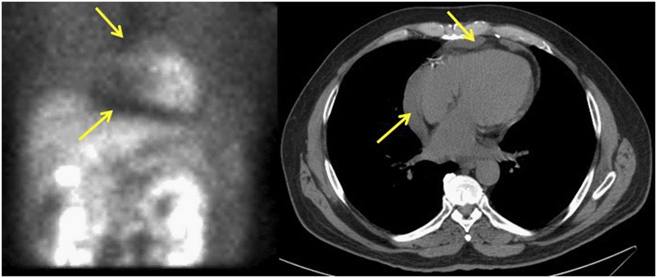
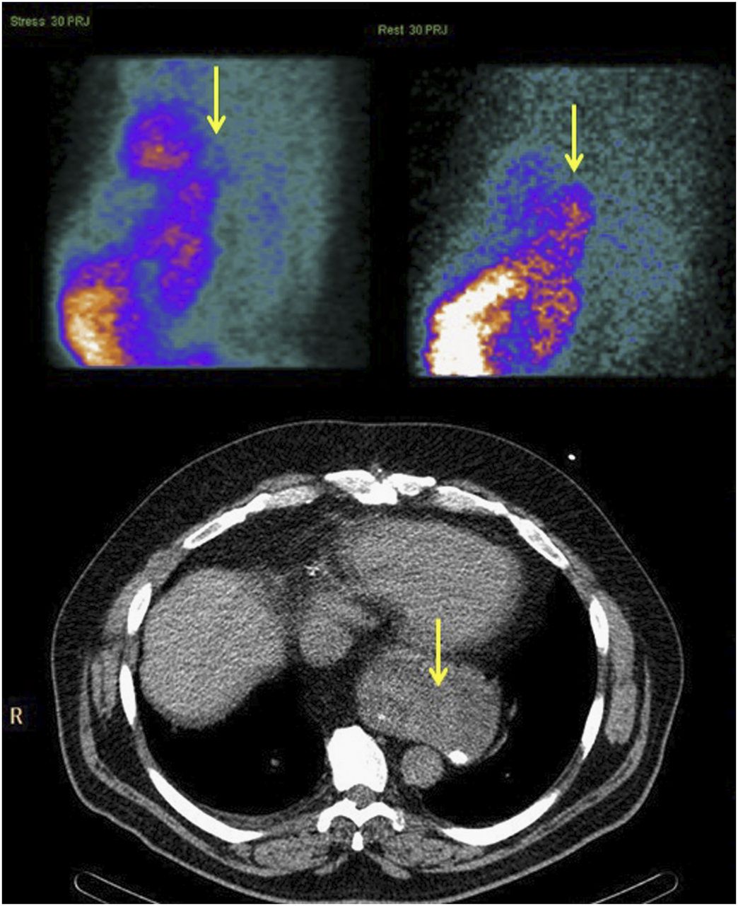
- FIGURE 8.
Patient with abnormal tracer accumulation posterior to heart on raw SPECT images, more pronounced at rest than at stress (arrows, top). CT image shows moderate hiatal hernia (arrow, bottom).
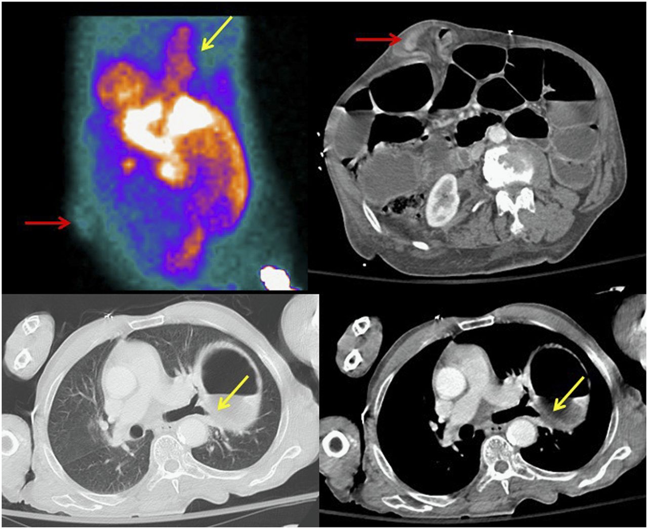
- FIGURE 9.
Patient who presented with chest pain. Raw SPECT image shows focus of uptake in anterior abdominal wall (red arrow, top left) and linear uptake in chest (yellow arrow, top left). CT images show ventral hernia (arrow, top right), as well as large hiatal hernia (yellow arrows, bottom).
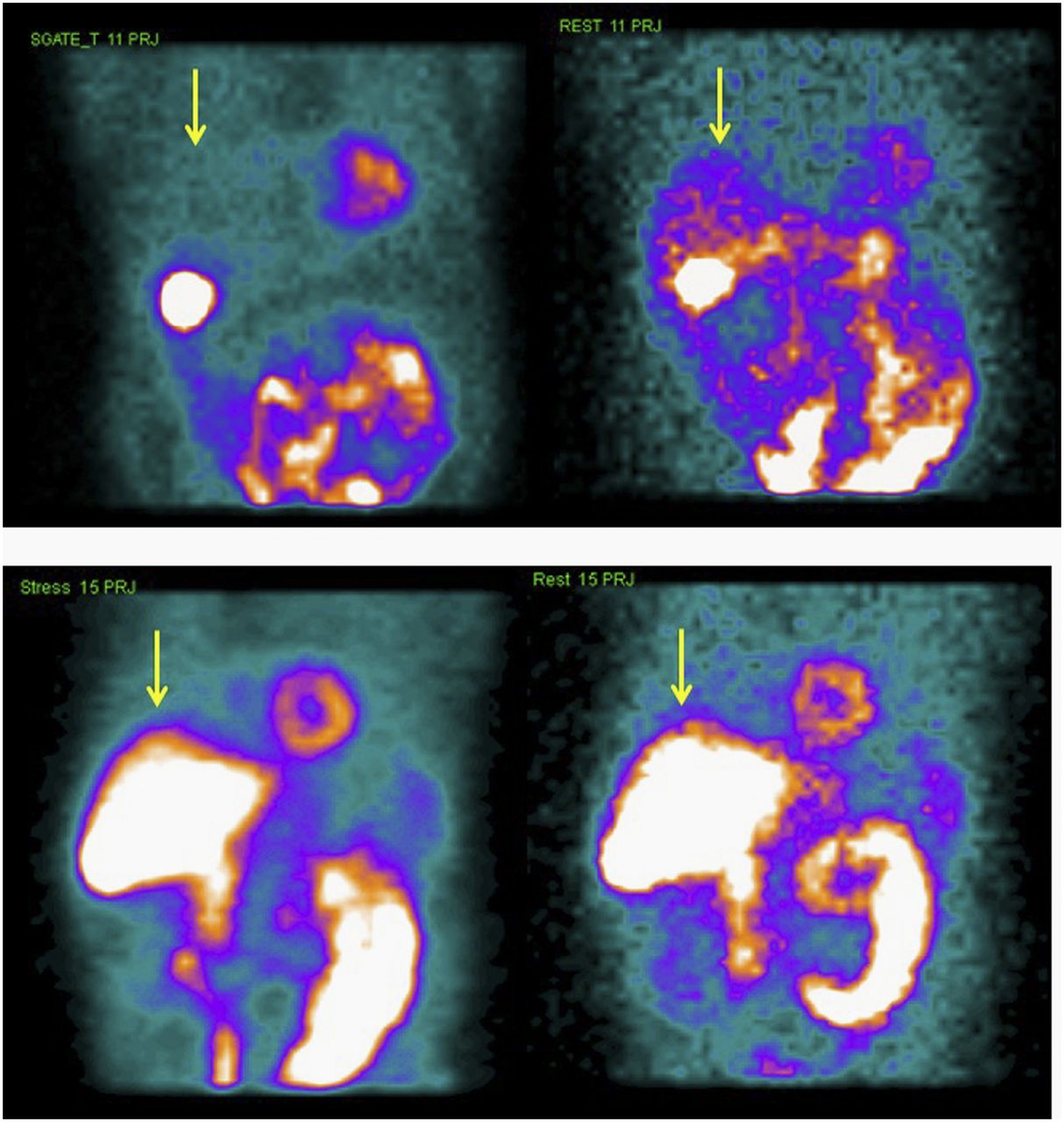
- FIGURE 10.
Patient who presented with chest pain. Raw SPECT images from treadmill stress test show minimal liver activity and greater distal bowel activity at stress (arrow, top left) than at rest (arrow, top right). Raw SPECT images from pharmacologic stress test show intense liver and bowel activity both at stress (arrow, bottom left) and at rest (arrow, bottom right).
- FIGURE 11.
Patient who presented with chest pain. Raw SPECT image shows round area of increased uptake in right upper chest (arrow, left). Right-mediastinum mass is seen on axial CT image (arrow, center) and coronal CT image (arrow, right). On biopsy, mass was found to be thymoma.
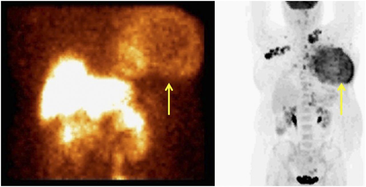
- FIGURE 12.
Female patient with recently diagnosed breast cancer who presented for preoperative risk assessment. Raw SPECT image shows large mass in left breast (arrow, left). Maximum-intensity-projection PET/CT image demonstrates large left-breast mass (arrow, right) with multiple small metastatic lymph nodes.
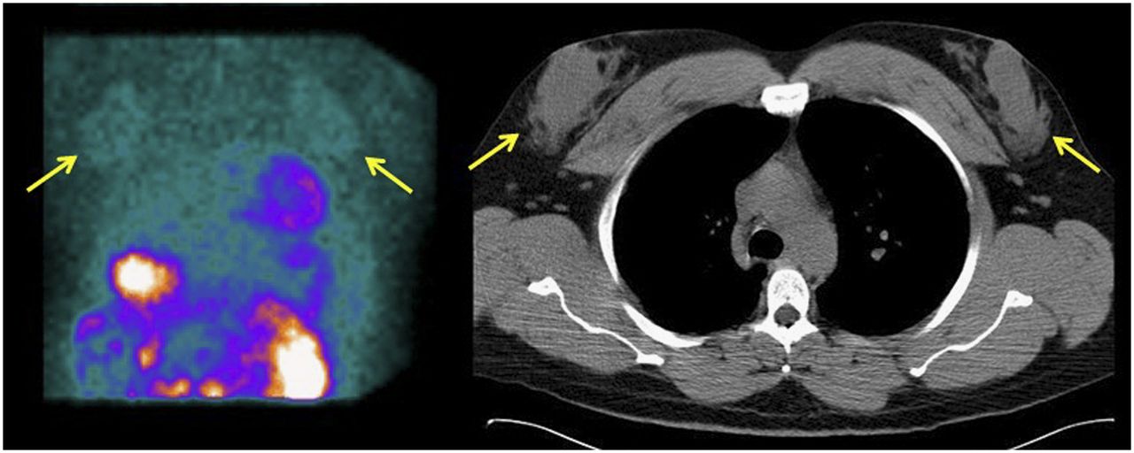
- FIGURE 13.
Male patient with end-stage liver disease who presented for pretransplantation evaluation. Raw SPECT image shows round areas of mildly increased uptake in chest (arrows, left) correlating with gynecomastia changes on CT image (arrows, right).
- FIGURE 14.
Female patient who presented with chest pain. Raw SPECT images show small areas of increased uptake bilaterally in chest (arrows). Clinical examination found abscesses bilaterally in lower breasts.
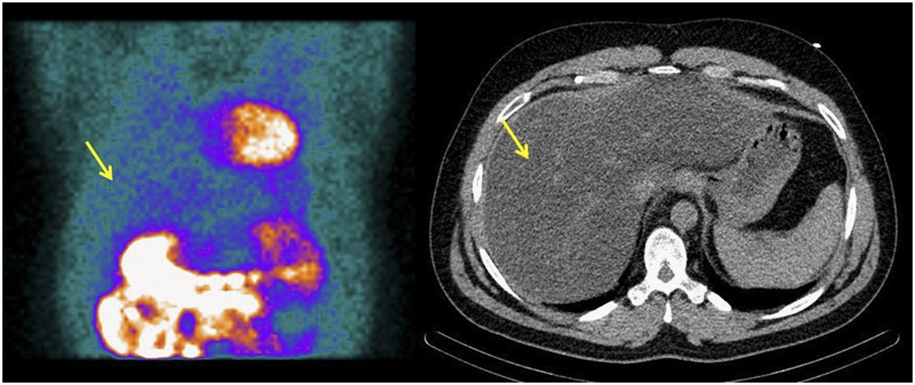
- FIGURE 15.
Patient with fatty liver disease. Raw SPECT image shows minimal uptake in liver (arrow, left). CT image shows diffuse fatty changes throughout liver (arrow, right).
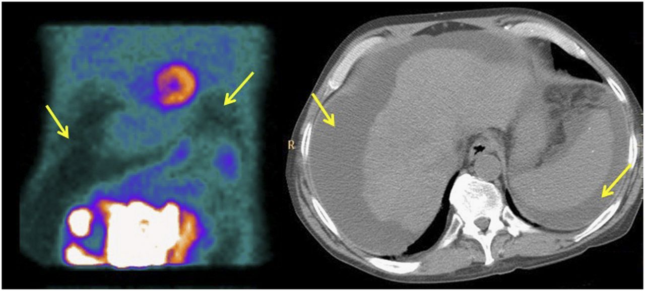
- FIGURE 16.
Patient with end-stage liver disease who presented for pretransplantation evaluation. Raw SPECT image shows areas of photopenia throughout abdomen (arrows, left). CT image shows diffuse ascites surrounding liver and spleen (arrows, right).
- FIGURE 17.
Patient with end-stage renal disease who presented for preoperative evaluation. Raw SPECT image demonstrates relative photopenia in liver (arrow, left). CT image shows polycystic hepatic disease (arrow, right) and renal disease.
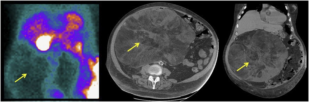
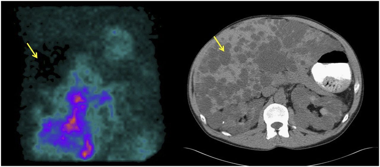
- FIGURE 18.
Patient with large abdominal mass who presented for preoperative evaluation. Raw SPECT image shows large area of photopenia within abdomen (arrow, left). CT images show large intraabdominal fatty mass (arrows, middle and right), which biopsy found to be well-differentiated liposarcoma.
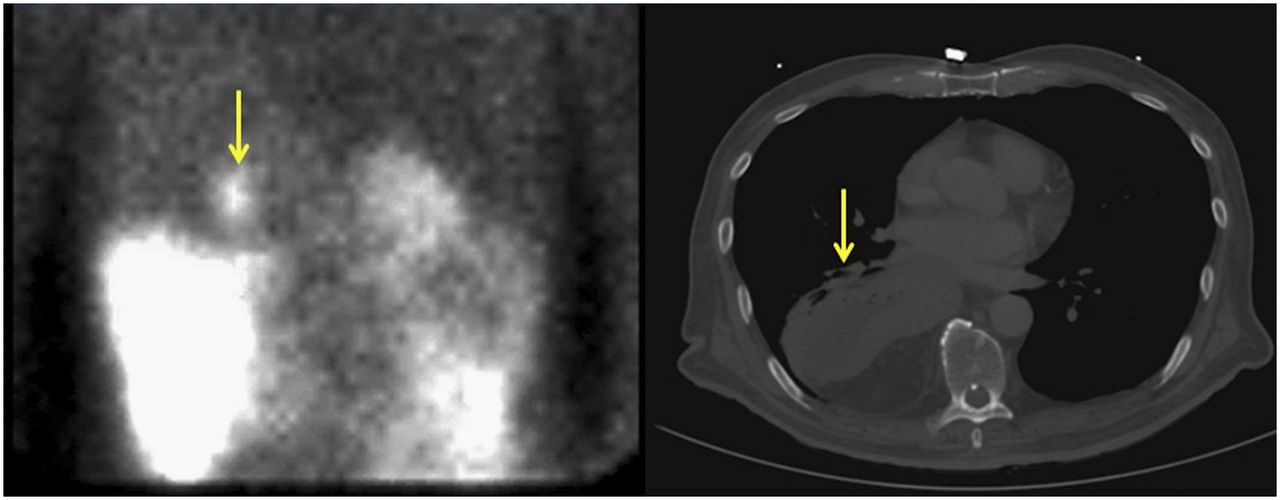
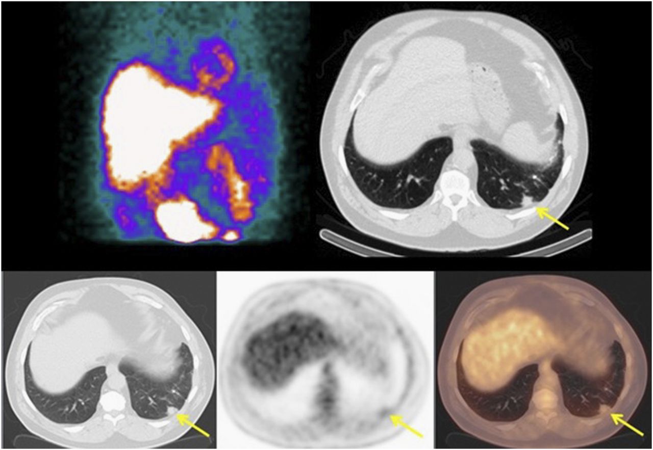
- FIGURE 19.
Patient who presented with chest pain. Raw SPECT image is unremarkable (top left). CT image obtained for attenuation correction shows subcentimeter spiculated nodule in left lung base (arrow, top right). PET/CT image shows mild 18F-FDG uptake in nodule (arrows, bottom row), with SUVmax of 2.5. Biopsy was performed and found adenocarcinoma.
- FIGURE 20.
Patient with history of achalasia who presented with chest pain. Raw SPECT image is unremarkable (left). CT images obtained for attenuation correction show significant esophageal dilation (arrows, middle and right), consistent with patient history.
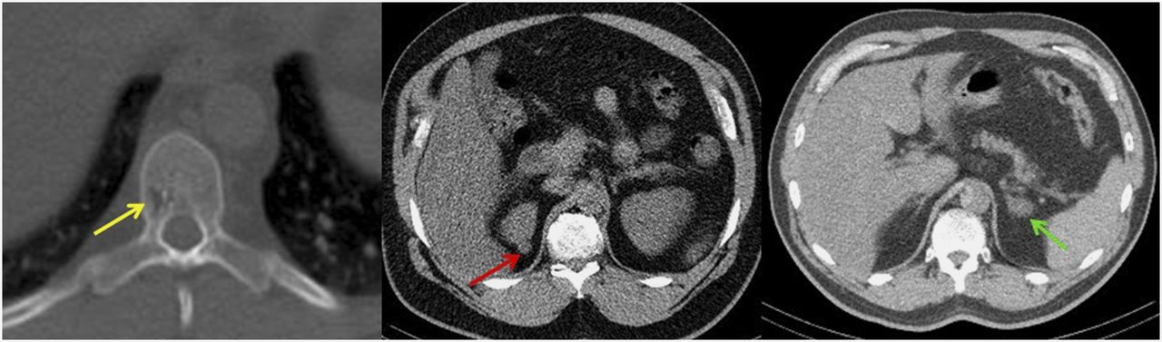
- FIGURE 21.
Various subtle abnormalities on CT images obtained for attenuation correction: small hemangioma in body of T12 (yellow arrow), subcentimeter exophytic isodensity in right kidney (red arrow), and small adenoma in left adrenal gland (green arrow).


