


Article Figures & Data
Figures
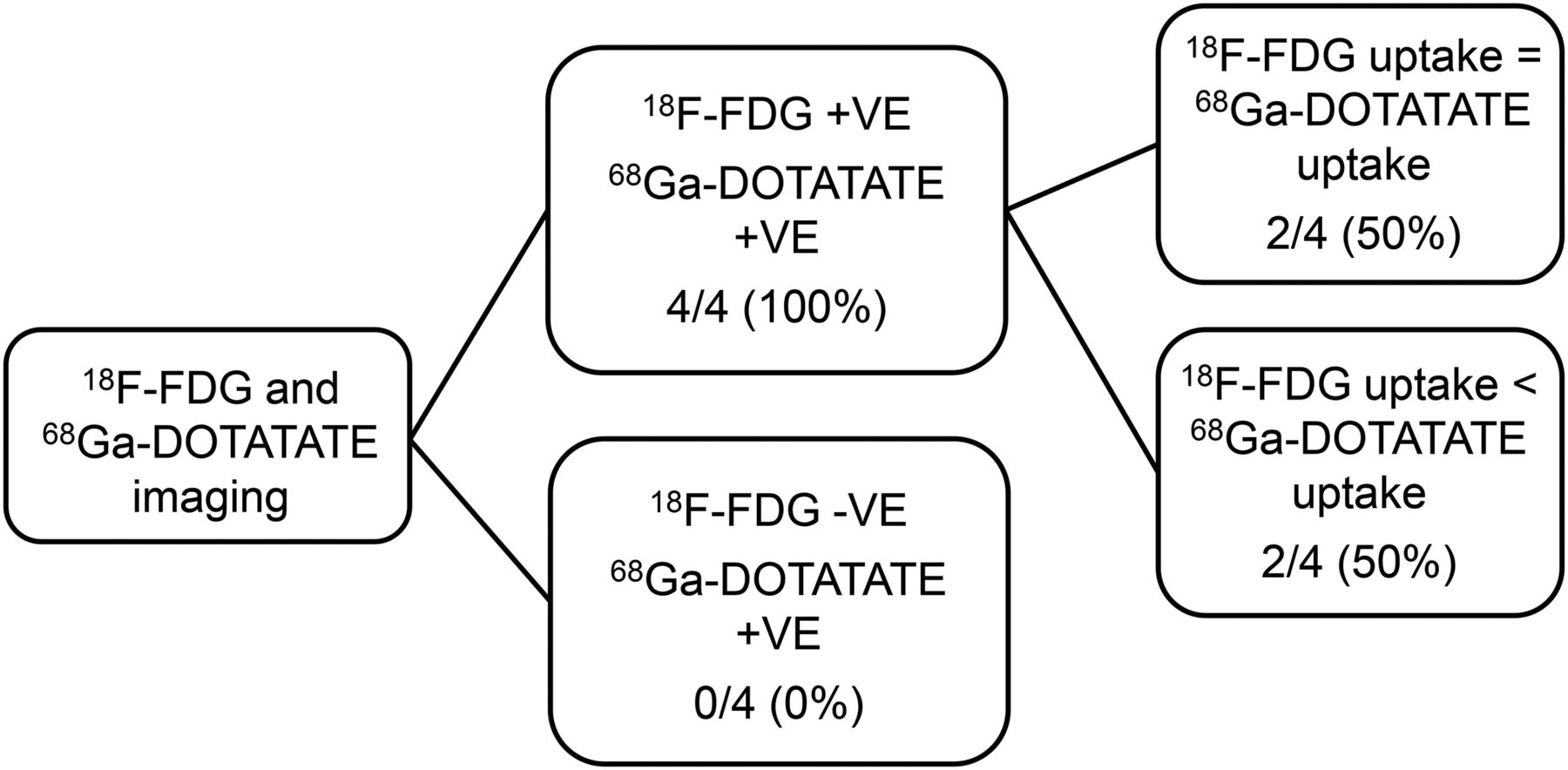
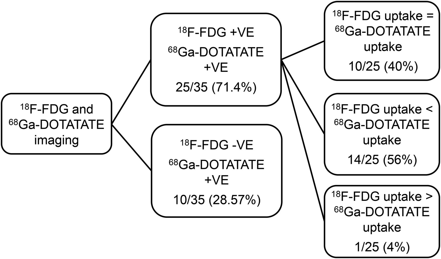
- FIGURE 1.
Flowchart demonstrating patient-specific analysis observed on 68Ga-DOTATATE and 18F-FDG PET/CT in group I patients (MIB-1 index, 1%–5%). +ve = positive; −ve = negative.
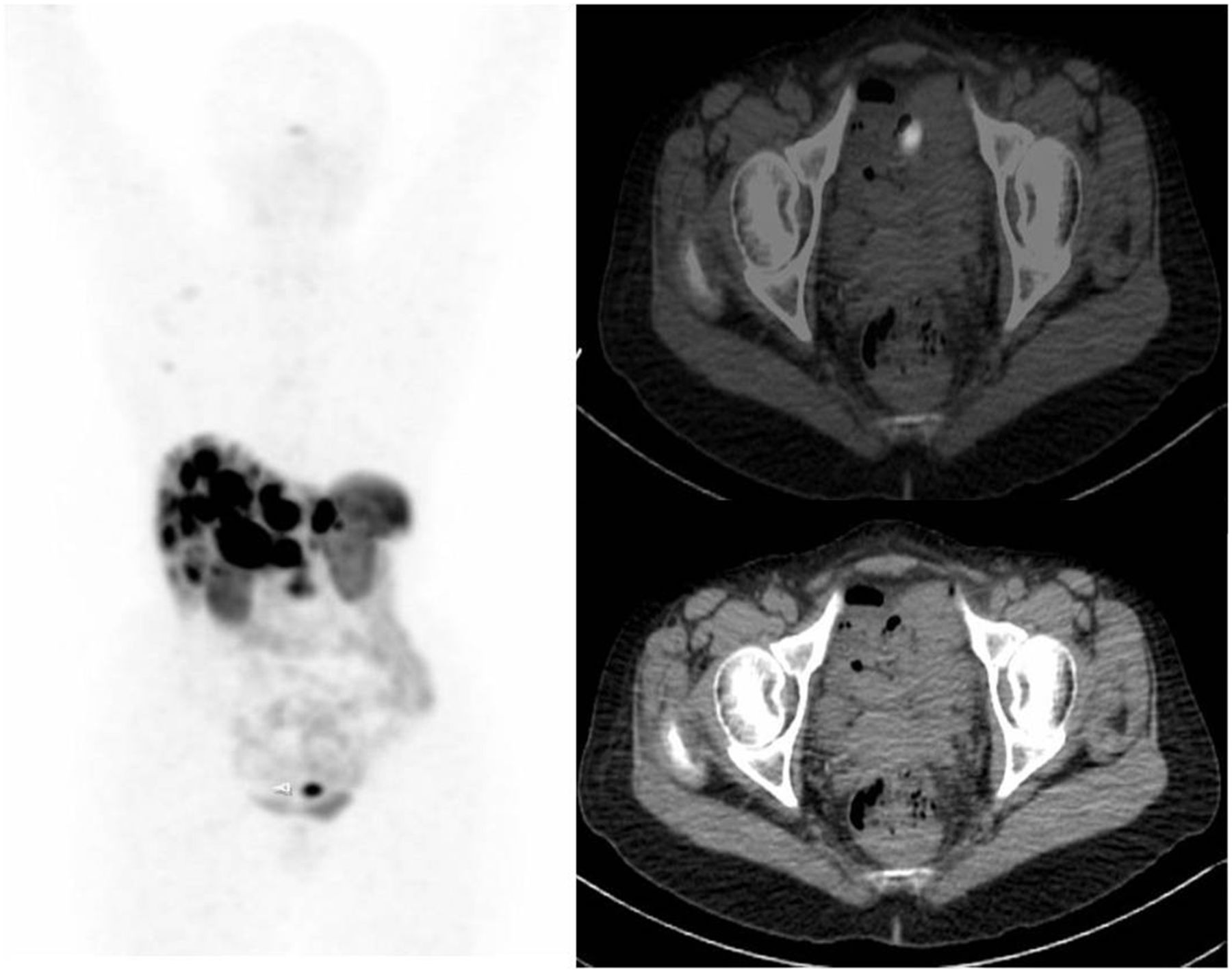
- FIGURE 2.
A 56-y-old woman with liver biopsy suggestive of metastatic NET of liver (MIB-1 index, <1%). Primary was undetected by conventional imaging. 68Ga-DOTATATE PET/CT scan showed multiple metastatic liver lesions and focal tracer concentration in the pelvic ileum. Final diagnosis was ileal NET with bilobar hepatic metastases. A color version of this figure is available as a supplemental file at http://tech.snmjournals.org.
- FIGURE 3.
Decision tree type ramification analysis to assess relative performance and uptake intensity in metastatic lesions by 68Ga-DOTATATE and 18F-FDG PET/CT in group II (MIB-1/Ki-67 index, 6%–10%). +ve = positive; −ve = negative.
- FIGURE 4.
Decision tree type ramification analysis to assess relative performance and uptake intensity in metastatic lesions by 68Ga-DOTATATE and 18F-FDG PET/CT in group III. +ve = positive; −ve = negative.
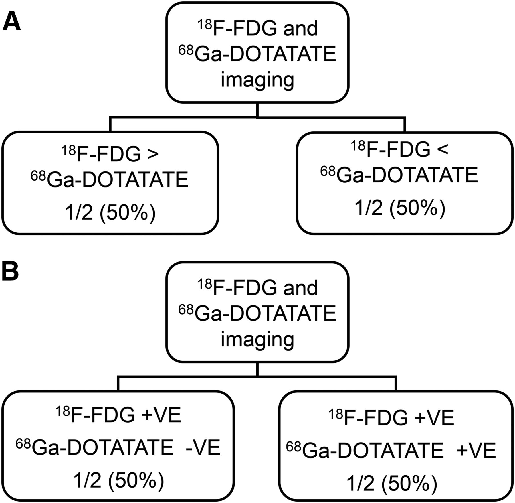
- FIGURE 5.
(A) Flowchart of assessment of relative positivity and uptake intensity in metastatic lesions by 68Ga-DOTATATE and 18F-FDG PET/CT in group IV. (B) Flowchart of assessment of relative positivity and uptake intensity in metastatic lesions by 68Ga-DOTATATE and 18F-FDG PET/CT in group V. +ve = positive; −ve = negative.
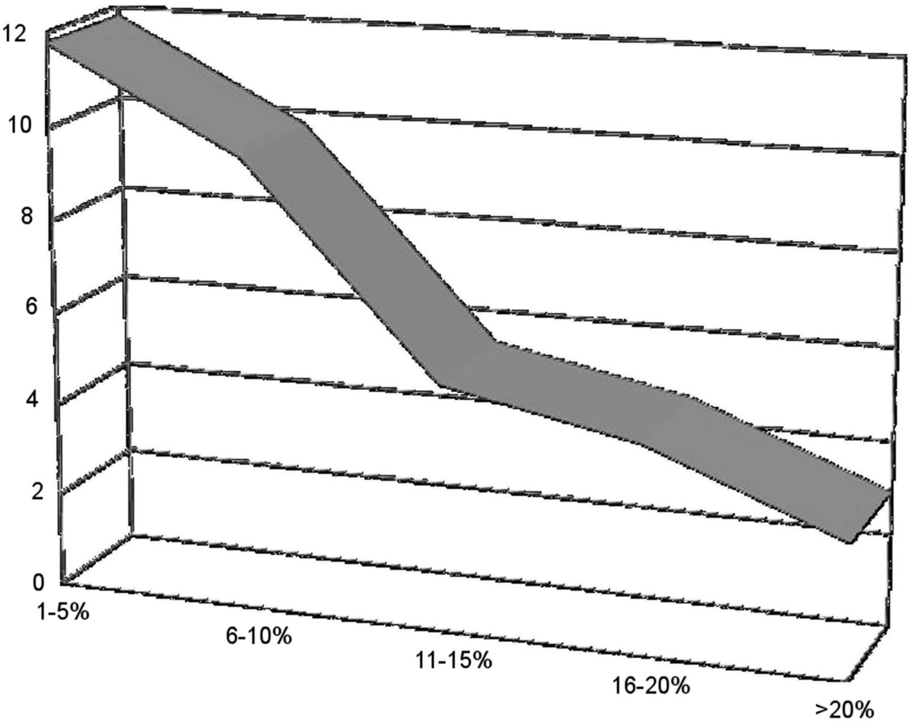
- FIGURE 6.
Graphical representation of variation in 68Ga-DOTATATE–to–18F-FDG uptake ratio in relation to increasing tumor proliferation (MIB-1) index.
- FIGURE 7.
(A) A 56-y-old woman, diagnosed as having metastatic NET. ceCT showed multiple hypodense liver lesions, abdominal nodes, multiple skeletal lesions, pararectal node, and right adnexal mass. Irregular wall thickening was seen in rectum, but was missed due to relative nonenhancement of lesion. In view of histopathology, patient was referred to us for 68Ga-DOTATATE scanning. (B) 68Ga-DOTATATE showed increased uptake in rectal wall, left adnexal mass, pararectal node, and multiple liver and skeletal lesions, thus confirming that this was a case of metastatic NET with rectal primary. A color version of this figure is available as a supplemental file at http://tech.snmjournals.org. (C) Corresponding images of MRI pelvis: confirming right adnexal mass and rectal primary.


Tables
Patient characteristic Value No. of patients presenting with these sites Age (y) Range 22–74 Mean 48 Sex (n = 51) Males 25 Females 26 Metastatic sites Liver 48 Lung/mediastinum 03 Skeletal 12 Abdominal nodes 25 Pericardium 01 Others (soft tissue) 03 - TABLE 2
Division of Patients into Groups of 5 Based on MIB-1/Ki-67–Labeling Index and Depiction of Number in Each Category
Group MIB-1/Ki-67 index No. of patients I 1%–5% 35 II 6%–10% 08 III 11%–15% 04 IV 16%–20% 02 V >20% 02 - TABLE 3
Semiquantitative Analysis of Uptake Intensity (SUVmax) in Cases Demonstrating Uptake of Both Tracers in Metastases
Group MIB-1/Ki-67 index 68Ga-DOTATATE mean SUVmax 18F-FDG mean SUVmax 68Ga-DOTATATE–to–18F-FDG uptake ratio I 1%–5% 43.5 3.66 11.89 II 6%–10% 38.90 3.98 9.77 III 11%–15% 29.80 5.60 5.32 IV 16%–20% 24.36 5.88 4.16 V > 20% 22.54 7.53 2.99 - TABLE 4
Performance of 68Ga-DOTATATE in Detecting Primary in Patients of CUP-NET by Conventional Imaging Modalities
Introduction N No. of cases in which primary was detected by 68Ga-DOTATATE PET/CT and not by any other conventional imaging modality 31 No. of cases in which primary could not be detected by 68Ga-DOTATATE PET/CT 20 Percentage sensitivity of 68Ga-DOTATATE PET/CT in detecting in CUP-NET (%) 60.78% Sites of primary Pancreas 10 (31.03) Duodenum 05 (17.24) Mesentery 05 (17.24) Ileum 03 (10.34) Jejunum 02 (06.89) Lung 02 (06.89) Stomach 01 (03.44) Rectum 02 (06.45) Data in parentheses are percentages.
- TABLE 5
Scan Positivity and Semiquantitative Analysis of Uptake intensity (SUVmax) in Cases Demonstrating Uptake of Both Tracers in Detected Sites of Primary: Comparison with MIB-1–Labeling Index Stratified Group
Group MIB-1/Ki-67 index 68Ga-DOTATATE mean SUVmax 18F-FDG mean SUVmax Sensitivity in detecting primary I 1%–5% 22.68 2.86 65.7% II 6%–10% 10.33 – 37.5% III 11%–15% 11.5 4.58 50% IV 16%–20% 20.22 5.95 50% V >20% 16.83 9.58 50% - TABLE 6
Comparison of All Imaging Modalities in Detecting Number of Lesions and Calculation of Their Sensitivity
Group Ultrasonography CT/ceCT 18F-FDG 68Ga-DOTATATE Total lesions I 37 (41.57%) 48 (53.93%) 40 (44.94%) 87 (97.75%) 89 II 9 (56.25%) 13 (81.25%) 9 (56.25%) 14 (87.5%) 16 III 4 (33.33%) 8 (66.66%) 9 (75%) 12 (100%) 12 IV 3 (60%) 3 (60%) 5 (100%) 5 (100%) 5 V 2 (33.33%) 2 (33.33%) 3 (50%) 4 (66.67%) 6 Total 55 (42.96%) 74 (57.81%) 66 (51.56%) 124 (96.87%) 128
Supplemental Data
Files in this Data Supplement:


