Abstract
Although the use of 13N-ammonia and 18F-FDG PET shows great promise as a tool for diagnosing heart involvement in inflammatory diseases, it can be equally powerful for following disease progression and treatment outcome. We describe a case in which 18F-FDG PET was effective in following up the treatment outcome of lupus myocarditis.
Although uncommon, lupus myocarditis carries a prevalence of 8%–25% (1). The condition is usually diagnosed through a combination of physical examination, electrocardiography and echocardiography. Unfortunately, the findings for lupus myocarditis are nonspecific and are the same as those for some other pathologic processes, such as drug-induced cardiomyopathy (antimalarial drugs, cyclophosphamide, phenothiazines), postpartum cardiomyopathy, and uremia. Under those circumstances, a myocardial biopsy is needed to confirm the cause of myocarditis (2). This case study demonstrates that 18F-FDG PET can be used to evaluate lupus myocarditis.
CASE REPORT
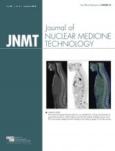
A 21-y-old woman presented with a history of worsening lupus myocarditis. Scanning with 13N-ammonia and 18F-FDG PET was ordered. The patient was instructed to follow a high-fat low-carbohydrate diet the day before undergoing imaging but did not have to fast overnight. One hour after injection of the tracers, multiple metabolic tomographic images of the myocardium were obtained. The rest 13N-ammonia perfusion images demonstrated defects in the basal inferior and basal inferior lateral regions, which also showed a corresponding increase in 18F-FDG uptake consistent with active infiltration of the myocardium (Figs. 1 and 2). The patient began treatment with high-dose prednisone, azathioprine, and colchicine. A follow-up myocardial PET study 3 mo later demonstrated findings similar to those of the prior study, with an ejection fraction of 33% (Figs. 2 and 3). These findings indicate a poor response to treatment. As a result, the patient’s treatment regimen was changed to mycophenolate mofetil (Cellcept; Genentech).
Rest 13N-ammonia perfusion images demonstrating defects in basal inferior region and basal inferior lateral region, with corresponding increase in 18F-FDG uptake.
Stress and rest 13N-ammonia perfusion images demonstrating decreased uptake in basal inferior lateral region, with corresponding increase in 18F-FDG uptake. Both stress and rest images also show severe hypoperfusion extending from mid inferior lateral region to mid inferior region. Lack of correspondingly enhanced 18F-FDG uptake suggests that the finding represents fibrosis or scar formation. HLA = horizontal long axis; SA = short axis; VLA = vertical long axis.
(A) Rest 13N-ammonia polar plot image before treatment. (B) Rest and stress 13N-ammonia polar plot images after treatment. (C) 18F-FDG polar plot image after treatment.
DISCUSSION
A literature search for common lupus myocarditis findings on 18F-FDG PET yielded no results; thus, it is hoped that this case study will popularize an underutilized diagnostic modality for this rare but devastating disease.
To evaluate for inflammatory processes in the heart, such as cardiac sarcoidosis and lupus myocarditis, 13N-ammonia or 82Rb radiotracers with 18F-FDG can be used (3). Cardiac MRI is a new, noninvasive method that has high spatial resolution; allows evaluation of morphology, function, and tissue characteristics; and requires no radiation (4). PET/MRI combines the strengths of both PET and MRI. PET is a noninvasive imaging technique that can detect active metabolic processes with high sensitivity. Hence, a combination with MRI using gadolinium–diethylenetriamine pentaacetic acid (providing high spatial resolution and lack of radiation exposure) seems reasonable (4). In the absence of coronary artery disease, perfusion defects seen on a PET scan may strongly point to an inflammatory condition such as cardiac sarcoidosis or lupus myocarditis (3). In healthy myocardium, PET images show normal perfusion and normal 18F-FDG uptake. However, inflamed myocardium shows decreased perfusion and increased 18F-FDG uptake (3). In our case, the results indicated a poor response to treatment, which corresponded to the unchanged clinical decline. Therefore, the treatment regimen was changed, and the effectiveness of the new treatment will be monitored by follow-up PET imaging for a few months.
Another option to distinguish between lupus myocarditis and other cardiomyopathies is endomyocardial biopsy.
CONCLUSION
Although the use of 13N-ammonia and 18F-FDG PET scanning shows great promise as a tool for diagnosing heart involvement in inflammatory diseases, it can be equally powerful for following disease progression and treatment outcome. In our case, we demonstrated that 18F-FDG PET can be effectively used to follow up the outcome of treatment for lupus myocarditis.
DISCLOSURE
No potential conflict of interest relevant to this article was reported.
Footnotes
Published online Jan. 14, 2016.
- Received for publication August 16, 2015.
- Accepted for publication November 10, 2015.


