Abstract
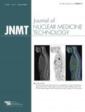
We present a proven case of pulmonary inflammatory pseudotumor that illustrates the 18F-FDG PET/CT imaging features of this “great mimicker of malignancy” both at initial diagnosis and during early monitoring of corticosteroid therapy, which is advocated as the first-line treatment. Although the patient showed some symptomatic response to corticosteroid therapy, as well as a modest reduction in SUVmax, complete surgical resection was eventually needed in view of the relative nonresponse. 18F-FDG avidity in untreated cases of pulmonary inflammatory pseudotumor has been quite characteristic and may potentially be used to evaluate early response of this IgG4-related disease to nonsurgical treatment and to detect residual disease or recurrence after therapeutic intervention.
Pulmonary inflammatory pseudotumor is a rare disease characterized by nonneoplastic proliferation of inflammatory cells. It is a close mimic of lung malignancy (1) and sometimes demonstrates an excellent response to corticosteroid therapy (2), the first-line agent usually applied to induce remission of IgG4-related diseases. Application of 18F-FDG PET/CT has been described in the literature only in recent years (1).
CASE REPORT
The patient, a 12-y-old boy, presented with symptoms of dry cough and right-sided chest pain of an 8-mo duration. A chest radiograph (Fig. 1) showed a mass in the right midzone. Contrast-enhanced CT of the chest (Fig. 2) showed a heterogeneously enhancing mass with calcification in the right middle lobe and obstruction of the right-middle-lobe bronchus (Fig. 3). CT-guided biopsy of the mass showed an immunoglobulin (IgG4)-expressing plasma cell infiltrate consistent with inflammatory pseudotumor. For further confirmation, serum IgG4 levels were determined and found to be significantly elevated, at 12.1 g/L (reference range, 0.049–1.985 g/L). 18F-FDG PET/CT (Fig. 4A) showed a metabolically active soft-tissue lesion with intense 18F-FDG uptake (SUVmax, 7.7) and coarse calcification in the right middle lobe. The patient began steroid therapy and was reassessed at 6 wk. He showed a clinical response in the form of symptom subsidence, but there was no radiologic resolution. Repeated PET/CT (Fig. 4B) showed some reduction in 18F-FDG uptake (SUVmax, 4.4), and the patient continued steroid therapy but demonstrated no further improvement. Six months after the last scan, the patient underwent surgical excision of a firm-to-hard 6 × 5 × 4.5 cm tumor abutting the overlying pleura. On immunohistochemistry, the ratio of IgG to IgG4 was almost 1:1. The findings were consistent with IgG4-related disease involving the lung. This case highlights the fact that IgG4-related inflammatory pseudotumor can be intensely 18F-FDG–avid, akin to lung malignancy, and that reduction in 18F-FDG uptake after steroid therapy can be observed early in the disease course.
Chest radiograph showing mass in right midzone.
Contrast-enhanced CT of chest showing heterogeneously enhancing mass with calcification in right middle lobe.
Histopathology with immunohistochemistry of CT-guided biopsy specimen showing IgG4-expressing plasma cell infiltrate.
(A) Baseline PET/CT scans showing metabolically active lesion with calcification in right middle lobe. (B) Follow-up PET/CT at 6 wk after initiation of corticosteroid therapy showing decreased uptake in previously active areas.
DISCUSSION
Although commonly located in the lungs, inflammatory pseudotumors can also involve a different site (e.g., liver, spleen, kidney, heart, brain, lymph node, salivary gland, breast, soft tissue, skin, mediastinum, mesentery, trachea, bronchus, orbit, sinonasal cavity, urinary bladder, epididymis, stomach, small intestine, large intestine, or esophagus) (1,3–6). Involvement of multiple organs has also been rarely reported (5). These tumorlike reactive masses of unknown etiology are now classified under the umbrella of IgG4-related diseases (1). Pulmonary inflammatory pseudotumors occur primarily in young patients, with more than half occurring in individuals younger than 40 y (3,5). IgG4-related diseases are a group of immune-mediated disorders characterized by mass-forming fibroinflammatory lesions involving multiple organs or locations (3). The characteristic histopathologic features include a dense lymphoplasmacytic infiltrate, a storiform pattern of fibrosis, and obliterative phlebitis (3). The consensus seems to be that histopathology is the primary characteristic for establishing the diagnosis and that tissue IgG4 counts and IgG4:IgG ratios are the secondary characteristics (3). Nonoperative treatment options such as corticosteroids and radiotherapy are sometimes used, though surgical resection has been described as the best treatment, with an excellent long-term prognosis following complete resection of the mass.
In a case report by Huellner et al., the pulmonary lesion also demonstrated intense uptake on initial 18F-FDG PET/CT, but because of inconclusive bronchoscopic biopsy results the patient underwent immediate surgical excision (7). Thus, the present report could be considered an initial endeavor to assess treatment response to corticosteroid therapy with 18F-FDG PET/CT.
CONCLUSION
18F-FDG avidity in untreated cases of pulmonary inflammatory pseudotumor has been quite characteristic and uniformly observed and may potentially be used to evaluate early response to nonsurgical treatment and to detect residual or recurrent disease after therapeutic intervention.
DISCLOSURE
No potential conflict of interest relevant to this article was reported.
Footnotes
Published online Feb. 4, 2016.
REFERENCES
- Received for publication October 17, 2015.
- Accepted for publication December 18, 2015.


