Abstract
A 52-y-old man presenting with dyspnea and a massive effusion in the right pleural cavity underwent wedge biopsies of pulmonary lesions found on thoracic CT, leading to a diagnosis of pleural angiosarcoma. Bleeding developed postoperatively and was investigated using pulmonary CT angiography, which failed to identify a site of active bleeding. 99mTc-labeled red blood cell (RBC) SPECT/CT of the chest was performed, and the site was rapidly located. To our knowledge, this is the first case reported in the literature of localization of intrathoracic bleeding using 99mTc-RBC SPECT/CT.
Scintigraphy with 99mTc-red blood cells (RBCs) is frequently used to locate a site of gastrointestinal bleeding (1) but can also be used to locate active bleeding or the formation of a hematoma at virtually any site (intraperitoneal, intraarticular, pericardial, vascular), especially when conventional investigations fail to identify the site. We present a case of intrathoracic bleeding localized with 99mTc-labeled RBC SPECT/CT.
CASE REPORT
A 52-y-old man with severe dyspnea and a massive effusion in the right pleural cavity was transferred to our center from a community hospital. Investigation with thoracic CT revealed pleura-based lesions in the right middle and right lower lobes of the lung, as well as a right adrenal lesion and multiple bone lesions. Wedge biopsies of the lung lesions were obtained, and the histopathologic findings were compatible with pleural angiosarcoma. Postoperatively, constant bleeding through the right pleural drain developed. Pulmonary CT angiography was performed but failed to identify a site of active bleeding. Because of a strong clinical suspicion of ongoing bleeding, the decision was made to investigate the source using 99mTc-labeled RBC scintigraphy.
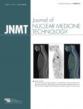
After injection of 99mTc-RBCs, dynamic flow scintigraphy of the thoracic region was performed for 8 min and demonstrated early extravasation of the labeled RBCs in the right perihilar region (Fig. 1A). A well-vascularized bone metastasis to the right scapula was also clearly visible. A SPECT/CT acquisition of the chest was then performed and revealed that the bleeding originated from the central portion of the right middle lobe (Figs. 1B–1F). The patient was transferred to the interventional radiology department for embolization, with resolution of the active thoracic bleeding.
Images revealing normal blood-pool activity and site of active bleeding in right middle lobe: frame from dynamic flow scintigraphic study after injection of 99mTc-labeled RBCs (A), selected SPECT/CT axial (B) and coronal (C) images, and corresponding nonfused SPECT images (E and F) and maximum-intensity-projection SPECT image (D).
DISCUSSION
To our knowledge, this is the first case report of localization of intrathoracic bleeding using 99mTc-RBC SPECT/CT. Only a few previous cases of thoracic bleeding localized using planar 99mTc-RBC scintigraphy or 99mTc-sulfur colloid have been reported (2–5). A dynamic 99mTc-RBC scintigraphic acquisition proves most useful for detecting bleeding that is active at the time of imaging, especially when conventional imaging fails to identify the culprit vessel. Indeed, conventional imaging may confirm that a bleed has occurred because of indirect signs such as a hematoma but may not localize the exact site of the bleeding. This case was particularly interesting as the 99mTc-RBC SPECT/CT directly guided an interventional procedure to treat the patient’s acute condition.
CONCLUSION
The advent of SPECT/CT and the fused images it produces has allowed even more precise localization of active bleeding sites (1), as well as providing anatomic correlates that can be used to guide interventional radiology procedures or surgery, as was successfully done in this case. This case shows that 99mTc-RBC SPECT/CT can be used to localize intrathoracic bleeding, but virtually any other site of active bleeding can also be identified with this useful and often underutilized nuclear medicine tool.
DISCLOSURE
No potential conflict of interest relevant to this article was reported.
Acknowledgments
We thank Dr. Louise Choiniere (Department of Thoracic Surgery) for her contribution to this case.
Footnotes
Published online Jun. 30, 2016.
REFERENCES
- Received for publication March 2, 2016.
- Accepted for publication April 20, 2016.


