  • Main menu
  • User menu
  • Search
  • English ▼
    • English
    • Afrikaans
    • Albanian
    • Amharic
    • Arabic
    • Armenian
    • Azerbaijani
    • Basque
    • Belarusian
    • Bengali
    • Bosnian
    • Bulgarian
    • Catalan
    • Cebuano
    • Chichewa
    • Chinese (Simplified)
    • Chinese (Traditional)
    • Corsican
    • Croatian
    • Czech
    • Danish
    • Dutch
    • Esperanto
    • Estonian
    • Filipino
    • Finnish
    • French
    • Frisian
    • Galician
    • Georgian
    • German
    • Greek
    • Gujarati
    • Haitian Creole
    • Hausa
    • Hawaiian
    • Hebrew
    • Hindi
    • Hmong
    • Hungarian
    • Icelandic
    • Igbo
    • Indonesian
    • Irish
    • Italian
    • Japanese
    • Javanese
    • Kannada
    • Kazakh
    • Khmer
    • Korean
    • Kurdish (Kurmanji)
    • Kyrgyz
    • Lao
    • Latin
    • Latvian
    • Lithuanian
    • Luxembourgish
    • Macedonian
    • Malagasy
    • Malay
    • Malayalam
    • Maltese
    • Maori
    • Marathi
    • Mongolian
    • Myanmar (Burmese)
    • Nepali
    • Norwegian
    • Pashto
    • Persian
    • Polish
    • Portuguese
    • Punjabi
    • Romanian
    • Russian
    • Samoan
    • Scottish Gaelic
    • Serbian
    • Sesotho
    • Shona
    • Sindhi
    • Sinhala
    • Slovak
    • Slovenian
    • Somali
    • Spanish
    • Sudanese
    • Swahili
    • Swedish
    • Tajik
    • Tamil
    • Telugu
    • Thai
    • Turkish
    • Ukrainian
    • Urdu
    • Uzbek
    • Vietnamese
    • Welsh
    • Xhosa
    • Yiddish
    • Yoruba
    • Zulu

Main menu

  • Home
  • Content
    • Current
      • JNMT Supplement
    • Ahead of print
    • Past Issues
    • Continuing Education
    • JNMT Podcast
    • SNMMI Annual Meeting Abstracts
  • Subscriptions
    • Subscribers
    • Rates
    • Journal Claims
    • Institutional and Non-member
  • Authors
    • Submit to JNMT
    • Information for Authors
    • Assignment of Copyright
    • AQARA Requirements
  • Info
    • Reviewers
    • Permissions
    • Advertisers
    • Corporate & Special Sales
  • About
    • About Us
    • Editorial Board
    • Contact Information
  • More
    • Alerts
    • Feedback
    • Help
    • SNMMI Journals
  • SNMMI
    • JNMT
    • JNM
    • SNMMI Journals
    • SNMMI

User menu

  • Subscribe
  • My alerts
  • Log in
  • My Cart

Search

  • Advanced search
Journal of Nuclear Medicine Technology
  • SNMMI
    • JNMT
    • JNM
    • SNMMI Journals
    • SNMMI
  • Subscribe
  • My alerts
  • Log in
  • My Cart
Journal of Nuclear Medicine Technology

Advanced Search

English ▼
  • English
  • Afrikaans
  • Albanian
  • Amharic
  • Arabic
  • Armenian
  • Azerbaijani
  • Basque
  • Belarusian
  • Bengali
  • Bosnian
  • Bulgarian
  • Catalan
  • Cebuano
  • Chichewa
  • Chinese (Simplified)
  • Chinese (Traditional)
  • Corsican
  • Croatian
  • Czech
  • Danish
  • Dutch
  • Esperanto
  • Estonian
  • Filipino
  • Finnish
  • French
  • Frisian
  • Galician
  • Georgian
  • German
  • Greek
  • Gujarati
  • Haitian Creole
  • Hausa
  • Hawaiian
  • Hebrew
  • Hindi
  • Hmong
  • Hungarian
  • Icelandic
  • Igbo
  • Indonesian
  • Irish
  • Italian
  • Japanese
  • Javanese
  • Kannada
  • Kazakh
  • Khmer
  • Korean
  • Kurdish (Kurmanji)
  • Kyrgyz
  • Lao
  • Latin
  • Latvian
  • Lithuanian
  • Luxembourgish
  • Macedonian
  • Malagasy
  • Malay
  • Malayalam
  • Maltese
  • Maori
  • Marathi
  • Mongolian
  • Myanmar (Burmese)
  • Nepali
  • Norwegian
  • Pashto
  • Persian
  • Polish
  • Portuguese
  • Punjabi
  • Romanian
  • Russian
  • Samoan
  • Scottish Gaelic
  • Serbian
  • Sesotho
  • Shona
  • Sindhi
  • Sinhala
  • Slovak
  • Slovenian
  • Somali
  • Spanish
  • Sudanese
  • Swahili
  • Swedish
  • Tajik
  • Tamil
  • Telugu
  • Thai
  • Turkish
  • Ukrainian
  • Urdu
  • Uzbek
  • Vietnamese
  • Welsh
  • Xhosa
  • Yiddish
  • Yoruba
  • Zulu
  • Home
  • Content
    • Current
    • Ahead of print
    • Past Issues
    • Continuing Education
    • JNMT Podcast
    • SNMMI Annual Meeting Abstracts
  • Subscriptions
    • Subscribers
    • Rates
    • Journal Claims
    • Institutional and Non-member
  • Authors
    • Submit to JNMT
    • Information for Authors
    • Assignment of Copyright
    • AQARA Requirements
  • Info
    • Reviewers
    • Permissions
    • Advertisers
    • Corporate & Special Sales
  • About
    • About Us
    • Editorial Board
    • Contact Information
  • More
    • Alerts
    • Feedback
    • Help
    • SNMMI Journals
  • Watch or Listen to JNMT Podcast
  • Visit SNMMI on Facebook
  • Join SNMMI on LinkedIn
  • Follow SNMMI on Twitter
  • Subscribe to JNMT RSS feeds
Review ArticleContinuing Education

A Bridge Not Too Far: Linking Disciplines Through Molecular Imaging Probes

John F. Valliant
Journal of Nuclear Medicine Technology September 2016, 44 (3) 173-183; DOI: https://doi.org/10.2967/jnumed.109.068312
John F. Valliant
Departments of Chemistry and Medical Physics and Centre for Probe Development and Commercialization, McMaster University, Hamilton, Ontario, Canada
  • Find this author on Google Scholar
  • Find this author on PubMed
  • Search for this author on this site
  • Article
  • Figures & Data
  • Info & Metrics
  • PDF
Loading

Article Figures & Data

Figures

  • FIGURE 1.
    • Download figure
    • Open in new tab
    • Download powerpoint
    FIGURE 1.

    Cadmium zinc telluride–based molecular breast imaging camera. (Reprinted with permission of (79).)
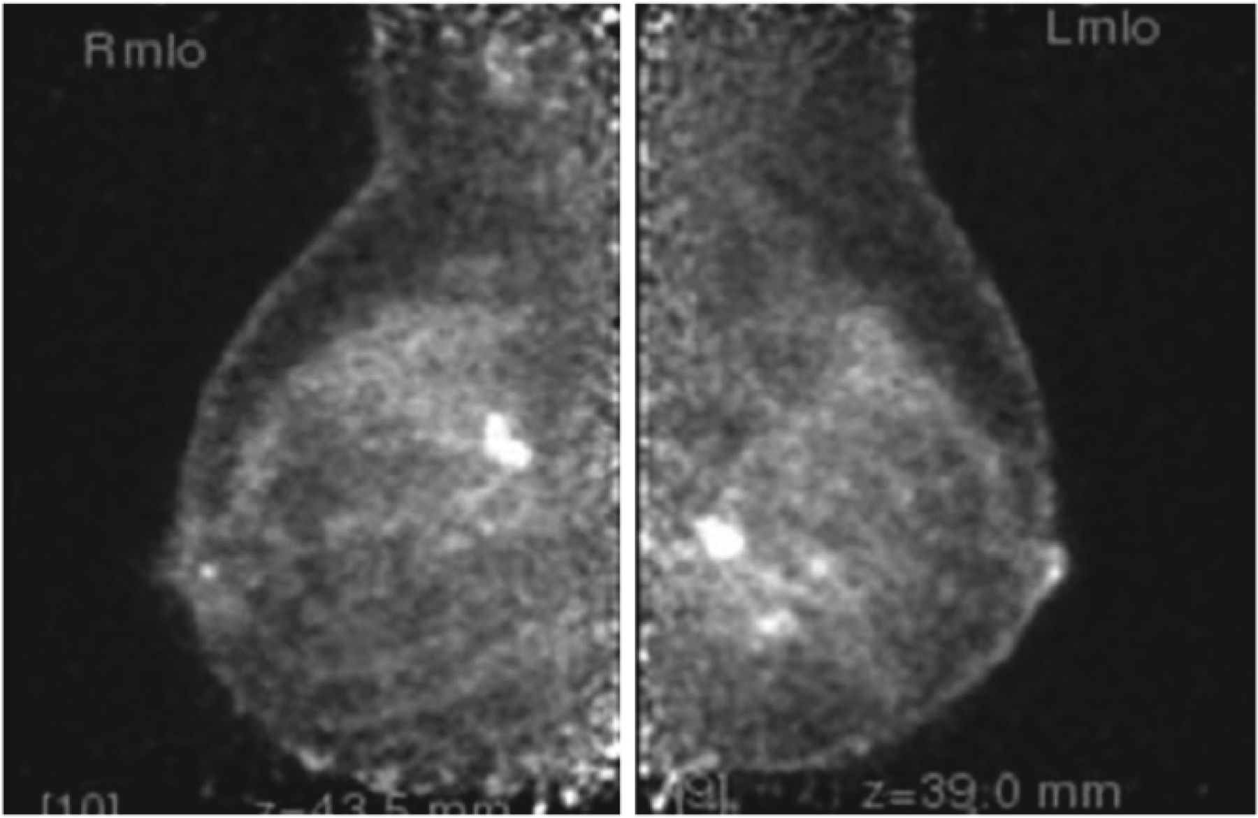
  • FIGURE 2.
    • Download figure
    • Open in new tab
    • Download powerpoint
    FIGURE 2.

    Mammogram (left) and molecular breast imaging scan (right) of same patient. Twenty-millimeter cancer (red box) can be seen on both images, but only molecular breast image (using sestamibi) shows additional 10-mm lesion. (Reprinted with permission of (80).)
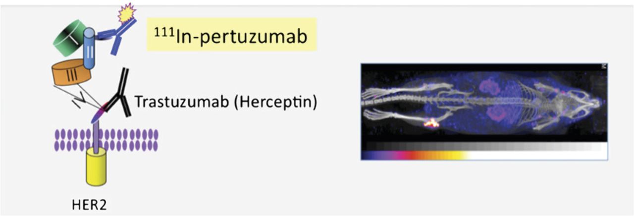
  • FIGURE 3.
    • Download figure
    • Open in new tab
    • Download powerpoint
    FIGURE 3.

    McLarty and Reilly showed that micro-SPECT/CT of HER2-overexpressing breast cancer could be achieved using 111In-labeled pertuzumab. Because this domain is different from that to which Herceptin binds, it should be possible to perform this type of molecular imaging of HER2 expression even in patients being treated with trastuzumab. (Courtesy of Raymond Reilly, University of Toronto.)
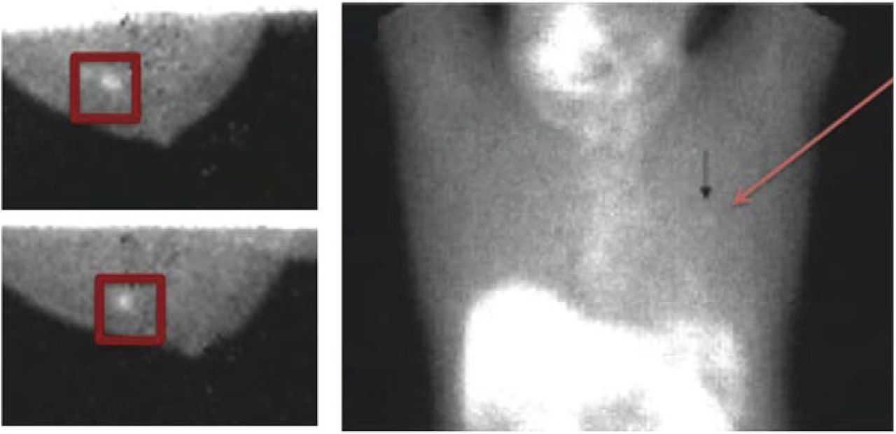
  • FIGURE 4.
    • Download figure
    • Open in new tab
    • Download powerpoint
    FIGURE 4.

    Images of peptide-based agent (99mTc-NC100692) using both dedicated breast imaging system and conventional scintimammography. Red boxes delineate 7-mm invasive ductal carcinoma that is barely visible on scintimammography (arrow). (Reprinted with permission of (16).)

  • FIGURE 5.
    • Download figure
    • Open in new tab
    • Download powerpoint
    FIGURE 5.

    18F-FDG positron emission mammography scan of 40-y-old woman whose first mammogram showed area of increased density in left inferior breast. Positron emission mammography scan shows invasive ductal carcinoma in both breasts. (Courtesy of Jacquelyn Jordan Gray, Naviscan, Inc.)
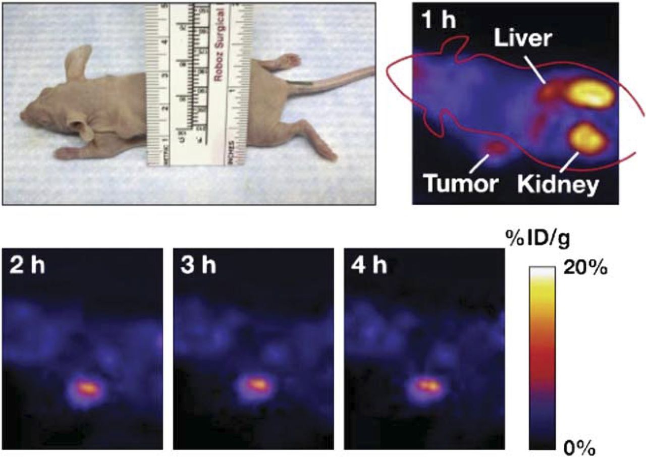
  • FIGURE 6.
    • Download figure
    • Open in new tab
    • Download powerpoint
    FIGURE 6.

    Small-animal PET images of 18F-labeled Affibody for HER2 in mice bearing SKOV-3 tumors. (Reprinted with permission of (33).)
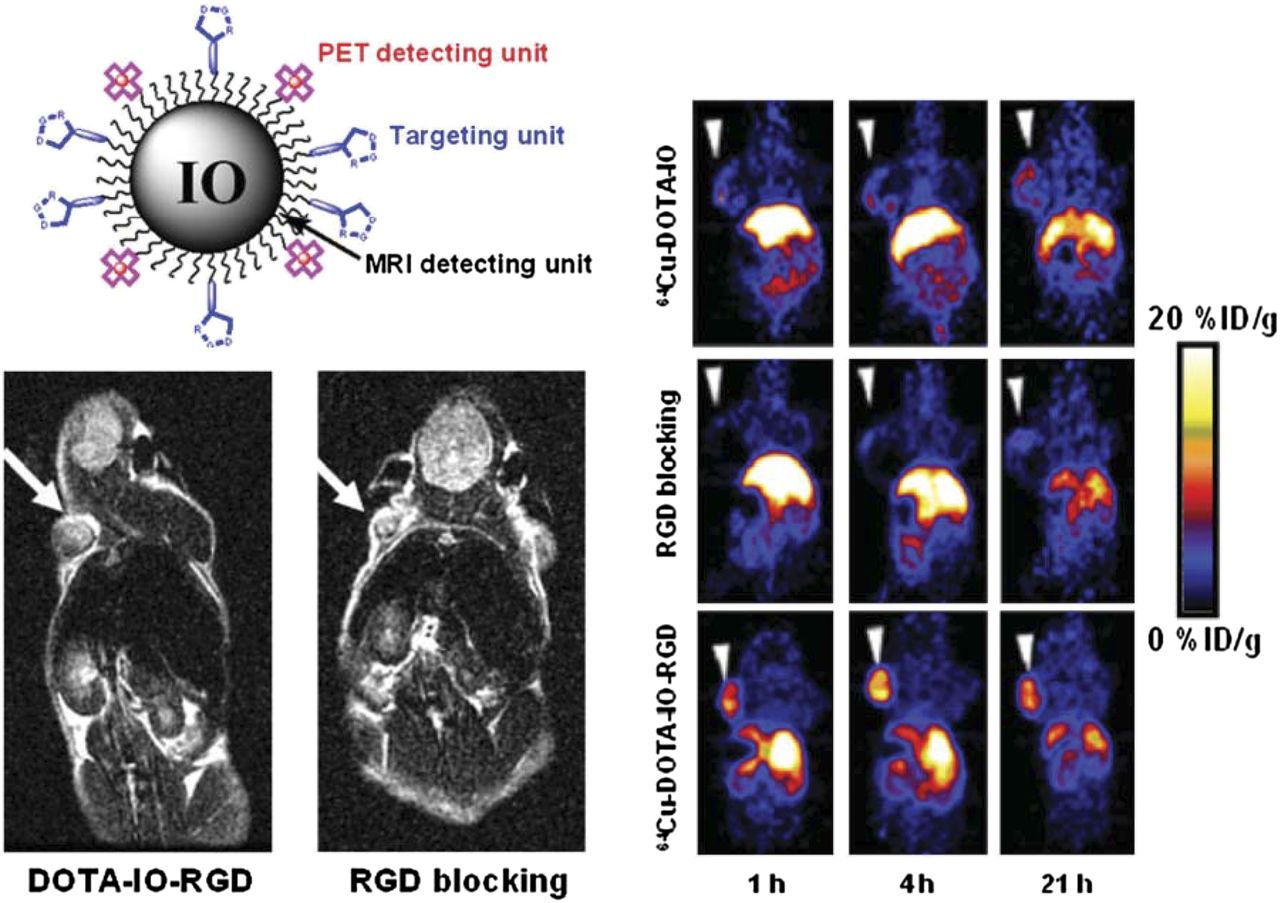
  • FIGURE 7.
    • Download figure
    • Open in new tab
    • Download powerpoint
    FIGURE 7.

    Small-animal MRI and PET scans of subcutaneous αvβ3-expressing glioma xenografts administered coated iron oxide multimodal probe derivatized with cyclic arginine-glycine-aspartic (RGD) acid peptides and DOTA chelating groups. Images involving targeted agent, control particles, and specific blocking experiments in nude mice are shown.
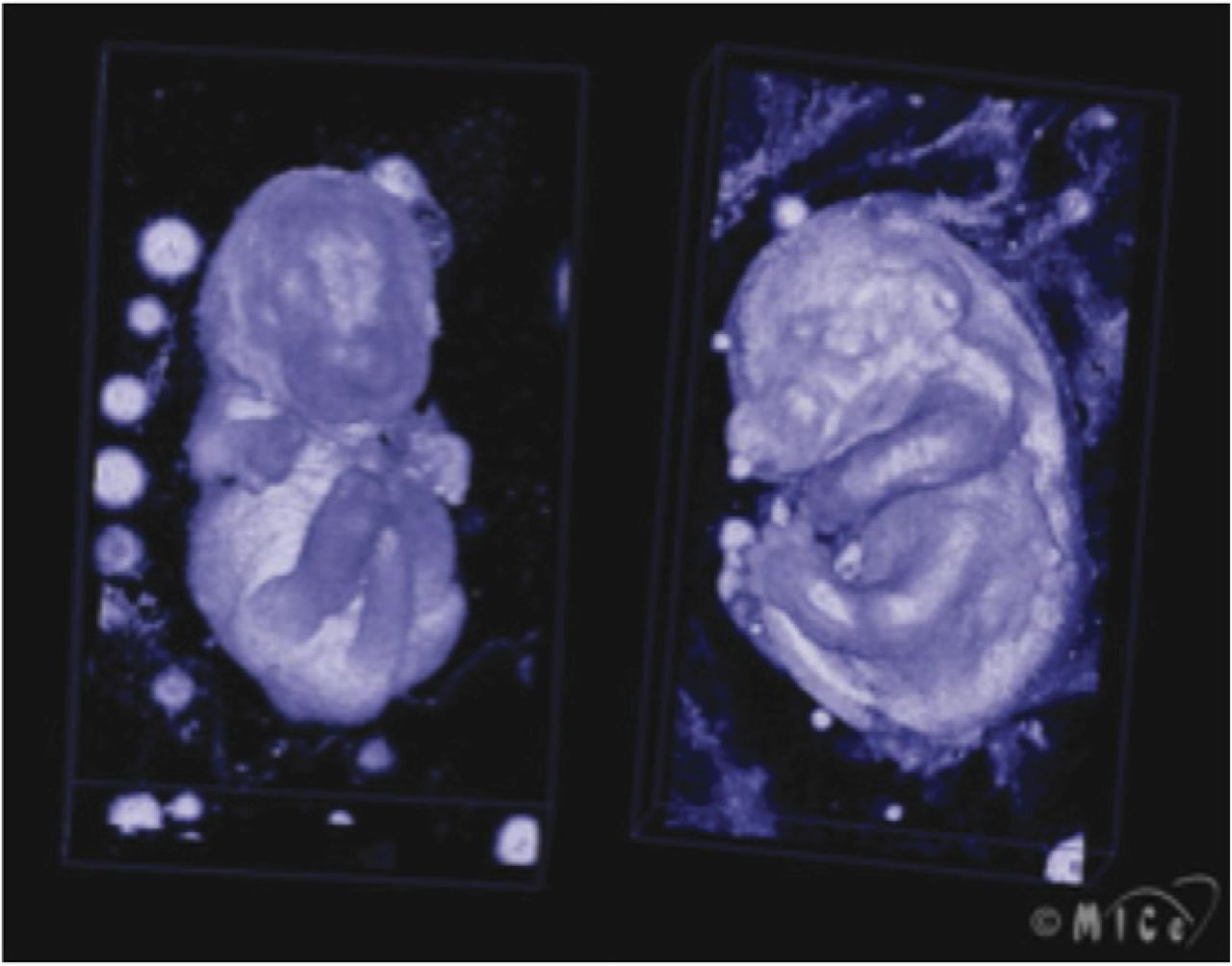
  • FIGURE 8.
    • Download figure
    • Open in new tab
    • Download powerpoint
    FIGURE 8.

    Ultrasound image of mouse embryo. (Courtesy of Stuart Foster, Sunnybrook Health Sciences Centre.)
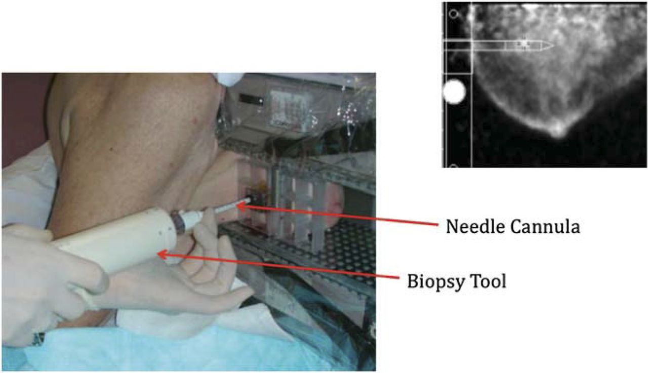
  • FIGURE 9.
    • Download figure
    • Open in new tab
    • Download powerpoint
    FIGURE 9.

    Positron emission mammography–guided biopsy based on uptake of 18F-FDG. (Courtesy of Jacquelyn Jordan Gray, Naviscan, Inc.)
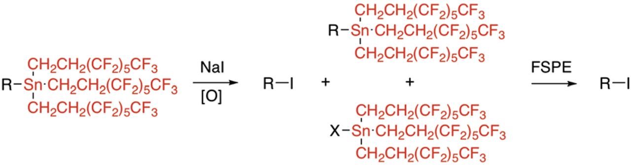
  • FIGURE 10.
    • Download figure
    • Open in new tab
    • Download powerpoint
    FIGURE 10.

    Fluorous labeling method. After treatment of fluorine-rich starting material with radioiodine and oxidant, unreacted starting material and fluorous byproducts can be separated from product using solid-phase extraction cartridge containing fluorine-rich bonded phase (fluorous solid-phase extraction, or FSPE). (Reprinted with permission of (70).)

PreviousNext
Back to top

In this issue

Journal of Nuclear Medicine Technology: 44 (3)
Journal of Nuclear Medicine Technology
Vol. 44, Issue 3
September 1, 2016
  • Table of Contents
  • About the Cover
  • Index by author
Print
Download PDF
Article Alerts
Email Article
Citation Tools
Share
A Bridge Not Too Far: Linking Disciplines Through Molecular Imaging Probes
John F. Valliant
Journal of Nuclear Medicine Technology Sep 2016, 44 (3) 173-183; DOI: 10.2967/jnumed.109.068312
Twitter logo Facebook logo LinkedIn logo Mendeley logo
  • Tweet Widget
Bookmark this article

Jump to section

  • Article
    • Abstract
    • ORGAN- AND DISEASE-SPECIFIC CAMERAS
    • HYBRID IMAGING SYSTEMS
    • LINKING DISCIPLINES THROUGH MOLECULAR IMAGING PROBES: ENHANCING BREAST CANCER TREATMENT
    • CHALLENGES IN PROBE DEVELOPMENT
    • CONCLUSION AND LOOKING TO THE FUTURE: A 2020 (20/20) VISION OF PROBE DEVELOPMENT
    • Acknowledgments
    • Footnotes
    • REFERENCES
  • Figures & Data
  • Info & Metrics
  • PDF

Related Articles

  • No related articles found.
  • PubMed
  • Google Scholar

Cited By...

  • No citing articles found.
  • Google Scholar

More in this TOC Section

  • Illuminating the Hidden: Standardizing Cardiac MIBG Imaging for Sympathetic Dysfunction
  • PET/CT Case Series: Unmasking the Mystery of Cardiac Sarcoidosis
  • Delivery Methods of Radiopharmaceuticals: Exploring Global Strategies to Minimize Occupational Radiation Exposure
Show more Continuing Education

Similar Articles

Keywords

  • probes
  • new technologies
  • breast cancer
  • collaboration
SNMMI

© 2025 SNMMI

Powered by HighWire
Alerts for this Article
Sign In to Email Alerts with your Email Address
Email this Article

Thank you for your interest in spreading the word on Journal of Nuclear Medicine Technology.

NOTE: We only request your email address so that the person you are recommending the page to knows that you wanted them to see it, and that it is not junk mail. We do not capture any email address.

Enter multiple addresses on separate lines or separate them with commas.
A Bridge Not Too Far: Linking Disciplines Through Molecular Imaging Probes
(Your Name) has sent you a message from Journal of Nuclear Medicine Technology
(Your Name) thought you would like to see the Journal of Nuclear Medicine Technology web site.
Citation Tools
A Bridge Not Too Far: Linking Disciplines Through Molecular Imaging Probes
John F. Valliant
Journal of Nuclear Medicine Technology Sep 2016, 44 (3) 173-183; DOI: 10.2967/jnumed.109.068312

Citation Manager Formats

  • BibTeX
  • Bookends
  • EasyBib
  • EndNote (tagged)
  • EndNote 8 (xml)
  • Medlars
  • Mendeley
  • Papers
  • RefWorks Tagged
  • Ref Manager
  • RIS
  • Zotero

We use cookies on this site to enhance your user experience

By clicking any link on this page you are giving your consent for us to set cookies.