
Main menu

  • Home
  • Content
    • Current
      • JNMT Supplement
    • Ahead of print
    • Past Issues
    • Continuing Education
    • JNMT Podcast
    • SNMMI Annual Meeting Abstracts
  • Subscriptions
    • Subscribers
    • Rates
    • Journal Claims
    • Institutional and Non-member
  • Authors
    • Submit to JNMT
    • Information for Authors
    • Assignment of Copyright
    • AQARA Requirements
  • Info
    • Reviewers
    • Permissions
    • Advertisers
    • Corporate & Special Sales
  • About
    • About Us
    • Editorial Board
    • Contact Information
  • More
    • Alerts
    • Feedback
    • Help
    • SNMMI Journals
  • SNMMI
    • JNMT
    • JNM
    • SNMMI Journals
    • SNMMI

User menu

  • Subscribe
  • My alerts
  • Log in
  • Log out
  • My Cart

Search

  • Advanced search
Journal of Nuclear Medicine Technology
  • SNMMI
    • JNMT
    • JNM
    • SNMMI Journals
    • SNMMI
  • Subscribe
  • My alerts
  • Log in
  • Log out
  • My Cart
Journal of Nuclear Medicine Technology

Advanced Search

  • Home
  • Content
    • Current
    • Ahead of print
    • Past Issues
    • Continuing Education
    • JNMT Podcast
    • SNMMI Annual Meeting Abstracts
  • Subscriptions
    • Subscribers
    • Rates
    • Journal Claims
    • Institutional and Non-member
  • Authors
    • Submit to JNMT
    • Information for Authors
    • Assignment of Copyright
    • AQARA Requirements
  • Info
    • Reviewers
    • Permissions
    • Advertisers
    • Corporate & Special Sales
  • About
    • About Us
    • Editorial Board
    • Contact Information
  • More
    • Alerts
    • Feedback
    • Help
    • SNMMI Journals
  • Watch or Listen to JNMT Podcast
  • Visit SNMMI on Facebook
  • Join SNMMI on LinkedIn
  • Follow SNMMI on Twitter
  • Subscribe to JNMT RSS feeds
Research ArticleImaging

Effect of Outflow Tract Contributions to 82Rb-PET Global Myocardial Blood Flow Computations

Andrew Van Tosh, Nathaniel Reichek, Christopher J. Palestro and Kenneth J. Nichols
Journal of Nuclear Medicine Technology June 2016, 44 (2) 78-84; DOI: https://doi.org/10.2967/jnmt.116.173005
Andrew Van Tosh
1Research Department, St. Francis Hospital, Roslyn, New York; and
  • Find this author on Google Scholar
  • Find this author on PubMed
  • Search for this author on this site
Nathaniel Reichek
1Research Department, St. Francis Hospital, Roslyn, New York; and
  • Find this author on Google Scholar
  • Find this author on PubMed
  • Search for this author on this site
Christopher J. Palestro
2Division of Nuclear Medicine and Molecular Imaging, Northwell Health, Manhasset and New Hyde Park, New York
  • Find this author on Google Scholar
  • Find this author on PubMed
  • Search for this author on this site
Kenneth J. Nichols
2Division of Nuclear Medicine and Molecular Imaging, Northwell Health, Manhasset and New Hyde Park, New York
  • Find this author on Google Scholar
  • Find this author on PubMed
  • Search for this author on this site
  • Article
  • Figures & Data
  • Info & Metrics
  • PDF
Loading

Article Figures & Data

Figures

  • Tables
  • FIGURE 1.
    • Download figure
    • Open in new tab
    • Download powerpoint
    FIGURE 1.

    American Heart Association/American College of Cardiology 17-segment map.
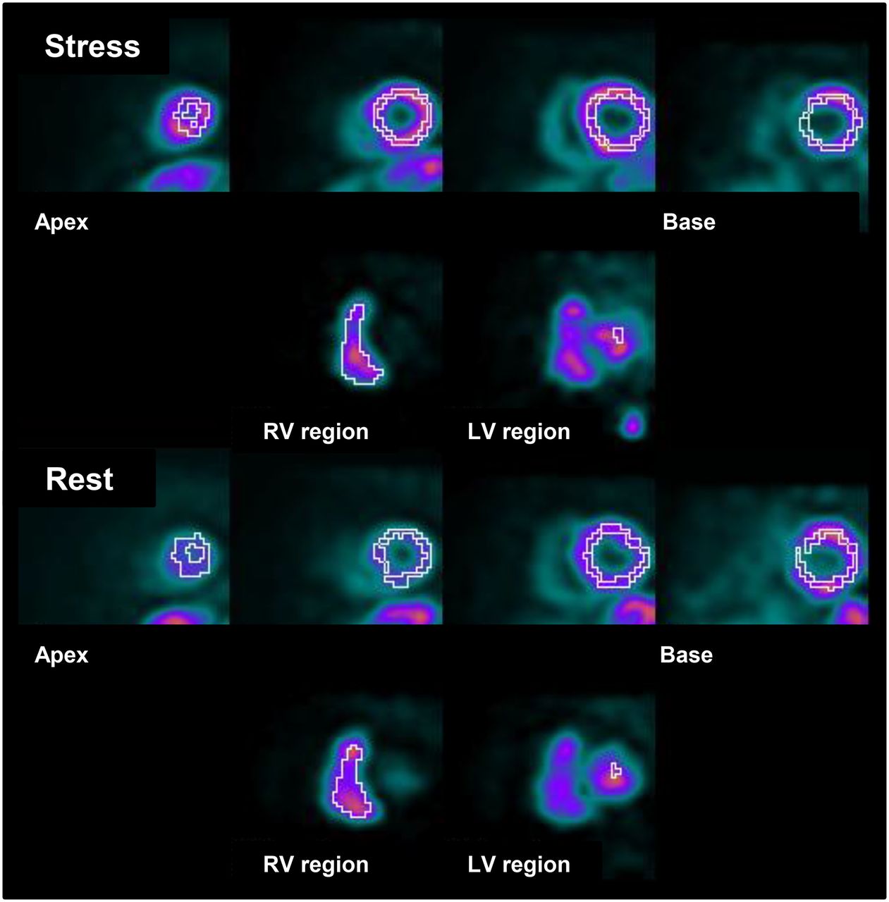
  • FIGURE 2.
    • Download figure
    • Open in new tab
    • Download powerpoint
    FIGURE 2.

    Segmentation and chamber identification. LV myocardial segments are identified (top row), as well as right ventricular (RV) and LV blood pools (bottom row).

  • FIGURE 3.
    • Download figure
    • Open in new tab
    • Download powerpoint
    FIGURE 3.

    Polar map display of MFRs. Values of segments 2 and 3 are markedly reduced compared with values of all other segments.
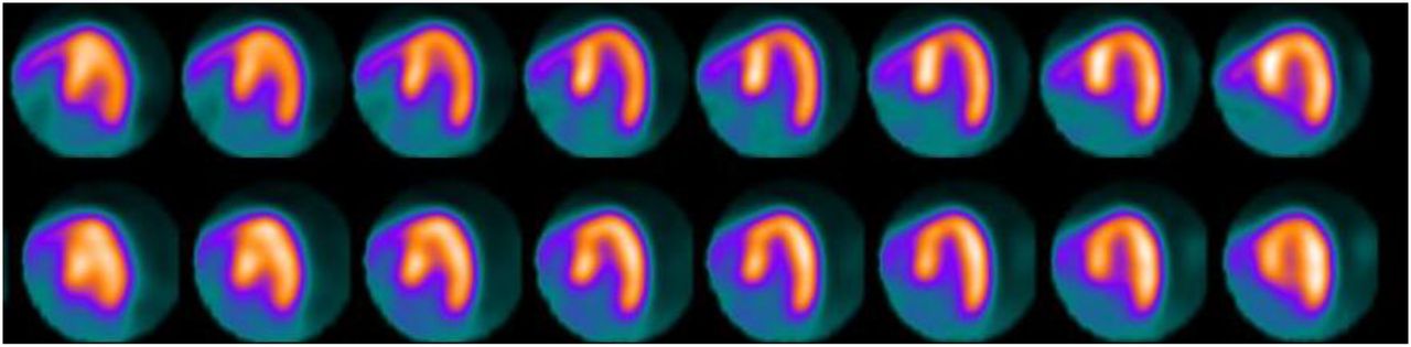
  • FIGURE 4.
    • Download figure
    • Open in new tab
    • Download powerpoint
    FIGURE 4.

    Stress (top) and rest (bottom) horizontal long-axis sections from septum (left) to lateral wall (right) for patient with essentially normal perfusion, all summed perfusion scores equal to 0, and normal function (ejection fraction, 70%). Respective flow inhomogeneities (ratio of SD to mean) were 15%, 23%, and 15% for rest MBF, stress MBF, and MFR when only segments 4–17 were included but increased to 31%, 34%, and 22% when all 17 segments were included.
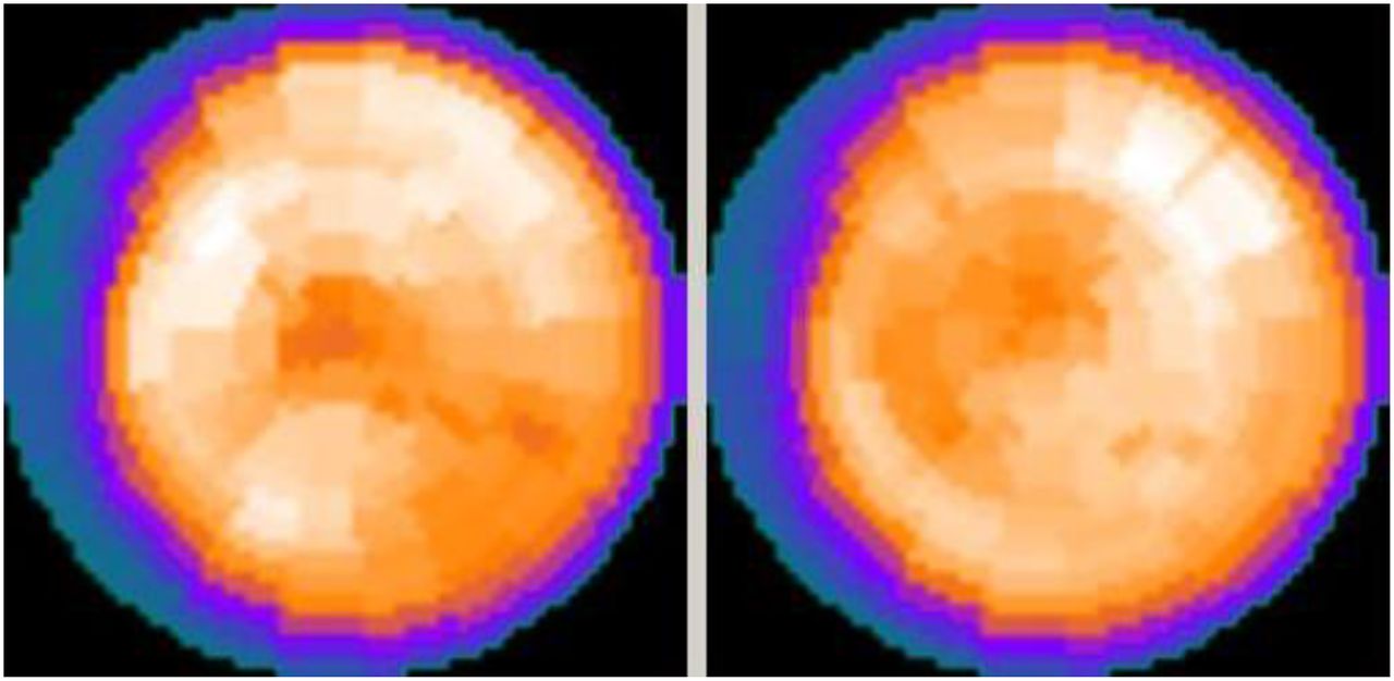
  • FIGURE 5.
    • Download figure
    • Open in new tab
    • Download powerpoint
    FIGURE 5.

    82Rb polar perfusion maps for stress (left) and rest (right) for the patient of Figure 4 display markedly reduced perfusion in basal–septal territories.
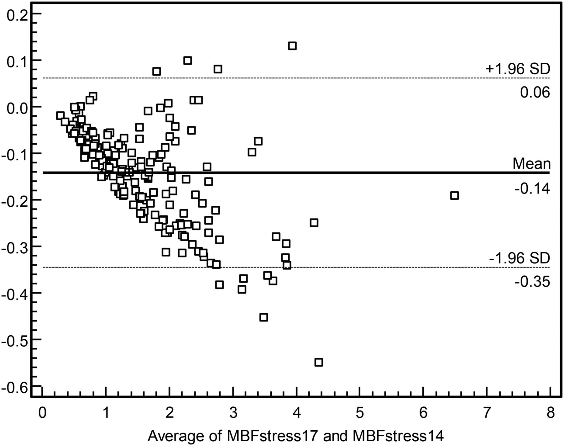
  • FIGURE 6.
    • Download figure
    • Open in new tab
    • Download powerpoint
    FIGURE 6.

    Bland–Altman plot of differences vs. mean values for 17-segment stress MBF (MBFstress17) and 14-segment stress MBF (MBFstress14), in units of mL/g/min.

Tables

  • Figures
    • View popup
    TABLE 1

    Comparison of MBF Parameters Obtained by Including All 17 LV Segments vs. Only Segments 4–17

    ParameterSegments 1–17Segments 4–17
    Rest MBF (mL/g/min)0.78 ± 0.50*0.85 ± 0.54
    Stress MBF (mL/g/min)1.50 ± 0.88*1.67 ± 0.96
    MFR2.11 ± 1.00*2.16 ± 1.00
    Rest CVR (mm Hg/mL/g/min)159 ± 86*147 ± 81
    Stress CVR (mm Hg/mL/g/min)85 ± 52*76 ± 48
    %SD of rest MBF39% ± 10%*31% ± 10%
    %SD of stress MBF42% ± 12%*32% ± 11%
    %SD of MFR28% ± 18%*25% ± 10%
    • ↵* Paired t test P < 0.0001 vs. segments 4–17.

    • View popup
    TABLE 2

    Differences and Percentage Differences Between 14- and 17-Segment Mean Values

    ParameterMean differenceMaximum differenceMean % differenceMaximum % difference
    Rest MBF (mL/g/min)0.06 ± 0.050.248.2% ± 4.2%16.8%
    Stress MBF (mL/g/min)0.14 ± 0.100.549.3% ± 4.5%16.3%
    MFR0.02 ± 0.070.261.1% ± 3.1%15.0%
    Rest CVR (mm Hg/mL/g/min)−12.7 ± 8.7−43.9−8.5% ± 4.2%−17.5%
    Stress CVR (mm Hg/mL/g/min)−8.2 ± 5.9−27.8−10.5% ± 5.2%−18.8%
    %SD of rest MBF−7.6% ± 5.1%−22.0%——
    %SD of stress MBF−9.4% ± 5.4%−20.0%——
    %SD of MFR−2.6% ± 14.1%12.0%——
    • View popup
    TABLE 3

    Comparison of MBF Parameters for Patients Divided into Groups for Whom 14-Segment MFR Was Below or Above Median

    MFR < 1.95MFR ≥ 1.95
    ParameterSegments 1–17Segments 4–17Segments 1–17Segments 4–17
    Rest MBF (mL/g/min)0.91 ± 0.58*0.98 ± 0.610.67 ± 0.40*0.72 ± 0.42
    Stress MBF (mL/g/min)1.23 ± 0.78*1.35 ± 0.841.81 ± 0.92*1.98 ± 0.97
    MFR1.40 ± 0.34*1.42 ± 0.352.86 ± 0.94*2.90 ± 0.91
    Rest CVR (mm Hg/mL/g/min)136 ± 79*125 ± 73182 ± 89*167 ± 83
    Stress CVR (mm Hg/mL/g/min)103 ± 60*94 ± 5664 ± 31*58 ± 28
    %SD of rest MBF39% ± 10%*31% ± 10%38% ± 9%*30% ± 9%
    %SD of stress MBF43% ± 12%*34% ± 13%38% ± 9%*30% ± 9%
    %SD of MFR26% ± 11%30% ± 23%24% ± 9%26% ± 9%
    • ↵* P < 0.0001 vs. segments 4–17.

PreviousNext
Back to top

In this issue

Journal of Nuclear Medicine Technology: 44 (2)
Journal of Nuclear Medicine Technology
Vol. 44, Issue 2
June 1, 2016
  • Table of Contents
  • About the Cover
  • Index by author
Print
Download PDF
Article Alerts
Sign In to Email Alerts with your Email Address
Email Article

Thank you for your interest in spreading the word on Journal of Nuclear Medicine Technology.

NOTE: We only request your email address so that the person you are recommending the page to knows that you wanted them to see it, and that it is not junk mail. We do not capture any email address.

Enter multiple addresses on separate lines or separate them with commas.
Effect of Outflow Tract Contributions to 82Rb-PET Global Myocardial Blood Flow Computations
(Your Name) has sent you a message from Journal of Nuclear Medicine Technology
(Your Name) thought you would like to see the Journal of Nuclear Medicine Technology web site.
Citation Tools
Effect of Outflow Tract Contributions to 82Rb-PET Global Myocardial Blood Flow Computations
Andrew Van Tosh, Nathaniel Reichek, Christopher J. Palestro, Kenneth J. Nichols
Journal of Nuclear Medicine Technology Jun 2016, 44 (2) 78-84; DOI: 10.2967/jnmt.116.173005

Citation Manager Formats

  • BibTeX
  • Bookends
  • EasyBib
  • EndNote (tagged)
  • EndNote 8 (xml)
  • Medlars
  • Mendeley
  • Papers
  • RefWorks Tagged
  • Ref Manager
  • RIS
  • Zotero
Share
Effect of Outflow Tract Contributions to 82Rb-PET Global Myocardial Blood Flow Computations
Andrew Van Tosh, Nathaniel Reichek, Christopher J. Palestro, Kenneth J. Nichols
Journal of Nuclear Medicine Technology Jun 2016, 44 (2) 78-84; DOI: 10.2967/jnmt.116.173005
Twitter logo Facebook logo LinkedIn logo Mendeley logo
  • Tweet Widget
  • Facebook Like
  • Google Plus One
Bookmark this article

Jump to section

  • Article
    • Abstract
    • MATERIALS AND METHODS
    • RESULTS
    • DISCUSSION
    • CONCLUSION
    • DISCLOSURE
    • Footnotes
    • REFERENCES
  • Figures & Data
  • Info & Metrics
  • PDF

Related Articles

  • No related articles found.
  • PubMed
  • Google Scholar

Cited By...

  • No citing articles found.
  • Google Scholar

More in this TOC Section

  • Early 10-Minute Postinjection [18F]F-FAPI-42 uEXPLORER Total-Body PET/CT Scanning Protocol for Staging Lung Cancer Using HYPER Iterative Reconstruction
  • Single- Versus Dual-Time-Point Imaging for Transthyretin Cardiac Amyloid Using 99mTc-Pyrophosphate
  • Does Arthrography Improve Accuracy of SPECT/CT for Diagnosis of Aseptic Loosening in Patients with Painful Knee Arthroplasty: A Systematic Review and Metaanalysis
Show more Imaging

Similar Articles

Keywords

  • PET/CT
  • myocardial perfusion imaging
  • PET
  • myocardial flow reserve
  • quantification
  • rubidium isotopes
SNMMI

© 2025 SNMMI

Powered by HighWire