
Main menu

  • Home
  • Content
    • Current
      • JNMT Supplement
    • Ahead of print
    • Past Issues
    • Continuing Education
    • JNMT Podcast
    • SNMMI Annual Meeting Abstracts
  • Subscriptions
    • Subscribers
    • Rates
    • Journal Claims
    • Institutional and Non-member
  • Authors
    • Submit to JNMT
    • Information for Authors
    • Assignment of Copyright
    • AQARA Requirements
  • Info
    • Reviewers
    • Permissions
    • Advertisers
    • Corporate & Special Sales
  • About
    • About Us
    • Editorial Board
    • Contact Information
  • More
    • Alerts
    • Feedback
    • Help
    • SNMMI Journals
  • SNMMI
    • JNMT
    • JNM
    • SNMMI Journals
    • SNMMI

User menu

  • Subscribe
  • My alerts
  • Log in
  • My Cart

Search

  • Advanced search
Journal of Nuclear Medicine Technology
  • SNMMI
    • JNMT
    • JNM
    • SNMMI Journals
    • SNMMI
  • Subscribe
  • My alerts
  • Log in
  • My Cart
Journal of Nuclear Medicine Technology

Advanced Search

  • Home
  • Content
    • Current
    • Ahead of print
    • Past Issues
    • Continuing Education
    • JNMT Podcast
    • SNMMI Annual Meeting Abstracts
  • Subscriptions
    • Subscribers
    • Rates
    • Journal Claims
    • Institutional and Non-member
  • Authors
    • Submit to JNMT
    • Information for Authors
    • Assignment of Copyright
    • AQARA Requirements
  • Info
    • Reviewers
    • Permissions
    • Advertisers
    • Corporate & Special Sales
  • About
    • About Us
    • Editorial Board
    • Contact Information
  • More
    • Alerts
    • Feedback
    • Help
    • SNMMI Journals
  • Watch or Listen to JNMT Podcast
  • Visit SNMMI on Facebook
  • Join SNMMI on LinkedIn
  • Follow SNMMI on Twitter
  • Subscribe to JNMT RSS feeds
Review ArticleTeaching Case Studies

Precise Localization of a Bile Leak with Hepatobiliary Scintigraphy

Sana Naeem, Hanna Li and Zhiyun Yang
Journal of Nuclear Medicine Technology March 2016, 44 (1) 44-45; DOI: https://doi.org/10.2967/jnmt.115.154997
Sana Naeem
Department of Radiology, Louisiana State University Health Sciences Center/University Health, Shreveport, Louisiana
  • Find this author on Google Scholar
  • Find this author on PubMed
  • Search for this author on this site
Hanna Li
Department of Radiology, Louisiana State University Health Sciences Center/University Health, Shreveport, Louisiana
  • Find this author on Google Scholar
  • Find this author on PubMed
  • Search for this author on this site
Zhiyun Yang
Department of Radiology, Louisiana State University Health Sciences Center/University Health, Shreveport, Louisiana
  • Find this author on Google Scholar
  • Find this author on PubMed
  • Search for this author on this site
  • Article
  • Figures & Data
  • Info & Metrics
  • PDF
Loading

Abstract

Hepatobiliary scintigraphy (HBS) is a widely used modality to diagnose biliary injury. In patients with a history of trauma or surgery, the common presentations of bile leaks on HBS are a progressive collection of radiotracer outside the biliary system. However, the precise localization of a bile leak is usually not conveyed by noninvasive techniques including CT or ultrasound or even HBS. This case study of a patient with liver trauma demonstrates a direct bile leak sign leading to recognition of an exact bile leakage site from the left intrahepatic duct. This diagnosis helped clinicians perform less-invasive management.

  • hepatobiliary scintigraphy (HBS)
  • bile leak
  • biliary leak
  • hepatobiliary iminodiacetic acid (HIDA) scan
  • cholescintigraphy
  • hepatic laceration

Hepatobiliary scintigraphy (HBS) is a widely used modality to diagnose biliary injury (1). In patients with a history of trauma or surgery, the common presentations of bile leaks on HBS are a progressive collection of radiotracer outside the biliary system (2). However, the precise localization of a bile leak is usually not conveyed by noninvasive techniques including CT or ultrasound or even HBS (3,4).

CASE REPORT

A 16-y-old male (who along with his parent gave permission to use the data from his studies for our article) presented with injuries from a motor vehicle accident. A CT scan of the abdomen/pelvis with contrast demonstrated central hepatic laceration with extension into gallbladder fossa and peritoneal fluid. HBS was required to determine whether a biliary injury was present or not.

After intravenous administration of 99mTc-mebrofenin, sequential dynamic abdominal images were obtained over 60 min (Fig. 1). Images showed an area with decreased activity over the region between the 2 hepatic lobes, corresponding with the known hepatic laceration seen on the CT (Fig. 2). Over time, there was a progressive accumulation of tracer outside the biliary system over the inferior left hepatic lobe. A couple of lines of tracer uptake connected this abnormal focal area to the left intrahepatic duct. Eventually, this abnormal tracer accumulation spread throughout the abdomen. These findings indicated an exact bile-leaking site at the left intrahepatic duct.

FIGURE 1.
  • Download figure
  • Open in new tab
  • Download powerpoint
FIGURE 1.

HBS over 60 min shows area with decreased activity over region between the 2 hepatic lobes, corresponding with known hepatic laceration seen on CT (black arrow). Over time, there is a progressive accumulation of tracer outside the biliary system over inferior left hepatic lobe (pink arrow). A couple of lines of tracer uptake (red arrows) connected this abnormal focal area to left intrahepatic duct. Eventually, this abnormal tracer accumulation spread throughout abdomen (blue arrows). These findings indicate exact bile-leaking site at left intrahepatic duct.

FIGURE 2.
  • Download figure
  • Open in new tab
  • Download powerpoint
FIGURE 2.

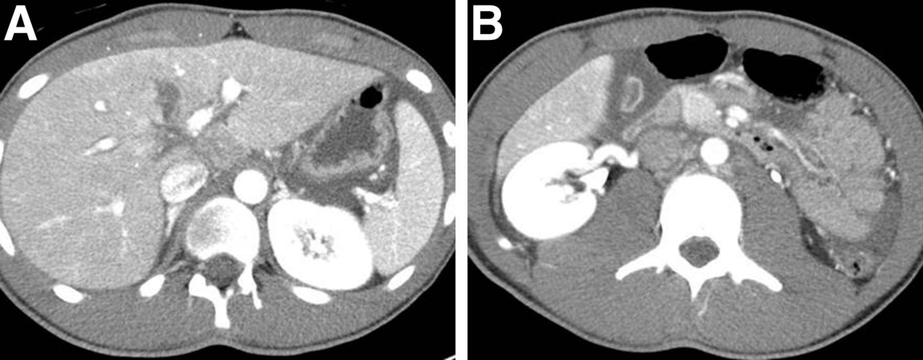
CT images of abdomen/pelvis with contrast demonstrate central hepatic laceration.

Subsequently an endoscopic retrograde cholangiopancreatography with initial cholangiogram confirmed the findings of the HBS (Fig. 3). Sphincterotomy and placement of 2 common bile duct stents were performed. Soon after, a diagnostic laparoscopy further confirmed a bile leak from the left hepatic duct, and a laparoscopic lavage and intraperitoneal drainage were performed. The patient did well postoperatively and was discharged with no extravasation of contrast in the follow-up endoscopic retrograde cholangiopancreatography with cholangiogram after the removal of common bile duct stents.

FIGURE 3.
  • Download figure
  • Open in new tab
  • Download powerpoint
FIGURE 3.

Endoscopic retrograde cholangiopancreatography with cholangiogram shows accumulation of contrast outside biliary system over inferior left hepatic lobe (yellow arrow). Line of contrast (red arrow) connects this abnormal focal area to left intrahepatic duct.

DISCUSSION

Bile leaks can occur after surgery or trauma. Imaging modalities used to detect bile leaks are CT, endoscopic retrograde cholangiopancreatography, MR cholangiopancreatography (MRCP), ultrasound, and HBS. Among these, only HBS and MRCP, noninvasive studies, can identify abnormal fluid collection as bile. However, there are limitations to emergency MRCP, including lack of readily available MR imaging equipment, technologists, personnel experienced with interpreting MRCP, and higher cost (4). Therefore, HBS is the commonly used modality to diagnose biliary injury (1).

HBS is a highly sensitive imaging modality and useful for early diagnosis of bile leaks. HBS can determine whether a fluid collection seen on ultrasound or CT is of biliary origin (1,2). HBS can determine the rate of leakage and can be used as a follow-up tool (1,2). A common finding of a bile leak is the radiopharmaceutical’s progressive accumulation outside the biliary system, either localized (bilomas) or diffuse. An accurate localization of a bile leak is difficult by HBS because of the lack of anatomic details. However, this localization is important because of the shift in management strategy from very invasive to minimally invasive (3).

This case demonstrates a direct bile leakage sign, bile communication tracks connecting a left intrahepatic duct to a biloma over the inferior left hepatic lobe. This direct sign leads to recognition of an exact bile-leaking site in the left hepatic duct and helps determine a bile leak type. A management approach can be selected on the basis of the bile leak’s type (5). In our case, a less-invasive management was used based on the HBS findings.

Accurate localization of the site of bile leak through a direct sign, bile communication tracks, can be obtained with HBS as shown in this case. Determining the site of a bile leak will further increase opportunities to detect an accurate bile leakage site in improved HBS with SPECT (SPECT/CT). The HBS SPECT/CT provides both anatomic and functional information and overcomes the lack of specificity of CT alone or detailed anatomic information of HBS alone (3).

CONCLUSION

Although the precise site of a bile leak is often difficult to detect with noninvasive techniques, it is possible with HBS. Knowledge of the bile leak site will increase opportunities to detect the direct sign of a bile leak in advanced HBS with SPECT/CT. This is vital given the change in approach regarding management of a bile leak from more invasive to less invasive.

DISCLOSURE

No potential conflict of interest relevant to this article was reported.

Footnotes

  • Published online Jun. 25, 2015.

REFERENCES

  1. 1.↵
    1. LeBedis CA,
    2. Anderson SW,
    3. Mercier G,
    4. et al
    . The utility of CT for predicting bile leaks in hepatic trauma. Emerg Radiol. 2015;22:101–107.
    OpenUrlCrossRefPubMed
  2. 2.↵
    1. Ziessman HA,
    2. O’Malley JP,
    3. Thrall JH,
    4. et al
    . Nuclear Medicine: The Requisites. 4th ed. Philadelphia, PA: Saunders; 2014.
  3. 3.↵
    Sharma P, Kumar R, Das KJ, et al. Detection and Localization of Post-Operative and Post-Traumatic Bile Leak: Hybrid SPECT/CT with 99m Tc-Mebrofenin. New York, NY: Springer Science + Business Media, LLC; 2012.
  4. 4.↵
    1. Mujoomdar M,
    2. Russell E,
    3. Dionne F,
    4. et al
    . CADTH Optimizing Health System Use of Medical Isotopes and Other Imaging Modalities. Ottawa, Ontario: Canadian Agency for Drugs and Technologies in Health; 2012.
  5. 5.↵
    1. Kapoor S,
    2. Nundy S
    . Bile duct leaks from the intrahepatic biliary tree: a review of its etiology, incidence, and management: a review article. HPB Surg. 2012;4:1–9.
    OpenUrl
  • Received for publication January 30, 2015.
  • Accepted for publication May 14, 2015.
View Abstract
PreviousNext
Back to top

In this issue

Journal of Nuclear Medicine Technology: 44 (1)
Journal of Nuclear Medicine Technology
Vol. 44, Issue 1
March 1, 2016
  • Table of Contents
  • About the Cover
  • Index by author
Print
Download PDF
Article Alerts
Sign In to Email Alerts with your Email Address
Email Article

Thank you for your interest in spreading the word on Journal of Nuclear Medicine Technology.

NOTE: We only request your email address so that the person you are recommending the page to knows that you wanted them to see it, and that it is not junk mail. We do not capture any email address.

Enter multiple addresses on separate lines or separate them with commas.
Precise Localization of a Bile Leak with Hepatobiliary Scintigraphy
(Your Name) has sent you a message from Journal of Nuclear Medicine Technology
(Your Name) thought you would like to see the Journal of Nuclear Medicine Technology web site.
Citation Tools
Precise Localization of a Bile Leak with Hepatobiliary Scintigraphy
Sana Naeem, Hanna Li, Zhiyun Yang
Journal of Nuclear Medicine Technology Mar 2016, 44 (1) 44-45; DOI: 10.2967/jnmt.115.154997

Citation Manager Formats

  • BibTeX
  • Bookends
  • EasyBib
  • EndNote (tagged)
  • EndNote 8 (xml)
  • Medlars
  • Mendeley
  • Papers
  • RefWorks Tagged
  • Ref Manager
  • RIS
  • Zotero
Share
Precise Localization of a Bile Leak with Hepatobiliary Scintigraphy
Sana Naeem, Hanna Li, Zhiyun Yang
Journal of Nuclear Medicine Technology Mar 2016, 44 (1) 44-45; DOI: 10.2967/jnmt.115.154997
Twitter logo Facebook logo LinkedIn logo Mendeley logo
  • Tweet Widget
  • Facebook Like
  • Google Plus One
Bookmark this article

Jump to section

  • Article
    • Abstract
    • CASE REPORT
    • DISCUSSION
    • CONCLUSION
    • DISCLOSURE
    • Footnotes
    • REFERENCES
  • Figures & Data
  • Info & Metrics
  • PDF

Related Articles

  • No related articles found.
  • PubMed
  • Google Scholar

Cited By...

  • No citing articles found.
  • Google Scholar

More in this TOC Section

  • High-Sensitivity Troponin Elevation in a Young Woman with Typical Chest Pain: The Heart of the Matter
  • Pulmonary Adenocarcinoma Revealed by Parathyroid Scintigraphy: An Incidental Case to Remember
  • Prominent Right Ventricular Tracer Uptake: A Harbinger of Multivessel Coronary Artery Disease
Show more Teaching Case Studies

Similar Articles

Keywords

  • hepatobiliary scintigraphy (HBS)
  • bile leak
  • biliary leak
  • hepatobiliary iminodiacetic acid (HIDA) scan
  • cholescintigraphy
  • hepatic laceration
SNMMI

© 2025 SNMMI

Powered by HighWire