


Article Figures & Data
Figures
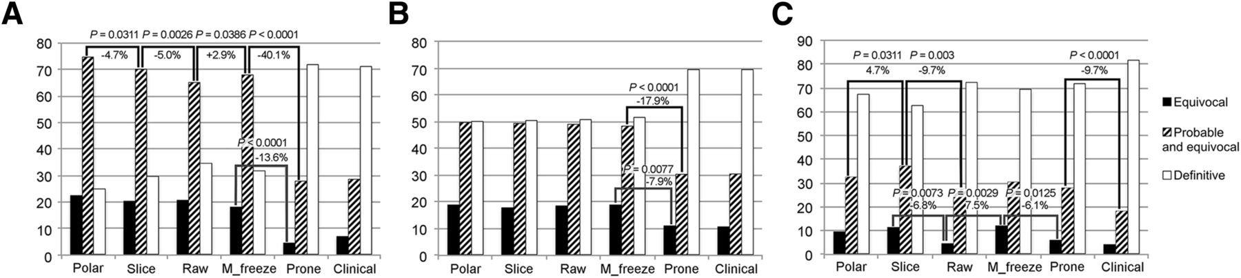
- FIGURE 1.
Percentages of equivocal, probable + equivocal, and definite studies in course of evaluation: physician 1 (A), physician 2 (B), and physician 3 (C).
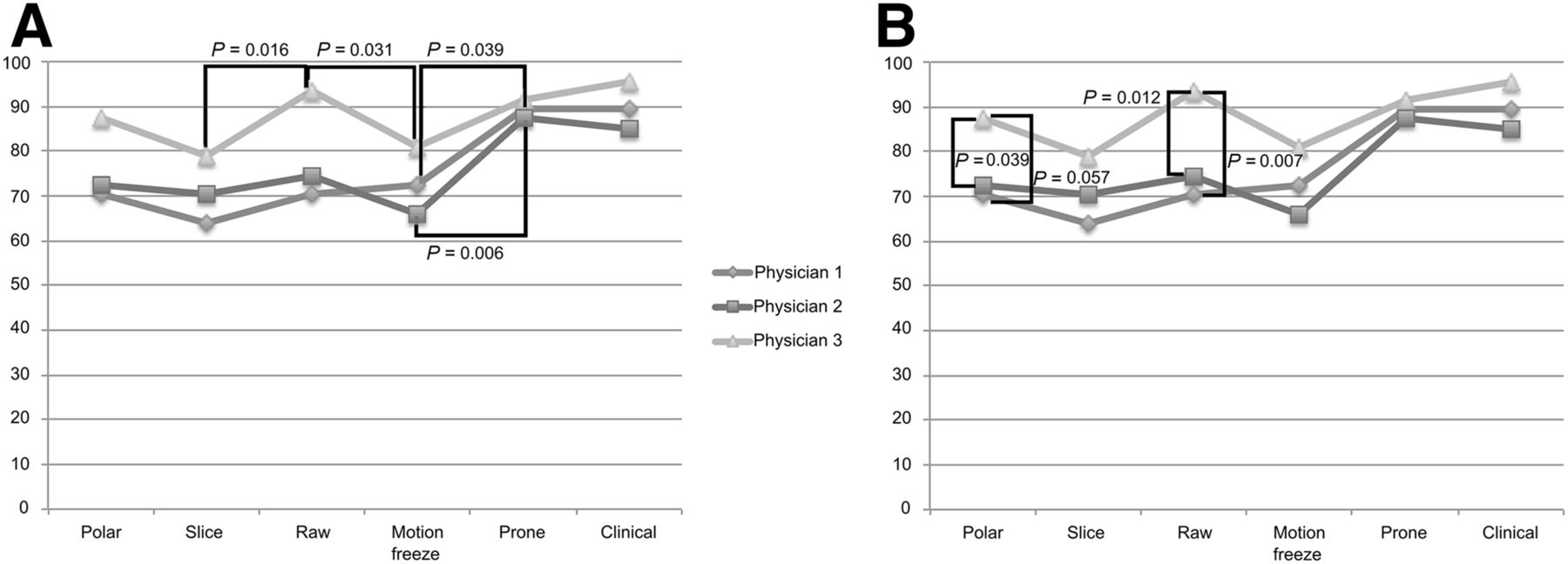
- FIGURE 2.
Normalcy rates at each evaluation, showing intraobserver changes (A) and interobserver differences (B).
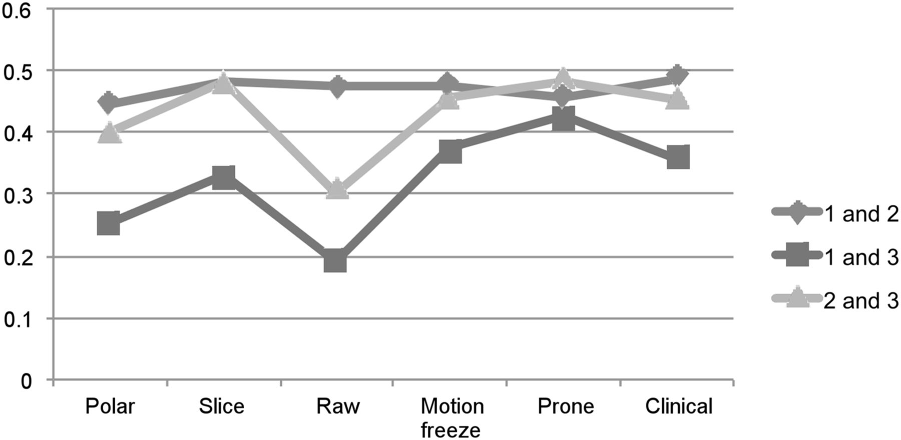
- FIGURE 3.
Interobserver agreement κ values.
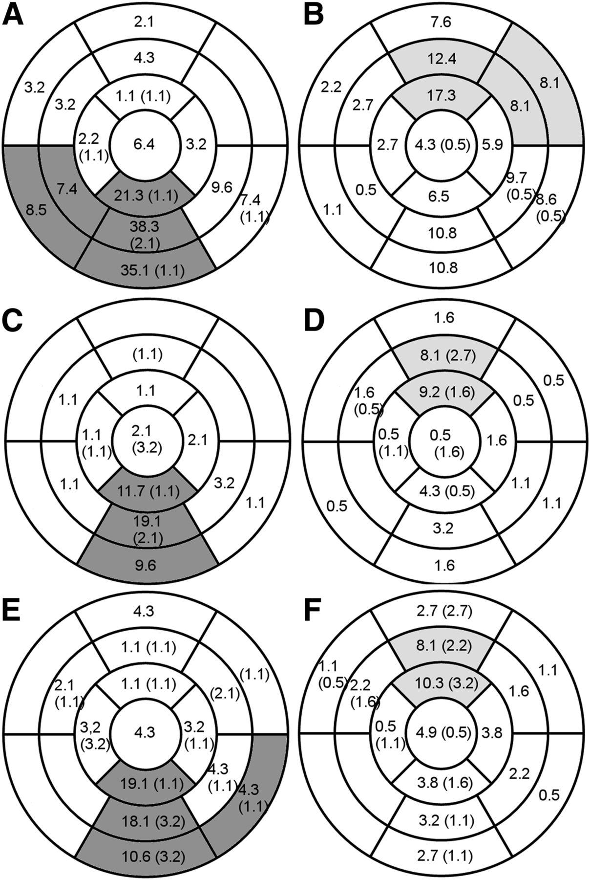
- FIGURE 4.
Attributions to attenuation after prone evaluation of each segment as percentage of total number of male and female patients, for each physician (A and B physician 1; C and D physician 2; E and F physician 3, male and female patient, respectively). Numbers in parentheses are new areas evaluated as abnormal after prone evaluation. Segments showing statistically significant difference between sexes are shaded.
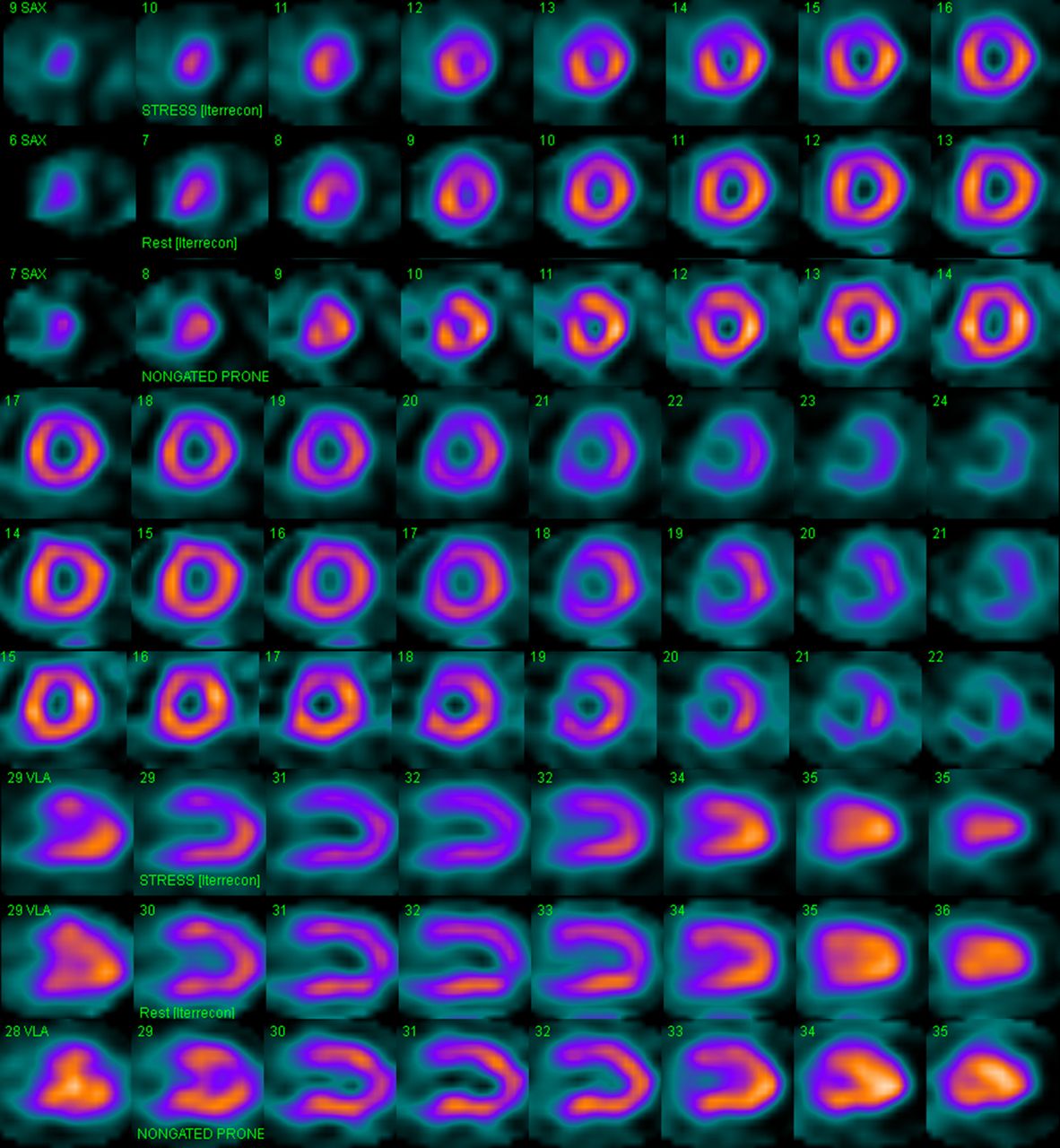
- FIGURE 5.
Example case. First 6 rows show stress, rest, and stress prone short-axis images; next 3 rows show stress, rest, and stress prone vertical long-axis images. A 55-y-old woman with atypical angina, with hypertension and family history as risk factors; 109% heart rate achieved at Bruce level 2. SSS, 6; SRS, 0; prone, SSS 3. All 3 interpreters used information in prone image to reverse their decisions from probably abnormal to definitely normal, citing breast attenuation. Apical perfusion defect in prone image is possibly due to changing position of breast and example of discordant finding in supine and prone that helps detect presence attenuation. SRS = summed rest score; SSS = summed stress score.
- FIGURE 6.
Example case. First 6 rows show stress, rest, and stress prone short-axis images; next 3 rows show stress, rest, and stress prone vertical long-axis images. A 56-y-old woman with atypical angina, with hypertension and hypercholesterolemia as risk factors; 92% heart rate achieved at Bruce level 3. SSS, 3; SRS, 0; prone SSS, 6. Mid inferior wall hypoperfusion is not seen in prone image, suggesting diaphragmatic attenuation, whereas perfusion defect in apical inferior and hypoperfusion in apical lateral walls persist. Physicians changed their probably abnormal evaluations to probably normal after seeing prone data. Coronary angiography showed no significant lesions.


Tables
Characteristic Data Age 57.4 ± 10.8 Sex (female) 66.7% (186) Hypertension 65.6% (183) Diabetes 35.8% (100) Hypercholesterolemia 52.0% (145) Smoking 27.6% (77) Family history 18.3% (51) Exercise stress test 88.1% (246) % Heart rate achieved 94.5% Average Bruce level 3.4 Typical chest pain 6% (17) History of coronary artery disease 11.8% (33) n = 279 patients in study.


