


Article Figures & Data
Figures
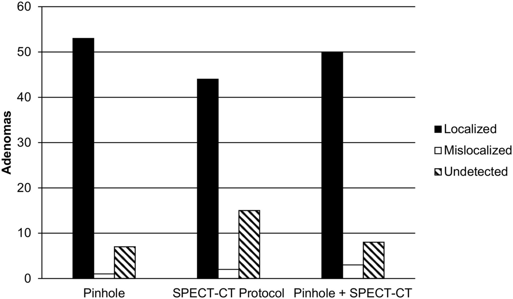
- FIGURE 1.
Performance of each of 3 protocols is shown for localizing 61 parathyroid adenomas in 59 patients. Data represent average of results for 2 observers. All 3 protocols included perfectly coregistered subtraction images created by subtracting 123I images from 99mTc-sestamibi images, plus anterior parallel-hole collimator image of neck and upper chest.
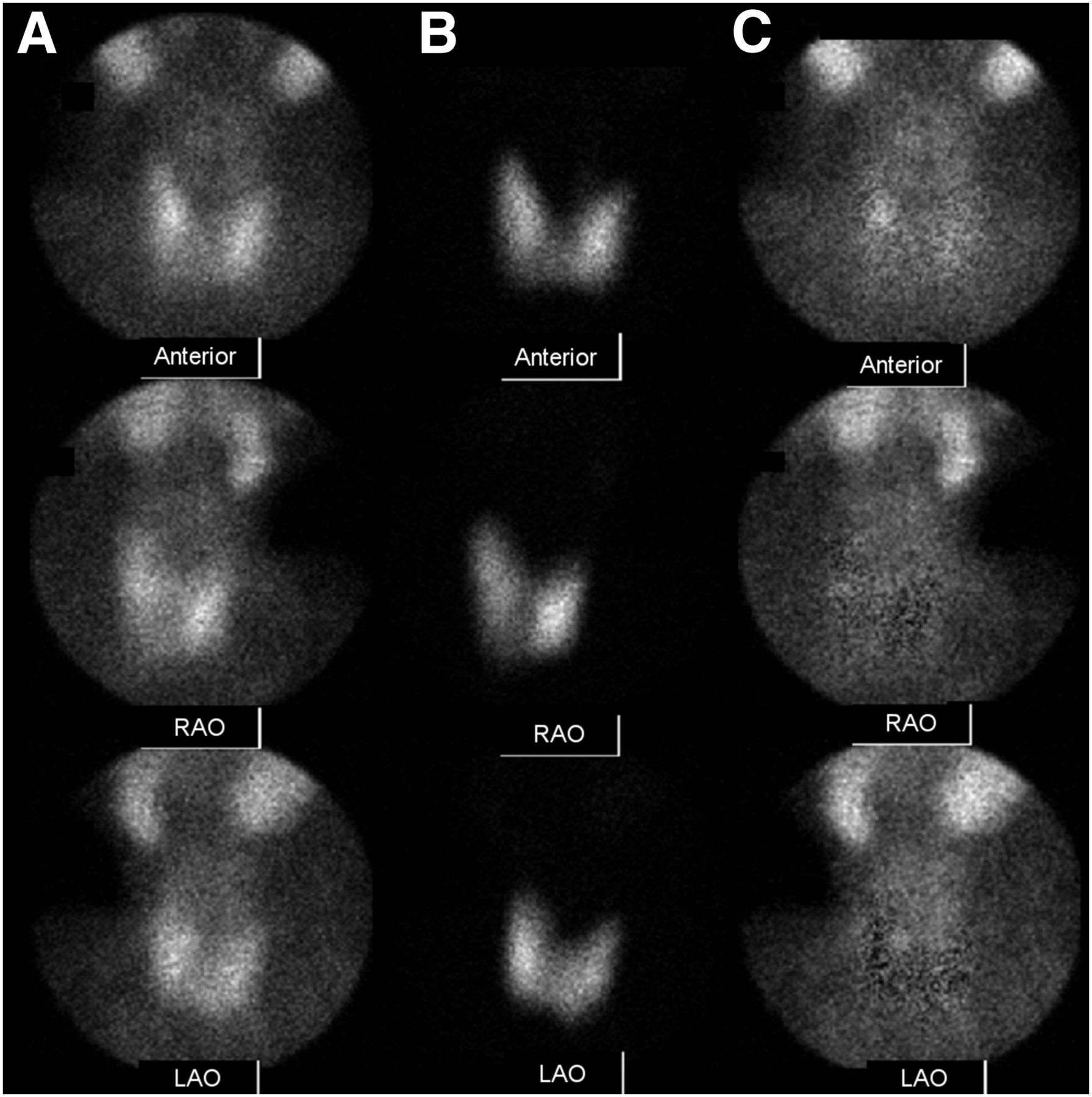
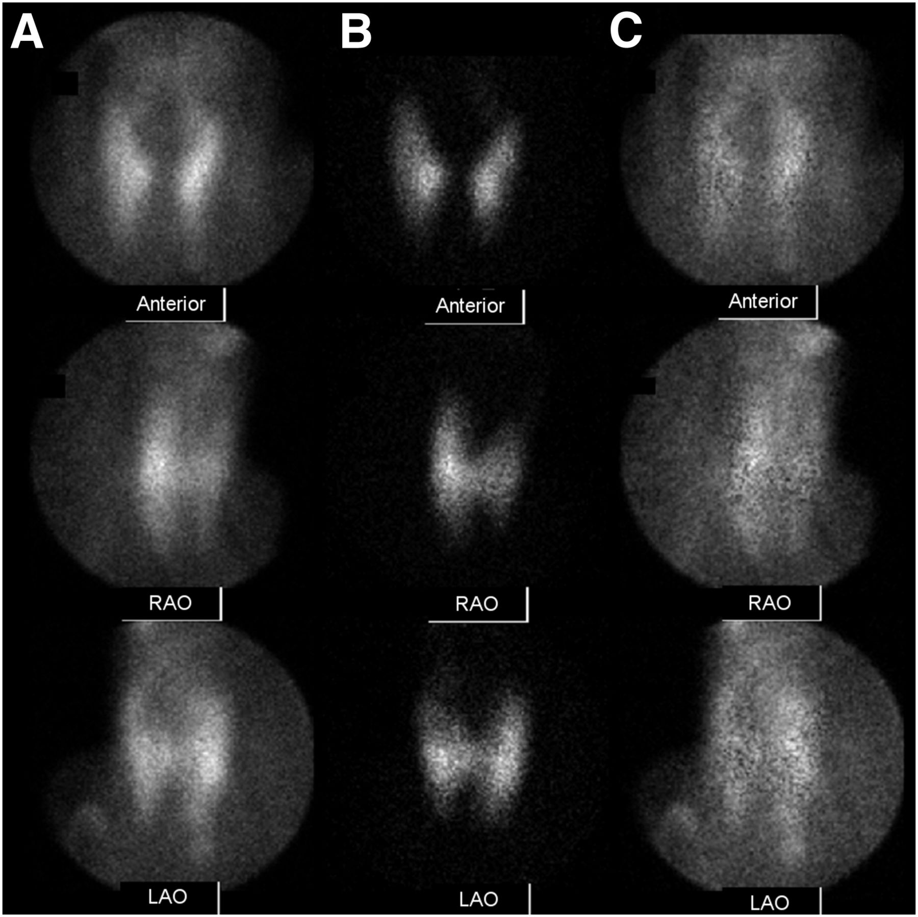
- FIGURE 2.
Anterior and anterior oblique pinhole images for 99mTc-sestamibi (A), 123I (B), and subtraction images (C) from pinhole protocol. Subtraction images show persistent focus of activity consistent with parathyroid adenoma just posterior to mid to superior pole of right lobe of thyroid. Both observers scored this finding as 2—that is, a probable adenoma.
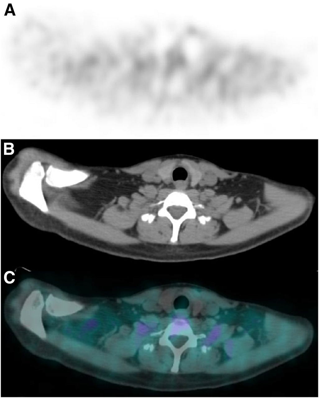
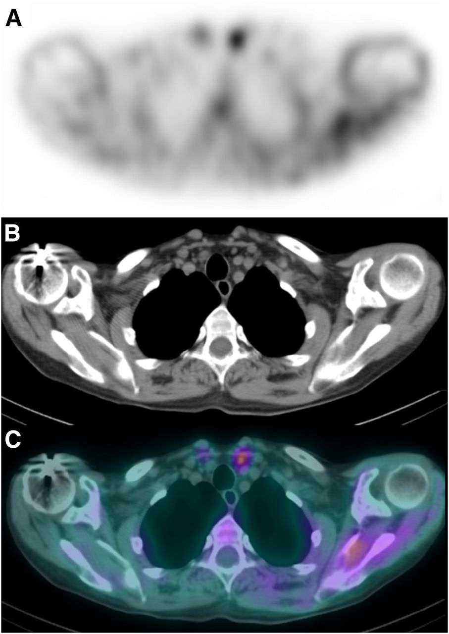
- FIGURE 3.
Selected perfectly coregistered subtraction SPECT image (A), corresponding CT image (B), and fused image (C) from SPECT/CT protocol. None of the maximum-intensity-projection images are shown. No focus of postsubtraction activity was seen in any image. Both observers scored this image set as 0—that is, no adenoma seen. However, in the CT image there is small low-density structure along posterior aspect of right lobe of thyroid consistent with location of focus of activity in pinhole protocol images. Surgery confirmed right superior parathyroid adenoma. This is an example of parathyroid adenoma that was better seen in pinhole protocol than in SPECT/CT protocol.
- FIGURE 4.
Anterior and anterior oblique pinhole images for 99mTc-sestamibi (A), 123I (B), and subtraction images (C) from pinhole protocol. Subtraction images show possible mild focus of activity consistent with parathyroid adenoma just inferior to inferior pole of left lobe of thyroid. The 2 observers scored this finding as 1 and 0—that is, possible adenoma and no adenoma seen.
- FIGURE 5.
Selected perfectly coregistered subtraction SPECT image (A), corresponding CT image (B), and fused image (C) from SPECT/CT protocol. None of the maximum-intensity-projection images are shown. Focus of postsubtraction activity was seen inferior to inferior tip of left thyroid. The 2 observers scored this finding as 3 and 2—that is, definite adenoma and probable adenoma. Surgery revealed ectopic parathyroid adenoma in superior left thymus. This is an example of parathyroid adenoma that was better seen in SPECT/CT protocol than in pinhole protocol. Patient had had 2 previous right parathyroidectomies.


Tables
Degree of certainty of location Protocol* 0 1 2 3 Pinhole Observer A 11 8 (2) 20 (1) 22 (1) Observer B 4 9 (10) 17 (2) 31 (1) Average 7.5 8.5 (6) 18.5 (1.5) 26.5 (1) SPECT/CT Observer A 21 7 (1) 8 25 Observer B 17 4 (1) 13 (1) 27 (2) Average 19 5.5 (1) 10.5 (0.5) 26 (1) Pinhole + SPECT/CT Observer A 17 5 (1) 10 (1) 29 Observer B 5 9 (6) 14 (4) 33 Average 11 7 (3.5) 12 (2.5) 31 ↵* Both pinhole and SPECT/CT protocols involved dual-tracer simultaneous acquisition with subtraction.
0= no adenomas seen; 1 = possible adenoma; 2 = probable adenoma; 3 = definite adenoma.
Numbers in parentheses indicate number of localizations that were incorrect (false-positives).
Localization success Protocol* Adenomas Correct 95% CI Pinhole Observer A 50/61 82% 73–91 Observer B 57/61 93% 90–99 Average 53.5/61 88% 84–92 SPECT/CT Observer A 40/61 66% 54–78 Observer B 44/61 72% 61–83 Average 42/61 69% 63–75 Pinhole + SPECT/CT Observer A 43/61 70% 64–76 Observer B 56/61 92% 89–95 Average 49.5/61 81% 76–86 ↵* Both pinhole and SPECT/CT protocols involved dual-tracer simultaneous acquisition with subtraction.
CI = exact binomial confidence interval.
Calculations are on per-adenoma basis. There were 61 adenomas in 59 patients.
Protocol rating Observer Pinhole better SPECT/CT better Equal A 19 (42%) 4 (9%) 22 (49%) B 14 (26%) 20 (34%) 23 (40%)


